. 2016 Oct 31;16(11):1826.
doi: 10.3390/s16111826.

New Flexible Silicone-Based EEG Dry Sensor Material Compositions Exhibiting Improvements in Lifespan, Conductivity, and Reliability

Affiliations

New Flexible Silicone-Based EEG Dry Sensor Material Compositions Exhibiting Improvements in Lifespan, Conductivity, and Reliability

Yi-Hsin Yu et al. Sensors (Basel). .

Abstract

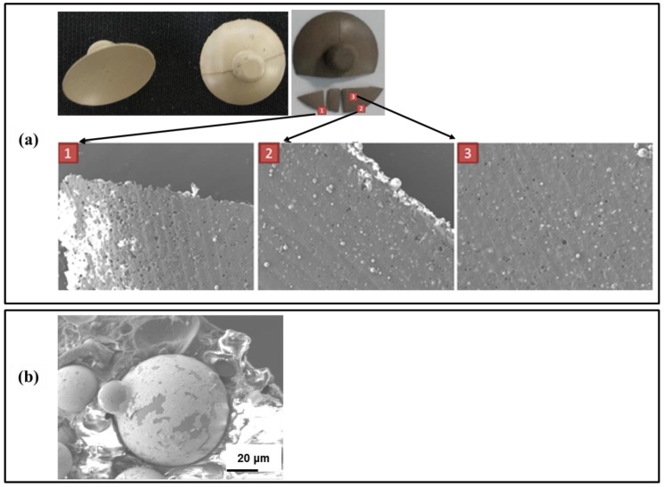
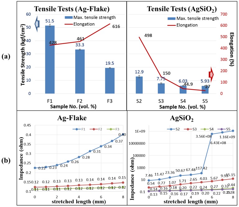
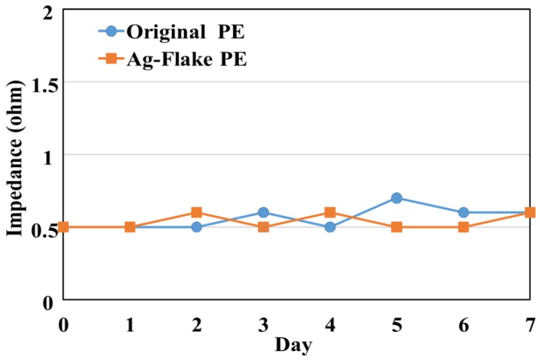
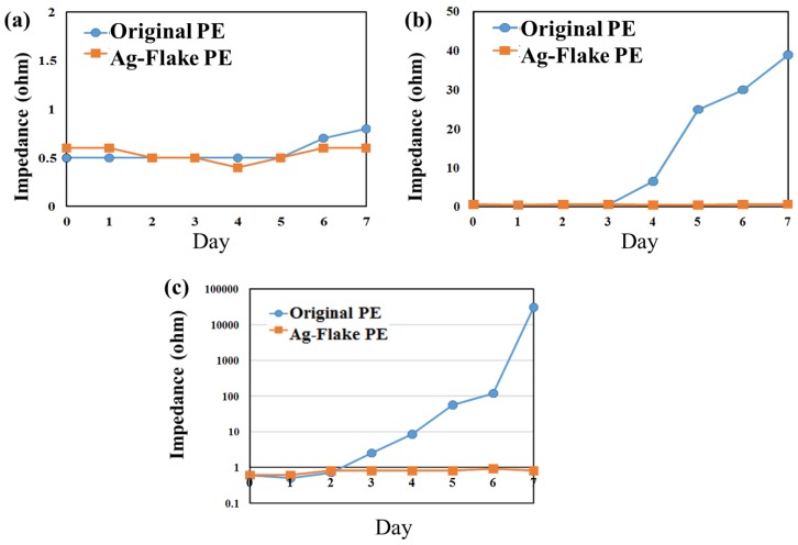
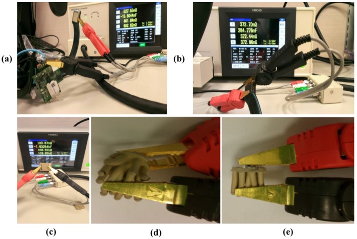
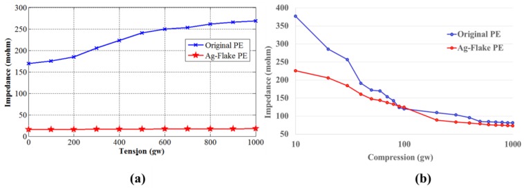
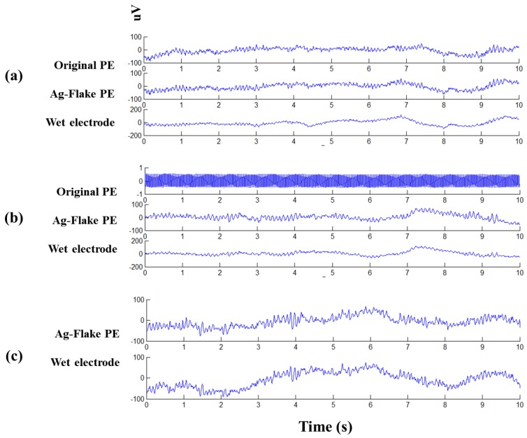
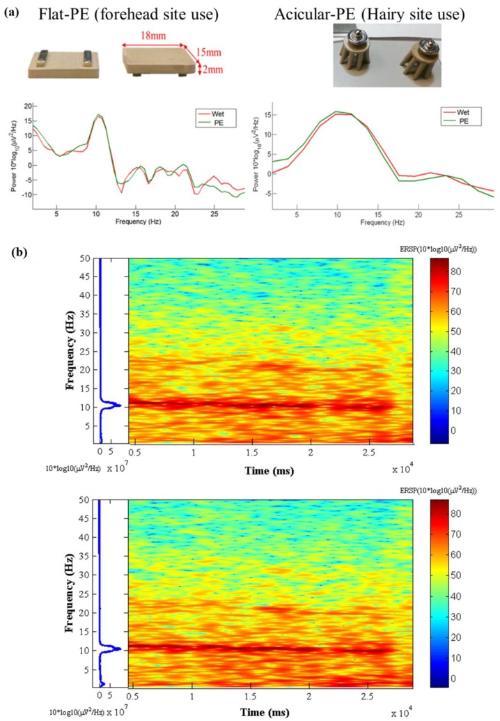
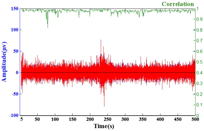
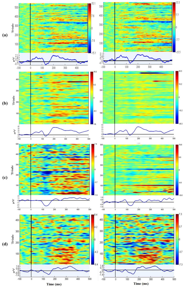
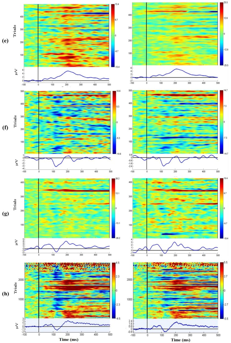
This study investigates alternative material compositions for flexible silicone-based dry electroencephalography (EEG) electrodes to improve the performance lifespan while maintaining high-fidelity transmission of EEG signals. Electrode materials were fabricated with varying concentrations of silver-coated silica and silver flakes to evaluate their electrical, mechanical, and EEG transmission performance. Scanning electron microscope (SEM) analysis of the initial electrode development identified some weak points in the sensors' construction, including particle pull-out and ablation of the silver coating on the silica filler. The newly-developed sensor materials achieved significant improvement in EEG measurements while maintaining the advantages of previous silicone-based electrodes, including flexibility and non-toxicity. The experimental results indicated that the proposed electrodes maintained suitable performance even after exposure to temperature fluctuations, 85% relative humidity, and enhanced corrosion conditions demonstrating improvements in the environmental stability. Fabricated flat (forehead) and acicular (hairy sites) electrodes composed of the optimum identified formulation exhibited low impedance and reliable EEG measurement; some initial human experiments demonstrate the feasibility of using these silicone-based electrodes for typical lab data collection applications.

Keywords: electroencephalography (EEG); scanning electron microscope (SEM); silicone-based dry sensors.


Conflict of interest statement

The authors declare no conflict of interest.

Figures

Figure 1
Figure 1
Pictures of the tensile testing apparatus using a pulley along with calibration weights. The impedance was measured by a HIOKI LCR Meter IM3533-01 (HIOKI, Nagano, Japan). Each sample was cut into the pieces 1 cm × 7 cm × 0.1 cm.
Figure 2
Figure 2
(a) Photographs of the top and reverse side of a failed electrode along with SEM images obtained from the labeled locations. The images reveal numerous holes appear on the polymer electrode surface, especially on the edge, after several applications. The nearly spherical holes and the presence of Ag/SiO2 particles on the surface seem to indicate particle pull-out; and (b) a high-magnification SEM image of the particle surface that seems to indicate that the silver coating is no longer uniform and that the silver is ablated from the silica surface during use.
Figure 3
Figure 3
(a) Mechanical properties as a function of composition for both Ag-flake and Ag/SiO2; (b) Strain-dependent impedance measurements obtained from compositions containing Ag-flake and Ag/SiO2.
Figure 4
Figure 4
The variation of surface impedance of dry PEs in the temperature and humidity test with 85% RH at 80 °C for one week.
Figure 5
Figure 5
The variations of dry PEs (original PE (Ag/SiO2) and Ag-flake PE) surface impedance after salt spray testing with the saline concentrations of (a) 0.3%; (b) 3.0%; and (c) 5.0% for one week.
Figure 6
Figure 6
(a,b) Testing of the impedance of flat Neurosky electrodes; (c) Flat silicone-based electrode; (d) The bent acicular silicone-based dry sensor; (e) The impedance of a normal acicular silicone-based dry sensor.
Figure 7
Figure 7
(a) Impedance tests of two PEs in different tensile stresses with a frequency of 40 Hz; (b) Top-bottom impedance tests of two PEs in different compressions with a frequency of 40 Hz.
Figure 8
Figure 8
Synchronic EEG tracings of eyes closed conditions for different electrodes (original PE, Ag-flake PE, and wet electrode) underwent the salt spray test in (a) 0.3%; (b) 3.0%; and (c) 5.0% saline solution.
Figure 9
Figure 9
(a) Alpha rhythm tests. The left diagram is the forehead EEG data and the right is the occipital EEG data. Wet sensors were located at Fp1and Oz and the proposed dry PEs were located near Fp1 and Oz; (b) Time-frequency analysis of the EEG signals (Oz) for 0~50 Hz spectrum. The top diagram is the wet sensor’s result located at Oz, and the bottom diagram is the proposed PE’s result located near Oz.
Figure 10
Figure 10
A comparison of the EEG signals recorded by a wet electrode (red curve) and the acicular Ag-flake PE (blue curve). The green curve shows the correlation of these two signals over a sliding one-second window and the black curve shows the root mean square error (RMSE) of these two signals. The average correlation between the blue and red lines is high (~97.85%), the maximum of the RMSE is 0.69 (μv) and the average of RMSE is 0.13 (μv). This test acquired 700 s of EEG data, and a sample of the EEG recorded data (5~500 s) was taken for analysis.
Figure 11
Figure 11
(ag) The P200 phenomena were presented in the ERPs for both wet and dry channels for each subject; (h) shows the grand mean P200 components were detected by the subjects’ EEG data.
Figure 11
Figure 11
(ag) The P200 phenomena were presented in the ERPs for both wet and dry channels for each subject; (h) shows the grand mean P200 components were detected by the subjects’ EEG data.

References

    1. Langenhove L.V. Smart Textiles for Medicine and Healthcare: Materials, Systems and Applications. Elsevier; Amsterdam, The Netherlands: 2007.
    1. Paul L., Nunez R.S. Electric Fields of the Brain: The Neurophysics of EEG. Oxford University Press; Oxford, UK: 2006.
    1. Moser E., Meyerspeer M., Fischmeister F.P.S., Grabner G., Bauer H., Trattnig S. Windows on the human body—In vivo high-field magnetic resonance research and applications in medicine and psychology. Sensors. 2010;10:5724–5757. doi: 10.3390/s100605724. - DOI - PMC - PubMed
    1. Gevins A., Le J., Martin N.K., Brickett P., Desmond J., Reutter B. High-resolution EEG-124-channel recording, spatial deblurring and mri integration methods. Electroen. Clin. Neuro. 1994;90:337–358. doi: 10.1016/0013-4694(94)90050-7. - DOI - PubMed
    1. Lin C.T., Chen S.A., Chiu T.T., Lin H.Z., Ko L.W. Spatial and temporal EEG dynamics of dual-task driving performance. J. Neuroeng. Rehabil. 2011;8:1. doi: 10.1186/1743-0003-8-11. - DOI - PMC - PubMed