. 2016 Nov 3;7(11):e2449.
doi: 10.1038/cddis.2016.329.

Extrasynaptic NMDA receptor-induced tau overexpression mediates neuronal death through suppressing survival signaling ERK phosphorylation

Affiliations

Extrasynaptic NMDA receptor-induced tau overexpression mediates neuronal death through suppressing survival signaling ERK phosphorylation

Xu-Ying Sun et al. Cell Death Dis. .

Abstract

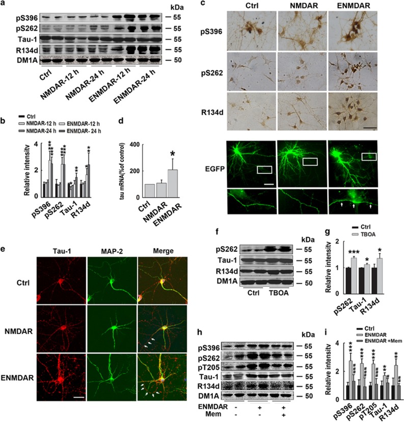
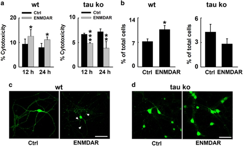
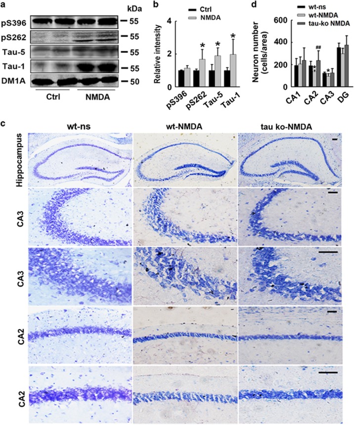
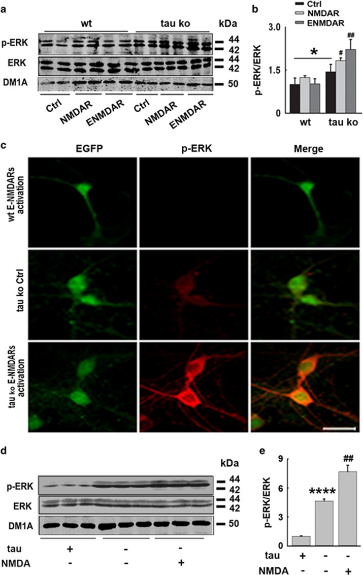
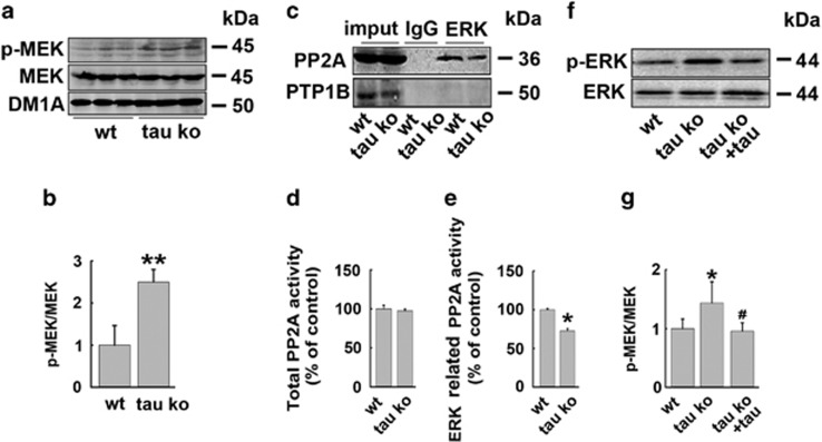
Intracellular accumulation of the hyperphosphorylated tau is a pathological hallmark in the brain of Alzheimer disease. Activation of extrasynaptic NMDA receptors (E-NMDARs) induces excitatory toxicity that is involved in Alzheimer's neurodegeneration. However, the intrinsic link between E-NMDARs and the tau-induced neuronal damage remains elusive. In the present study, we showed in cultured primary cortical neurons that activation of E-NMDA receptors but not synaptic NMDA receptors dramatically increased tau mRNA and protein levels, with a simultaneous neuronal degeneration and decreased neuronal survival. Memantine, a selective antagonist of E-NMDARs, reversed E-NMDARs-induced tau overexpression. Activation of E-NMDARs in wild-type mouse brains resulted in neuron loss in hippocampus, whereas tau deletion in neuronal cultures and in the mouse brains rescued the E-NMDARs-induced neuronal death and degeneration. The E-NMDARs-induced tau overexpression was correlated with a reduced ERK phosphorylation, whereas the increased MEK activity, decreased binding and activity of ERK phosphatase to ERK, and increased ERK phosphorylation were observed in tau knockout mice. On the contrary, addition of tau proteins promoted ERK dephosphorylation in vitro. Taking together, these results indicate that tau overexpression mediates the excitatory toxicity induced by E-NMDAR activation through inhibiting ERK phosphorylation.


Figures

Figure 1
Figure 1
Activation of extrasynaptic but not synaptic NMDA receptors increases tau expression with neurodegeneration in cortical neurons. (a) Rat primary cortical neurons (12–14 DIV) were incubated with Bic (50 μM) /4-AP (2.5 mM) to activate synaptic NMDA receptors for 12 or 24 h (herein used as NMDAR). To specifically induce extrasynaptic NMDA receptors (E-NMDAR) activation, neurons were incubated with Bic (50 μM)/4-AP (2.5 mM) for 2 min, after wash, open NMDA receptors blocker MK-801 (10 μM) was administrated for another 2 min to block synaptic NMDA receptors, at last NMDA (30 μM) and glycine (10 μM) were used to selectively activate E-NMDARs for 12 or 24 h (herein used as E-NMDAR). Total tau and phosphorylated tau at Ser396 and Ser262 sites, and dephosphorylated tau level at Tau-1(Ser198/199/202) sites were detected by western blotting. (b) Quantitative analysis of the blots in (a). Total, phosphorylated and dephosphorylated tau levels were normalized with DM1A. *P<0.05, **P<0.01 and ***P<0.001 versus control neurons, n=8, N=4 independent cultures. (c) Upper panel: primary cortical rat neurons (12–14 DIV) were treated with synaptic or extrasynaptic NMDA receptors activation protocols as previously described for 24 h. Images from DAB immunocytochemistry staining with pS396, pS262, R134d (total tau) antibodies were acquired under a confocal microscope. Scale bar=100 μm; Bottom panel: primary cortical rat neurons (11 DIV) were transfected with surface EGFP to visualize the neuronal morphology, 48 h later, neurons were treated with synaptic or extrasynaptic NMDAR activation protocols for 24 h. At the end of treatment, cells were fixed with 4% paraformaldehyde and observed under the fluorescence microscope. Amplified images of axons in the rectangle were showed at the bottom. White arrows showed puncta-like enhanced EGFP fluorescence in the axon. Scale bar=20 μm. (d) Primary cortical rat neurons (12–14 DIV) were treated with synaptic or extrasynaptic NMDA receptors activation protocols for 6 h, total RNA was extracted from neuronal cultures. Real-time PCR analysis was performed to quantify relative expression of tau mRNA in the different groups. The expression level of tau gene was analyzed according to the Ct method (comparative Ct method), in which Ct is the threshold cycle value and normalized by β-actin. Histograms represent means±S.D., and statistical analysis was performed by ANOVA (n=6, N=3 independent cultures; *P<0.05 versus control). (e) Primary cortical mouse neurons (12–14 DIV) were treated with synaptic or extrasynaptic NMDA receptors activation protocols for 24 h, immunofluorescence staining images with Tau-1 (red) and MAP-2 (dendrite marker, green) were acquired under a confocal microscope. Scale bar=50 μm. White arrows showed the axons that were only stained by Tau-1 antibody. (f) Rat primary cortical neurons (12–14 DIV) were incubated with Bic/4-AP for 2 min to activate synaptic NMDA receptors, then incubated with NMDA receptors blocker MK-801 (10 μM) for another 2 min to block synaptic NMDAR, at last glutamate transporters blocker DL-TBOA (30 μM) was used to selectively induce extrasynaptic NMDA receptors activation for 24 h. Total (R134d), phosphorylated (pS262) and dephosphorylated tau (Tau-1) levels were detected by western blotting. (g) Quantitative analysis of the blots in (f). Total, phosphorylated and dephosphorylated tau levels were normalized with DM1A. *P<0.05, ***P<0.001 versus control group, n=6, N=3 independent cultures. (h) Rat primary cortical neurons were treated as previously described to activate E-NMDARs for 24 h, with or without pretreatment of extrasynaptic NMDA receptor antagonist memantine (1 μM) for 30 min, total tau and phosphorylated tau levels at Ser396, Ser262 and Thr205, and dephosphorylated tau level at Tau-1 (Ser198/199/202) sites were detected by western blotting. (i) Quantitative analysis of the blots in (h). Total, phosphorylated and dephosphorylated tau levels were normalized with DM1A. **P<0.01, ***P<0.001 versus control group; #P<0.05, ##P<0.01 and ###P<0.001 versus extrasynaptic NMDAR activation group, n=6, N=3 independent cultures
Figure 2
Figure 2
Tau deletion protects neurons from E-NMDAR-triggered neuronal death and degeneration in cortical neurons. (a) Primary cultured mouse cortical neurons at DIV 12–14 were subjected to extrasynaptic NMDAR activation for 12 or 24 h. The level of LDH released into the culture medium was determined by measuring the decrease in absorbance at 490 nm resulting from the oxidation of NADH. LDH levels in wildtype (Wt, left) or tau knockout (tau Ko, right) neurons treated with DMSO (Ctrl) or extrasynaptic NMDAR activation protocols (E-NMDAR) during the indicated times (12 or 24 h). *P<0.05, **P<0.01 and ***P<0.001 versus control group, n=20, N=3 independent cultures. (b) Quantification of apoptotic cells in Wt (left) or tau Ko (right) neuron cultures exposed to extrasynaptic treatment for 24 h. Quantification of the cell populations was achieved on four independent cultures. About 6000 cells were evaluated in each group. *P<0.05 versus control group. (c) Wt mouse primary cortical neurons at 5 DIV were transfected with EGFP by lentivirus. At 12 DIV, neurons were subjected to extrasynaptic NMDAR activation for 24 h. Morphological changes of EGFP-labeled neurons treated with DMSO (Ctrl) or the extrasynaptic NMDA receptor activating protocol for 24 h (E-NMDAR). Images were acquired by confocal microscopy. White arrows showed abnormal neurodegeneration. (d) Representative neuron images from tau Ko mouse cortical neurons treated with DMSO (Ctrl) or E-NMDARs activation protocol (E-NMDAR) for 24 h, neurons were directly fixed and visualized under the fluorescence microscope. Scale bar=50 μm
Figure 3
Figure 3
Tau deletion protects neurons from E-NMDARs-triggered neuronal death in mouse hippocampus. (a) Wild-type (Wt) C57 mice were injected with saline (Ctrl) or NMDA (60 mM, 2 μl) into the hippocampus, 24 h later, hippocampi were isolated and homogenized, total tau (Tau-5), dephosphorylated tau levels at Tau-1 (Ser198/199/202) sites and phosphorylated tau levels at Ser396 and Ser262 sites were detected by western blotting. (b) Quantitative analysis of the blots in (a). Total, phosphorylated and dephosphorylated tau levels were normalized with DM1A. *P<0.05 versus saline-injected control mice, n=3 per group. (c) Wt or tau Ko mice were injected with saline (NS) or NMDA (60 mM, 2 μl) into the hippocampus. Twenty-four hours later, half brain of the mice was fixed. Neurons in the hippocampus were stained by Nissl staining. Representative images from the hippocampus, scale bars=50 μm. (d) Quantitative analysis of the neuron number in CA1, CA2, CA3 and DG regions of hippocampus, *P<0.05 versus Wt NS group. ##P<0.01 versus NMDA-treated Wt mice (n=3 mice per group, cells in 10 brain slices were counted for each animal)
Figure 4
Figure 4
Tau deletion restores ERK activation in E-NMDAR-activated primary mouse cortical neurons and hippocampus. (a) Wt- or tau-deleted mouse neurons were cultured and treated with synaptic or extrasynaptic NMDAR activating protocols for 24 h, respectively. The total protein levels and active forms of ERK were detected by western blotting. (b) Quantitative analysis of the protein levels in (a), *P<0.05 versus wild-type neurons, #P<0.05 versus tau Ko control neurons, ##P<0.01 versus tau Ko control neurons, n=6, N=3 independent cultures. (c) Wt or tau Ko mouse neurons were cultured and treated with or without E-NMDAR-activating protocols for 24 h, p-ERK and EGFP staining were acquired by confocal microscopy, for Wt neurons, the cells were transfected with EGFP lentivirus to be visualized. Scale bar=50 μm. (d) Wt or tau Ko mice were injected with saline (NS) or NMDA (60 mM, 2 μl) into the hippocampus. Twenty-four hours later, left part of hippocampi was isolated and homogenized, total protein levels and active forms of ERK were detected by western blotting. (e) Quantitative analysis of the ERK levels in (d), ****P<0.0001 versus Wt neurons, ##P<0.01 versus tau Ko control neurons, n=3 mice per group
Figure 5
Figure 5
Tau protein suppresses ERK signaling pathway through inhibiting MEK and promoting ERK dephosphorylation by recruiting ERK phosphatase. (a) Wt and tau Ko mice hippocampi were homogenized. The total protein levels and active forms of MEK were detected by western blotting. (b) Quantitative analysis of the p-MEK level in (a), **P<0.01 versus Wt mice, n=3 mice per group. (c) Wt and tau Ko mice hippocampal lysates were immunoprecipitated with anti-ERK antibody, then detected with anti-PP2A and PTP1B antibodies through western blotting. n=3 mice per group. (d) Wt and tau Ko mice hippocampi were homogenized and subjected to PP2A activity assay, n=3 mice per group. (e) Wt and tau Ko mice hippocampal lysates were co-immunoprecipitated with anti-ERK antibody, and then were subjected to PP2A activity assay, *P<0.05 versus Wt mice, n=3 mice per group. (f) His-tagged phosphorylated ERK protein was incubated for 10 min with Wt or tau Ko hippocampal homogenates in the presence of U0126 (10 μM), with or without the addition of recombinant human tau441 protein. Then anti-his antibody and protein G were added to the lysates overnight at 4 °C. The phosphorylated p-ERK and total ERK were analyzed by western blotting. (g) Quantitative analysis of the ERK phosphorylation levels in (f), *P<0.05 versus Wt mouse brain homogenates, #P<0.05 versus tau Ko mouse brain homogenates without tau441 protein addition, n=5 mouse hippocampi per group

Similar articles

Cited by

References

    1. Khatoon S, Grundke-Iqbal I, Iqbal K. Levels of normal and abnormally phosphorylated tau in different cellular and regional compartments of Alzheimer disease and control brains. FEBS Lett 1994; 351: 80–84. - PubMed
    1. Khatoon S, Grundke-Iqbal I, Iqbal K. Brain levels of microtubule-associated protein tau are elevated in Alzheimer's disease: a radioimmuno-slot-blot assay for nanograms of the protein. J Neurochem 1992; 59: 750–753. - PubMed
    1. Barton AJ, Harrison PJ, Najlerahim A, Heffernan J, McDonald B, Robinson JR et al. Increased tau messenger RNA in Alzheimer's disease hippocampus. Am J Pathol 1990; 137: 497–502. - PMC - PubMed
    1. Yasojima K, McGeer EG, McGeer PL. Tangled areas of Alzheimer brain have upregulated levels of exon 10 containing tau mRNA. Brain Res 1999; 831: 301–305. - PubMed
    1. Obulesu M, Venu R, Somashekhar R. Tau mediated neurodegeneration: an insight into Alzheimer's disease pathology. Neurochem Res 2011; 36: 1329–1335. - PubMed

Publication types

MeSH terms

Substances