. 2016 Nov 3;7(11):e2454.
doi: 10.1038/cddis.2016.361.

Downregulation of microRNA-27b-3p enhances tamoxifen resistance in breast cancer by increasing NR5A2 and CREB1 expression

Affiliations

Downregulation of microRNA-27b-3p enhances tamoxifen resistance in breast cancer by increasing NR5A2 and CREB1 expression

Jiang Zhu et al. Cell Death Dis. .

Abstract

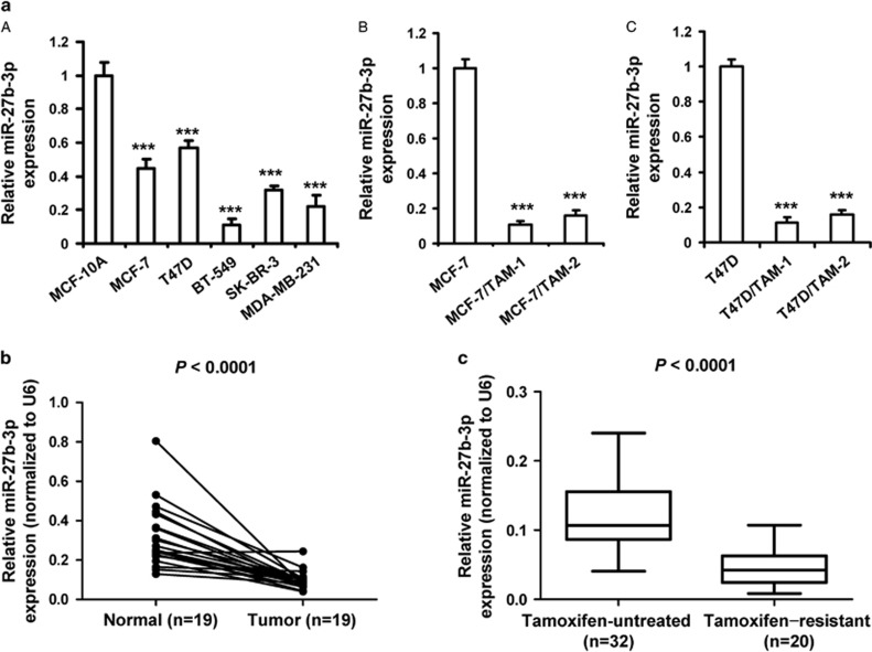
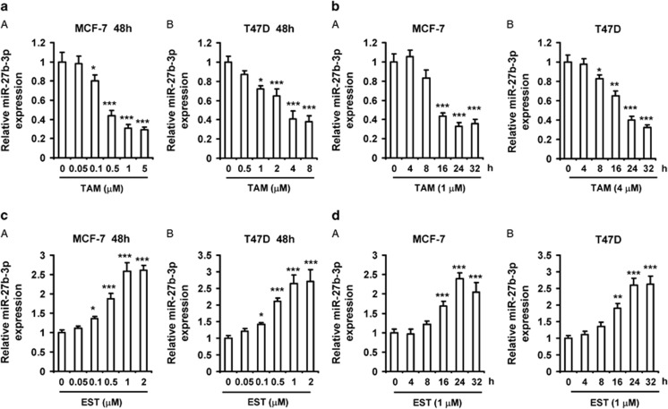
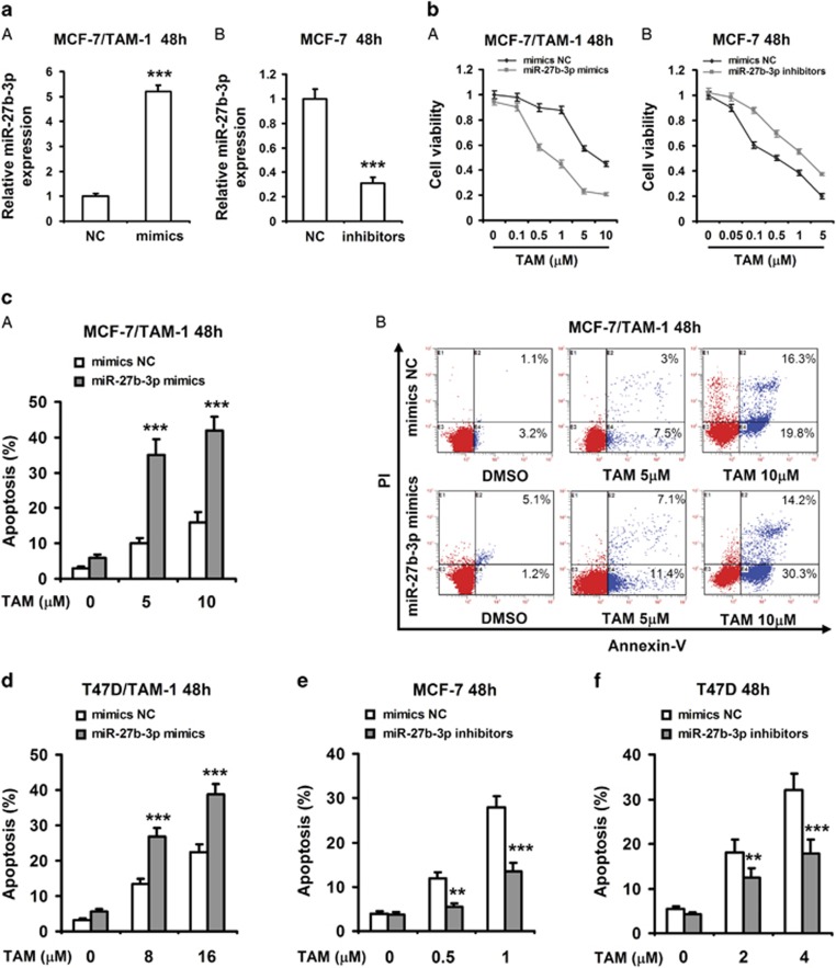
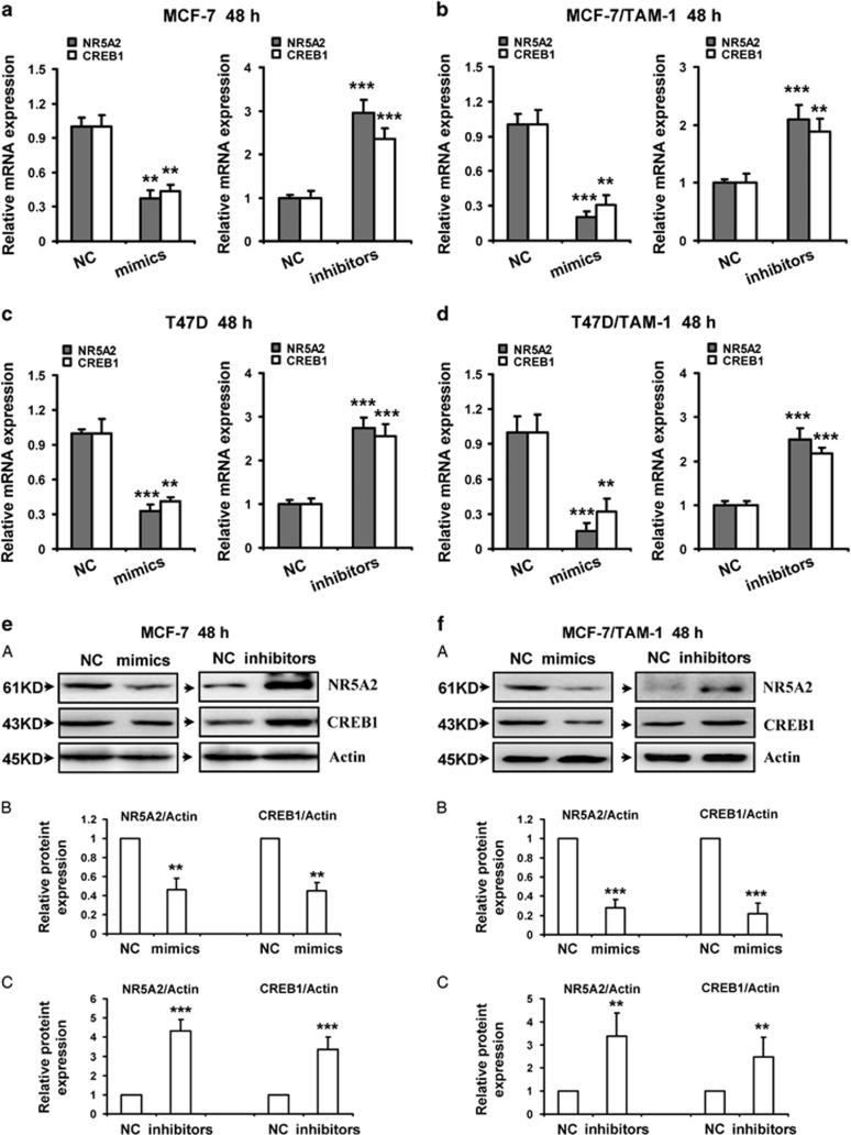
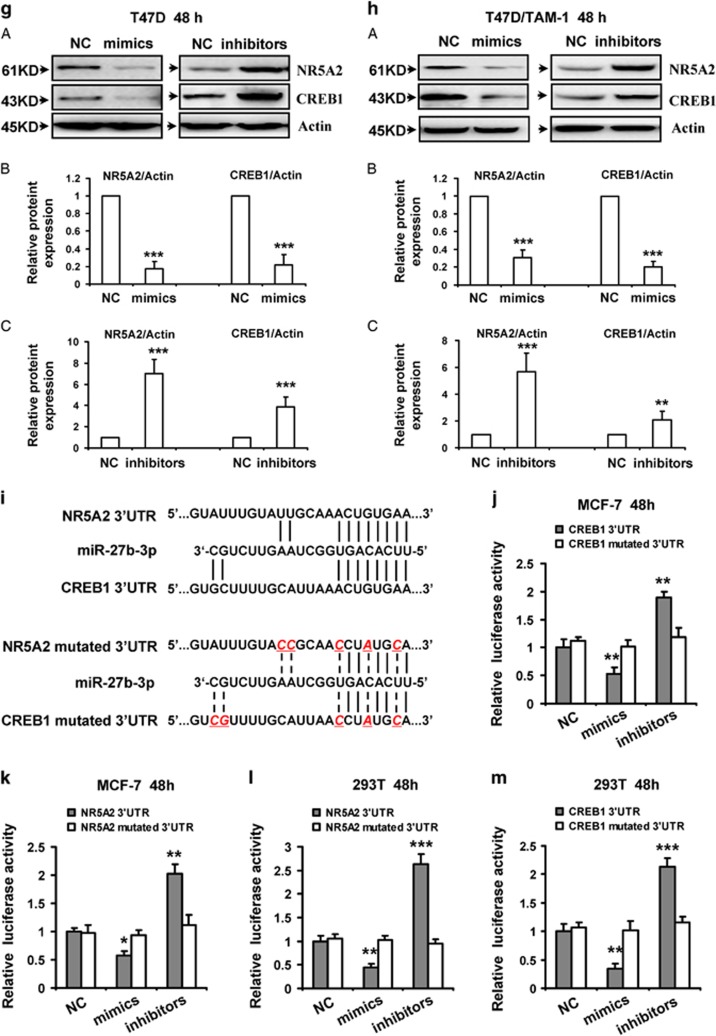
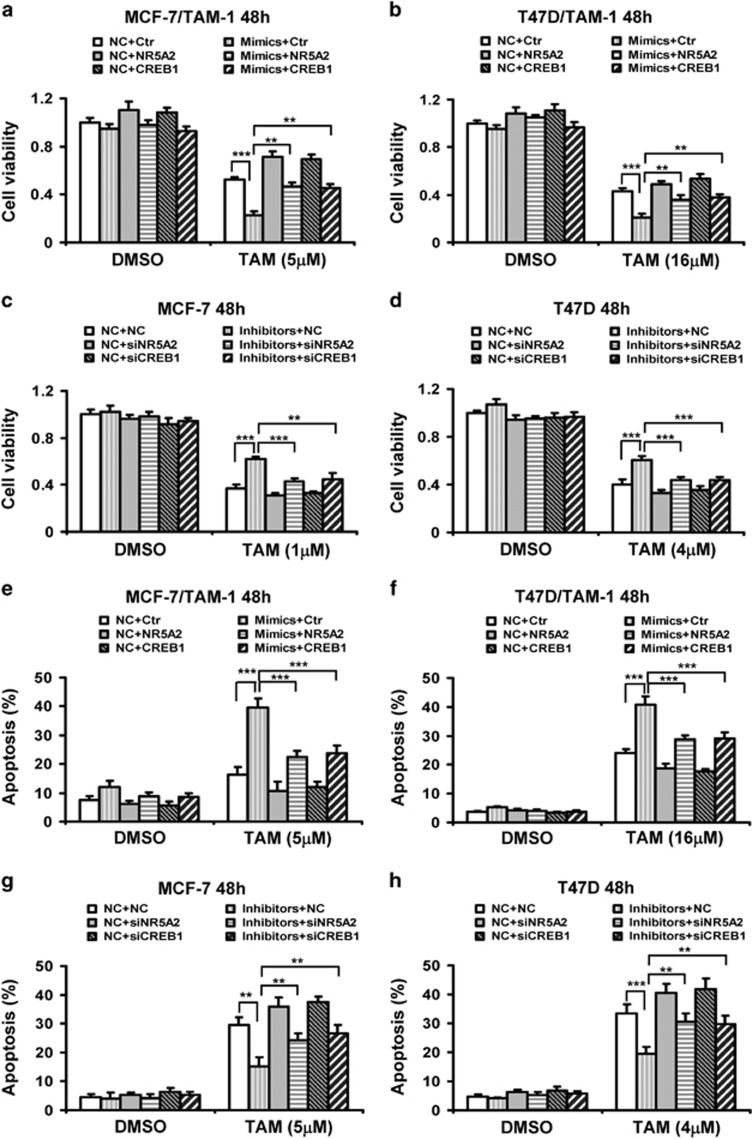
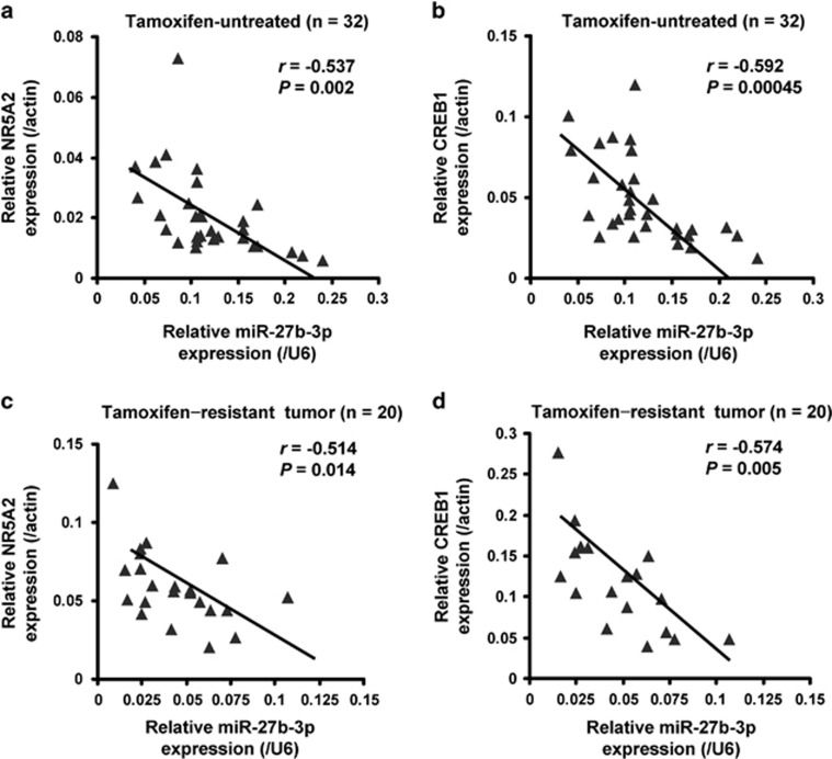
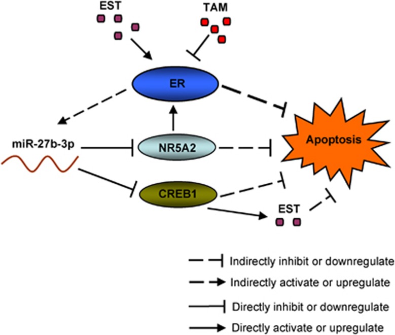
Estrogen-dependent breast cancer is often treated with the aromatase inhibitors or estrogen receptor (ER) antagonists. Tamoxifen as a major ER antagonist is usually used to treat those patients with ERα-positive breast cancer. However, a majority of patients with ERα positive fail to respond to tamoxifen due to the presence of intrinsic or acquired resistance to the drug. Altered expression and functions of microRNAs (miRNAs) have been reportedly associated with tamoxifen resistance. In this study, we investigated the role of miR-27b-3p in resistance of breast cancer to tamoxifen. MiR-27b-3p levels were remarkably reduced in the tamoxifen-resistant breast cancer cells compared with their parental cells. In addition, miR-27b-3p was also significantly downregulated in breast tumor tissues relative to adjacent non-tumor tissues. Moreover, the expression levels of miR-27b-3p were lower in the breast cancer tissues from tamoxifen-resistant patients compared with that from untreated-tamoxifen patients. Notably, tamoxifen repressed miR-27b-3p expression, whereas estrogen induced miR-27b-3p expression in breast cancer cells. Besides, we provided experimental evidences that miR-27b-3p enhances the sensitivity of breast cancer cells to tamoxifen in vitro and in vivo models. More importantly, we validated that miR-27b-3p directly targeted and inhibited the expression of nuclear receptor subfamily 5 group A member 2 (NR5A2) and cAMP-response element binding protein 1 (CREB1) and therefore augmented tamoxifen-induced cytotoxicity in breast cancer. Lastly, miR-27b-3p levels were found to be significantly negatively correlated with both NR5A2 and CREB1 levels in breast cancer tissues. Our findings provided further evidence that miR-27b-3p might be considered as a novel and potential target for the diagnosis and treatment of tamoxifen-resistant breast cancer.


Figures

Figure 1
Figure 1
Downregulation of miR-27b-3p in tamoxifen-resistant breast cancer. (a) (A) MiR-27b-3p expression in different breast cell lines as measured by RT-PCR analysis and relative to their expression levels in MCF-10A cells. (B and C) MiR-27b-3p expression in tamoxifen-resistant MCF-7 and T47D cells as measured by RT-PCR analysis and relative to their expression levels in parental MCF-7 and T47D cells. The experiments were repeated three times. Data represent mean±S.D. ***P<0.001. (b) The expression levels of miR-27b-3p in tumor tissues compared with adjacent normal tissues in 19 breast cancer patients, P<0.001 by paired t-test. (c) The expression levels of miR-27b-3p in 32 tamoxifen untreated breast cancer tissues and 20 tamoxifen-resistant breast cancer tissues
Figure 2
Figure 2
MiR-27b-3p is repressed by tamoxifen and induced by estrogen in breast cancer cells. (a) Tamoxifen inhibited miR-27b-3p expression in a dose-dependent manner. MCF-7 and T47D cells were treated with increasing amounts of 4-hydroxytamoxifen (TAM) for 48 h. (b) Tamoxifen inhibits miR-27b-3p expression in a time-dependent manner. MCF-7 and T47D cells were treated with 1 and 4 μM of TAM for different time, respectively. (c) Estrogen induced miR-27b-3p expression in a dose-dependent manner. MCF-7 and T47D cells were treated with increasing amounts of 17β-estradiol (EST) for 48 h. (d) Estrogen induced miR-27b-3p expression in a time-dependent manner. MCF-7 and T47D cells were treated with 1 μM of EST for different time, respectively. RNA were then collected and subjected to RT-PCR analysis of gene expression. The levels of miR-27b-3p were presented as fold change compared with the levels in control cells. Columns, means of three determinations; bars, S.D. Results shown are representative of three independent experiments. *P<0.05; **P<0.01; ***P<0.001, compared with DMSO-treated cells
Figure 3
Figure 3
MiR-27b-3p enhances breast cancer cells apoptosis induced by tamoxifen. (a) MCF-7/TAM-1 and MCF-7 cells were transfected with miR-27b-3p mimics (A) or miR-27b-3p inhibitors (B) and the negative control (NC) for 48 h, respectively. RT-PCR was performed to detect the expression of miR-27b-3p. (b) MCF-7/TAM-1 and MCF-7 cells were transfected with miR-27b-3p mimics (A) or miR-27b-3p inhibitors (B) and NC for 8 h, and then cells were treated with the indicated dose of 4-hydroxytamoxifen (TAM) for additional 48 h. MTT assay was performed to examine cell viability. (c–f) MCF-7/TAM-1 and T47D/TAM-1 cells were transfected with miR-27b-3p mimics and NC (c and d); MCF-7 and T47D cells were transfected with miR-27b-3p inhibitors and NC (e and f). After transfection for 8 h, and then cells were treated with indicated dose of TAM for additional 48 h. Cell apoptosis was assessed by Annexin V-FITC/PI staining assay by flow cytometry. Columns, means of three determinations; bars, S.D. **P <0.01; ***P<0.001, compared with NC-treated cells
Figure 4
Figure 4
NR5A2 and CREB1 are direct targets of miR-27b-3p. The mRNA expression (a–d) and protein levels (e–h) of NR5A2 and CREB1 were downregulated by miR-27b-3p in breast cancer cells. MCF-7 (a and e), MCF-7/TAM-1 (b and f), T47D (c and g) and T47D/TAM-1 (d and h) cells were transfected with miR-27b-3p mimics or inhibitors and the negative control (NC), respectively. RT-PCR was performed to detect the mRNA expression of NR5A2 and CREB1. Western blot was performed to detect the protein expression of NR5A2 and CREB1. Actin was used as a loading control. Data were from three independent experiments. (e–h) (B) and (C) Relative protein levels of NR5A2/Actin and CREB1/Actin were quantified using Image J software. Data are mean±S.D. from three independent experiments. **P<0.01; ***P<0.001, compared with the control group. (i) The predicted miR-27b-3p target sites in the 3′UTR of NR5A2 and CREB1 mRNA and their mutated version. (j–m) Luciferase activity assays in MCF-7 and 293T cells showed that miR-27b-3p inhibited the expression of NR5A2 and CREB1. MCF-7 and 293T cells were cotransfected with pGL3 vector containing the wild type or mutated 3′UTR of NR5A2 and CREB1, pGL3-Control vector, along with miR-27b-3p mimics and inhibitors or NC. After 48 h, luciferase activity was detected. Data were normalized to luciferase activity in the corresponding cells transfected with NC and are represented as the mean±S.D. of three replicates
Figure 4
Figure 4
NR5A2 and CREB1 are direct targets of miR-27b-3p. The mRNA expression (a–d) and protein levels (e–h) of NR5A2 and CREB1 were downregulated by miR-27b-3p in breast cancer cells. MCF-7 (a and e), MCF-7/TAM-1 (b and f), T47D (c and g) and T47D/TAM-1 (d and h) cells were transfected with miR-27b-3p mimics or inhibitors and the negative control (NC), respectively. RT-PCR was performed to detect the mRNA expression of NR5A2 and CREB1. Western blot was performed to detect the protein expression of NR5A2 and CREB1. Actin was used as a loading control. Data were from three independent experiments. (e–h) (B) and (C) Relative protein levels of NR5A2/Actin and CREB1/Actin were quantified using Image J software. Data are mean±S.D. from three independent experiments. **P<0.01; ***P<0.001, compared with the control group. (i) The predicted miR-27b-3p target sites in the 3′UTR of NR5A2 and CREB1 mRNA and their mutated version. (j–m) Luciferase activity assays in MCF-7 and 293T cells showed that miR-27b-3p inhibited the expression of NR5A2 and CREB1. MCF-7 and 293T cells were cotransfected with pGL3 vector containing the wild type or mutated 3′UTR of NR5A2 and CREB1, pGL3-Control vector, along with miR-27b-3p mimics and inhibitors or NC. After 48 h, luciferase activity was detected. Data were normalized to luciferase activity in the corresponding cells transfected with NC and are represented as the mean±S.D. of three replicates
Figure 5
Figure 5
Overexpression of NR5A2 and CREB1 reverses reduction of cell viability and induction of apoptosis by miR-27b-3p mimics, and depletion of NR5A2 and CREB1 reverses induction of cell viability and reduction of apoptosis by miR-509-5p inhibitors in tamoxifen-treated cells. (a–h) MCF-7/TAM-1 (a) and T47D/TAM-1 (b) cells were cotransfected with negative control (NC) or miR-27b-3p mimics along with control (Ctr) or NR5A2 or CREB1 vectors. MCF-7 (c) and T47D (d) cells were cotransfected with NC or miR-27b-3p inhibitors along with NC or NR5A2 or CREB1 siRNA. After 8 h, cells were treated with indicated dose of 4-hydroxytamoxifen (TAM) for additional 48 h. (a–d) MTT assay was performed to examine cell viability. (e–h) Cell apoptosis was assessed by Annexin-V-FITC/PI staining assay by flow cytometry. Columns, means of three determinations; bars, S.D.; **P<0.01; ***P<0.001, compared with NC-treated cells
Figure 6
Figure 6
MiR-27b-3p enhances sensitivity of breast tumor to tamoxifen in xenograft tumor models. (a and b) MCF-7/TAM-1 cells stably expressing miR-27b-3p mimics or control were injected into nude mice. Nude mice were administered with unitary dose of tamoxifen (120 mg/kg per day). (a) Tumor volume was measured two times a week by using calipers (as indicated at each time point) for 25 days. (b) Average body weight changes were measured over the course of the study. (c and d) MCF-7 cells stably expressing miR-27b-3p inhibitors (anti-miR-27b-3p) or control were injected into nude mice. Nude mice were administered with a unitary dose of tamoxifen (30 mg/kg per day). (c) Tumor volume was measured two times a week by using calipers (as indicated at each time point) for 25 days. (d) Average body weight changes were measured over the course of the study. Data are shown as mean±S.D. (n=6 per group). ***P<0.001
Figure 7
Figure 7
MiR-27b-3p is negatively correlated with NR5A2 and CREB1 mRNA levels in breast cancer. (a and b) Relative expression of NR5A2 (a) and CREB1 (b) along with miR-27b-3p was determined by RT-PCR in 32 breast cancer tissues from patients with untreated-tamoxifen. (c and d) Relative expression of NR5A2 (c) and CREB1 (d) along with miR-27b-3p was determined by RT-PCR in 20 breast cancer tissues from tamoxifen-resistant patients. For NR5A2 and CREB1, β-Actin was used as an internal control; for miR-27b-3p, U6 was used as an internal control. Their expression correlation was analyzed by correlation coefficient and t-test
Figure 8
Figure 8
A schematic model depicting miR-27b-3p downregulation promotes tamoxifen resistance in breast cancer cells. Estrogen receptor (ER) inhibitor tamoxifen (TAM) represses miR-27b-3p levels, and estrogen (EST) increases miR-27b-3p levels in breast cancer cells. TAM promotes cell apoptosis by inhibiting ER. On the other hand, miR-27b-3p downregulation by TAM blocks cell apoptosis by upregulating CREB1 and NR5A2. NR5A2 increases ER mRNA levels. CREB1 induces the expression of aromatase, and aromatase promotes the biosynthesis of EST and enhances breast cancer cells resistance to tamoxifen

Similar articles

Cited by

References

    1. Thomas C, Gustafsson JA. The different roles of ER subtypes in cancer biology and therapy. Nat Rev Cancer 2011; 11: 597–608. - PubMed
    1. Lee O, Page K, Ivancic D, Helenowski I, Parini V, Sullivan ME et al. A randomized phase II presurgical trial of transdermal 4-hydroxytamoxifen gel versus oral tamoxifen in women with ductal carcinoma in situ of the breast. Clin Cancer Res 2014; 20: 3672–3682. - PMC - PubMed
    1. Davies C, Godwin J, Gray R, Clarke M, Cutter D, Darby S et al. Relevance of breast cancer hormone receptors and other factors to the efficacy of adjuvant tamoxifen: patient-level meta-analysis of randomised trials. Lancet 2011; 378: 771–784. - PMC - PubMed
    1. Huang B, Warner M, Gustafsson JA. Estrogen receptors in breast carcinogenesis and endocrine therapy. Mol Cell Endocrinol 2015; 418(Pt 3): 240–244. - PubMed
    1. Jordan VC. The new biology of estrogen-induced apoptosis applied to treat and prevent breast cancer. Endocr Relat Cancer 2015; 22: R1–R31. - PMC - PubMed

Publication types

MeSH terms