. 2016 Nov 3;18(1):108.
doi: 10.1186/s13058-016-0769-2.

Evidence of two distinct functionally specialized fibroblast lineages in breast stroma

Affiliations

Evidence of two distinct functionally specialized fibroblast lineages in breast stroma

Mikkel Morsing et al. Breast Cancer Res. .

Abstract

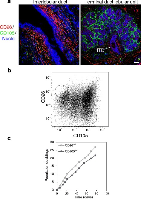
Background: The terminal duct lobular unit (TDLU) is the most dynamic structure in the human breast and the putative site of origin of human breast cancer. Although stromal cells contribute to a specialized microenvironment in many organs, this component remains largely understudied in the human breast. We here demonstrate the impact on epithelium of two lineages of breast stromal fibroblasts, one of which accumulates in the TDLU while the other resides outside the TDLU in the interlobular stroma.

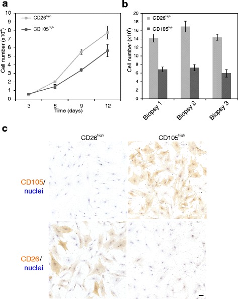
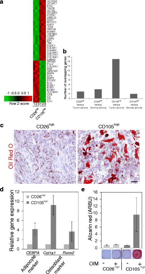
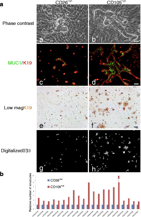
Methods: The two lineages are prospectively isolated by fluorescence activated cell sorting (FACS) based on different expression levels of CD105 and CD26. The characteristics of the two fibroblast lineages are assessed by immunocytochemical staining and gene expression analysis. The differentiation capacity of the two fibroblast populations is determined by exposure to specific differentiating conditions followed by analysis of adipogenic and osteogenic differentiation. To test whether the two fibroblast lineages are functionally imprinted by their site of origin, single cell sorted CD271low/MUC1high normal breast luminal epithelial cells are plated on fibroblast feeders for the observation of morphological development. Epithelial structure formation and polarization is shown by immunofluorescence and digitalized quantification of immunoperoxidase-stained cultures.

Results: Lobular fibroblasts are CD105high/CD26low while interlobular fibroblasts are CD105low/CD26high. Once isolated the two lineages remain phenotypically stable and functionally distinct in culture. Lobular fibroblasts have properties in common with bone marrow derived mesenchymal stem cells and they specifically convey growth and branching morphogenesis of epithelial progenitors.

Conclusions: Two distinct functionally specialized fibroblast lineages exist in the normal human breast, of which the lobular fibroblasts have properties in common with mesenchymal stem cells and support epithelial growth and morphogenesis. We propose that lobular fibroblasts constitute a specialized microenvironment for human breast luminal epithelial progenitors, i.e. the putative precursors of breast cancer.

Keywords: Breast; Epithelial morphogenesis; Fibroblasts; Mesenchymal stem cells.


Figures

Fig. 1
Fig. 1
Characterization, isolation and cultivation of interlobular and intralobular fibroblasts. a Multicolor imaging of cryostat sections of normal breast tissue showing an interlobular duct (left) and a terminal duct lobular unit (TDLU, right) stained for CD105 (green), CD26 (red) and nuclei (blue). An intralobular terminal duct (ITD) connects the TDLU to the interlobular duct. Phenotypically distinct fibroblasts surround the two anatomical structures. b Fluorescence activated cell sorting diagram of serially passaged fibroblasts stained with CD26 and CD105. Circles indicate gates selected for sorting. c Time course of population doublings of CD26high (light squares) and CD105high (dark squares) fibroblasts in serial passage subculture (scale bar = 50 μm)
Fig. 2
Fig. 2
CD26high and CD105high fibroblasts are inherently different with respect to growth and staining pattern. a Representative growth curves of CD26high (light squares) and CD105high (dark squares) fibroblasts from eighth-passage cells in triplicate (error bars represent mean +/−SD). b Endpoint number at day 12 per 24-well in triplicate of CD26high (light bars) and CD105high (dark bars) fibroblasts from three different biopsies (error bars represent mean +/−SD). CD26high fibroblasts consistently grow faster than CD105high fibroblasts and reach a higher cell density at day 9 and 12, respectively (unpaired Student’s t test, p < 0.05). c Immunoperoxidase staining and nuclei counterstain in passage 9, showing that distinct phenotypes are passed on (scale bar = 50 μm)
Fig. 3
Fig. 3
CD105high fibroblasts exhibit a transforming growth factor (TGF)β profile and mesenchymal stem-like properties. a Heat map representation of microarray analysis of 44 selected, differentially expressed genes in CD26high and CD105high fibroblasts (passage 9). Color key indicates centered and row-scaled normalized intensity values. b Overlap between the top 302 median ranked significantly and differentially expressed genes and previously published profiles of breast normal and tumor stroma, respectively. Bars indicate the number of overlapping genes in CD26high or CD105high cells compared to tumor or normal stroma, respectively. The overlap between genes expressed by CD105high cells and tumor stroma was statistically significant on analysis by Fisher’s exact test (p < 0.001). c Oil Red O staining of lipid droplets at day 15 of adipocyte differentiation in CD26high and CD105high cells, respectively, with nuclei stained with hematoxylin. d RT-qPCR of osteoblast and adipocyte marker gene expression (CCAAT/Enhancer binding protein, alpha (CEBPA), Collagen type 1 alpha 1 (Col1a1), and Runt-Related Transcription Factor 2 (Runx2)) in CD26high (light bars) and CD105high (dark bars) cells at day 3 of differentiation in passage 11 presented as gene expression relative to the geometric mean of two reference genes (UBC and B2m). The difference was statistically significant for CEBPA and Col1a1 in three different biopsies on analysis by unpaired Student’s t test at p < 0.05. e Alizarin Red staining and quantification of the mineralized matrix at day 15 after exposure to osteogenic induction medium (OIM, +) or control conditions without inducing factors (−). On analysis by unpaired Student’s t test the difference in Alizarin Red staining in samples representing four biopsies, two in passage 11 and two in passage 13, was not statistically significant in CD26high with and without osteogenic induction, but was significant at p < 0.05 in CD105high with and without induction (scale bar = 100 μm). Error bars represent mean +/− SD
Fig. 4
Fig. 4
CD105high stroma is a specialized microenvironment for branching morphogenesis of luminal breast epithelial cells. a Primary cultures of purified luminal breast epithelial cells plated at clonal density on confluent feeders of CD26high (left column) or CD105high (right column) fibroblasts: phase contrast micrographs of co-cultures twenty days after plating showing branching morphogenesis primarily on CD105high fibroblasts (a’, b’); dual-color imaging of co-cultures stained with keratin K19 (red) and MUC1 (green). Note the correctly polarized staining pattern and the elaborate structures on CD105high fibroblasts (c’, d’); low-magnification micrographs of co-cultures ten days after plating and immunoperoxidase staining for keratin K19 (e’, f’); digitalized images of keratin K19-stained epithelial structures (g’, h’). b Quantitative representation of K19-stained morphological structures in multiple recombinant cultures representing eight biopsies showing consistent growth advantage of luminal epithelial cells on CD105high fibroblasts (red bars) as normalized in each set of samples to structures formed on CD26high fibroblasts (blue bars) (scale bar = 50 μm (a’, b’); 100 μm (c’, d’); 500 μm (e’-h’))

References

    1. Ball EMA, Risbridger GP. Activins as regulators of branching morphogenesis. Dev Biol. 2001;238:1–12. doi: 10.1006/dbio.2001.0399. - DOI - PubMed
    1. Driskell RR, Lichtenberger BM, Hoste E, Kretzschmar K, Simons BD, Charalambous M, Ferrori SR, Herault Y, Pavlovic G, Ferguson-Smith AC, et al. Distinct fibroblast lineages determine dermal architecture in skin development and repair. Nature. 2013;504:277–81. doi: 10.1038/nature12783. - DOI - PMC - PubMed
    1. Rønnov-Jessen L, Petersen OW, Bissell MJ. Cellular changes involved in conversion of normal to malignant breast: The importance of the stromal reaction. Physiol Rev. 1996;76:69–125. - PubMed
    1. Cardiff RD, Wellings SR. The comparative pathology of human and mouse mammary glands. J Mammary Gland Biol Neoplasia. 1999;4(1):105–22. doi: 10.1023/A:1018712905244. - DOI - PubMed
    1. Villadsen R, Fridriksdottir AJ, Rønnov-Jessen L, Gudjonsson T, Rank F, LaBarge MA, Bissell MJ, Petersen OW. Evidence of a stem cell hierarchy in the adult human breast. J Cell Biol. 2007;177:87–101. doi: 10.1083/jcb.200611114. - DOI - PMC - PubMed