. 2016 Nov 3;9(1):117.
doi: 10.1186/s13045-016-0348-0.

LncRNAs H19 and HULC, activated by oxidative stress, promote cell migration and invasion in cholangiocarcinoma through a ceRNA manner

Affiliations

LncRNAs H19 and HULC, activated by oxidative stress, promote cell migration and invasion in cholangiocarcinoma through a ceRNA manner

Wen-Tao Wang et al. J Hematol Oncol. .

Abstract

Background: Long non-coding RNAs (lncRNAs) are known to play important roles in different cell contexts, including cancers. However, little is known about lncRNAs in cholangiocarcinoma (CCA), a cholangiocyte malignancy with poor prognosis, associated with chronic inflammation and damage to the biliary epithelium. The aim of the study is to identify if any lncRNA might associate with inflammation or oxidative stress in CCA and regulate the disease progression.

Methods: In this study, RNA-seqs datasets were used to identify aberrantly expressed lncRNAs. Small interfering RNA and overexpressed plasmids were used to modulate the expression of lncRNAs, and luciferase target assay RNA immunoprecipitation (RIP) was performed to explore the mechanism of miRNA-lncRNA sponging.

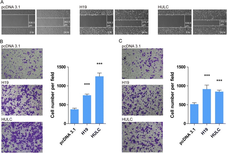
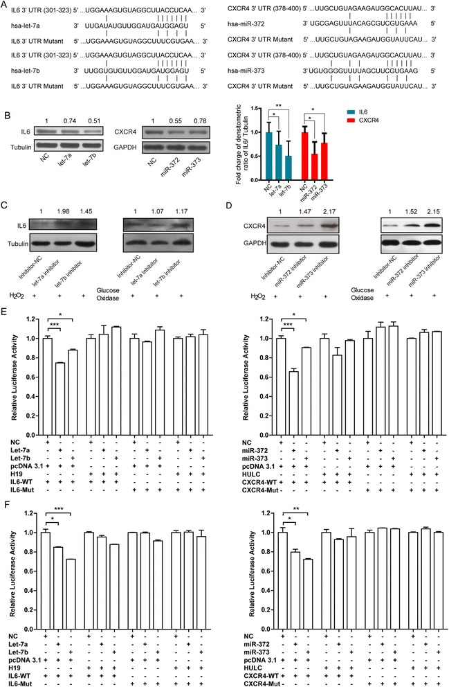
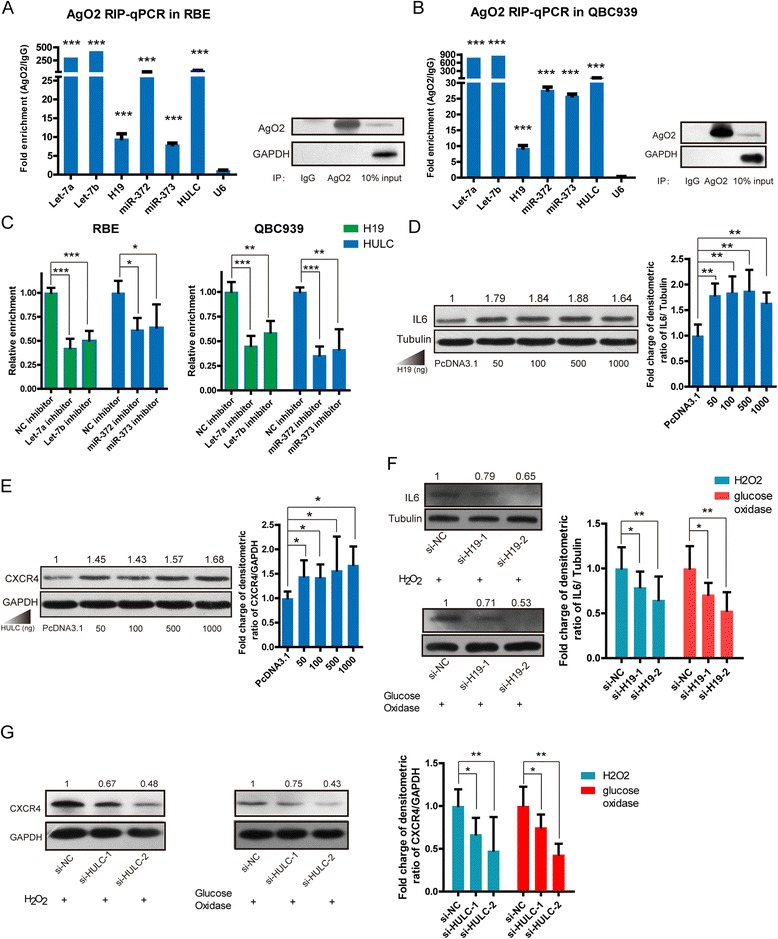
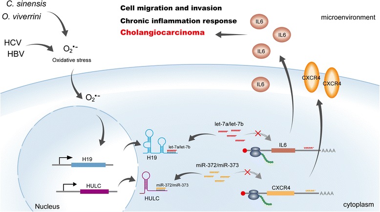
Results: We firstly analyzed five available RNA-seqs datasets to investigate aberrantly expressed lncRNAs which might associate with inflammation or oxidative stress. We identified that two lncRNAs, H19 and HULC, were differentially expressed among all the samples under the treatment of hypoxic or inflammatory factors, and they were shown to be stimulated by short-term oxidative stress responses to H2O2 and glucose oxidase in CCA cell lines. Further studies revealed that these two lncRNAs promoted cholangiocyte migration and invasion via the inflammation pathway. H19 and HULC functioned as competing endogenous RNAs (ceRNAs) by sponging let-7a/let-7b and miR-372/miR-373, respectively, which activate pivotal inflammation cytokine IL-6 and chemokine receptor CXCR4.

Conclusions: Our study revealed that H19 and HULC, up-regulated by oxidative stress, regulate CCA cell migration and invasion by targeting IL-6 and CXCR4 via ceRNA patterns of sponging let-7a/let-7b and miR-372/miR-373, respectively. The results suggest that these lncRNAs might be the chief culprits of CCA pathogenesis and progression. The study provides new insight into the mechanism linking lncRNA function with CCA and may serve as novel targets for the development of new countermeasures of CCA.

Keywords: Cholangiocarcinoma; Inflammation response; Migration and invasion; Oxidative stress; ceRNA.


Figures

Fig. 1
Fig. 1
Analysis of Inflammation-associated lncRNAs. a Heat maps of differential lncRNA profiles are based on studies upon lncRNAs responded to hypoxic and inflammatory factor. Each of the GEO data has suggested the aberrantly lncRNA related with inflammation and/or oxidative stress. For example, GSE76743 identified endothelial cell response to hypoxia. Others: GSE57539 (PBS_VS_IL6 treated MDA-MB-231 cells), GSE67106 (Non_ VS_ inflamed Crohn’s disease), GSE55146 (Non_ VS_ cystic fibrosis bronchial epithelium), and GSE70544 (Normoxia_vs_Hypoxia in proximal tubular epithelial cells (PTECs)). b LncRNAs stimulated by short-term oxidative stress using H2O2 in QBC939, SK-cha-1, and RBE cells. c LncRNAs stimulated by long-term oxidative stress using glucose oxidase in QBC939, SK-cha-1, and RBE cells
Fig. 2
Fig. 2
Enforced H19 and HULC expression increased the ability of migration and invasion in CCA cells. a Scratch wound healing assays of CCA cells. b Migration assays of CCA cells. c Invasion assays of CCA cells
Fig. 3
Fig. 3
H19 and HULC regulated genes via ceRNA behaviors. a Schematic representation of the interaction between miRNAs and the 3'-UTR of IL-6 or CXCR4. b Let-7a/let-7b and miR-372/miR-373 reduced the abundance of IL-6 and CXCR4 proteins, respectively in CCA cells. The right histogram analyzed the relative fold charges of protein levels. Triplicate experiments were analyzed by mean ± SD, p value <0.01, **, <0.05, *. MiRNA down-regulation by transfecting the miRNA inhibitors into RBE cells under oxidative stress, and the expression levels of IL-6 (c) and CXCR4 (d) increased significantly. Enforced expressed of H19 and HULC rescued luciferase activity for vectors with the 3'-UTR of IL-6 or CXCR4, which were suppressed by let-7a/let-7b and miR-372/miR-373, respectively, in RBE cells (e), and in HEK-293 T cells (f)
Fig. 4
Fig. 4
The mechanism of H19 and HULC that regulated genes. a AGO2 rip-qPCR of miRNAs and lncRNAs in RBE cells. The enrichment of miRNAs binding with AGO2 were used as the positive controls and U6 as the negative. b AGO2 rip-qPCR of miRNAs and lncRNAs in QBC939 cells. Triplicate experiments were analyzed by mean ± SD, p value <0.001, ***. c Relative enrichment of H19 (green) and HULC (blue) were analyzed in AGO2 rip-qPCR with let-7a, let-7b inhibitor or miR-372, miR-373 that compared to inhibitor NC. Enforced expression of different concentration of H19 (d) and HULC (e) plasmid increased IL-6 and CXCR4 protein levels, respectively, in RBE cells. The expression of IL-6 (f) and CXCR4 (g) had decreased significantly when we knocked down H19 and HULC, respectively, under the oxidative stress condition. The right histogram of gray density to quantitatively analyze the relative fold charges of protein levels. Triplicate experiments were analyzed by mean ± SD, p value < 0.001, ***, <0.01, **, <0.05, *
Fig. 5
Fig. 5
Schematic representations of pathways modulated by H19 and HULC in CCA cells. H19 and HULC are up-regulated by oxidative stress induced by viruses or liver flukes and sponging miRNAs, such as let-7a/let-7b and miR-372/miR-373. This increases the expression of inflammation-related genes, including IL-6 and CXCR4, resulting in abnormal inflammation responses and pathogenesis of CCA

References

    1. Fava G, Lorenzini I. Molecular pathogenesis of cholangiocarcinoma. Int J Hepatol. 2012;2012:630543. doi: 10.1155/2012/630543. - DOI - PMC - PubMed
    1. Karin M, Clevers H. Reparative inflammation takes charge of tissue regeneration. Nature. 2016;529:307–15. doi: 10.1038/nature17039. - DOI - PMC - PubMed
    1. Pinlaor S, Ma N, Hiraku Y, Yongvanit P, Semba R, Oikawa S, et al. Repeated infection with Opisthorchis viverrini induces accumulation of 8-nitroguanine and 8-oxo-7,8-dihydro-2′-deoxyguanine in the bile duct of hamsters via inducible nitric oxide synthase. Carcinogenesis. 2004;25:1535–1542. doi: 10.1093/carcin/bgh157. - DOI - PubMed
    1. Rizvi S, Gores GJ. Pathogenesis, diagnosis, and management of cholangiocarcinoma. Gastroenterology. 2013;145:1215–1229. doi: 10.1053/j.gastro.2013.10.013. - DOI - PMC - PubMed
    1. Kawanishi S, Hiraku Y, Pinlaor S, Ma N. Oxidative and nitrative DNA damage in animals and patients with inflammatory diseases in relation to inflammation-related carcinogenesis. Biol Chem. 2006;387:365–372. doi: 10.1515/BC.2006.049. - DOI - PubMed

Publication types

LinkOut - more resources