. 2017 Jan;220(Pt B):807-817.
doi: 10.1016/j.envpol.2016.10.057. Epub 2016 Oct 31.

Embryonic exposures to perfluorooctanesulfonic acid (PFOS) disrupt pancreatic organogenesis in the zebrafish, Danio rerio

Affiliations

Embryonic exposures to perfluorooctanesulfonic acid (PFOS) disrupt pancreatic organogenesis in the zebrafish, Danio rerio

Karilyn E Sant et al. Environ Pollut. 2017 Jan.

Abstract

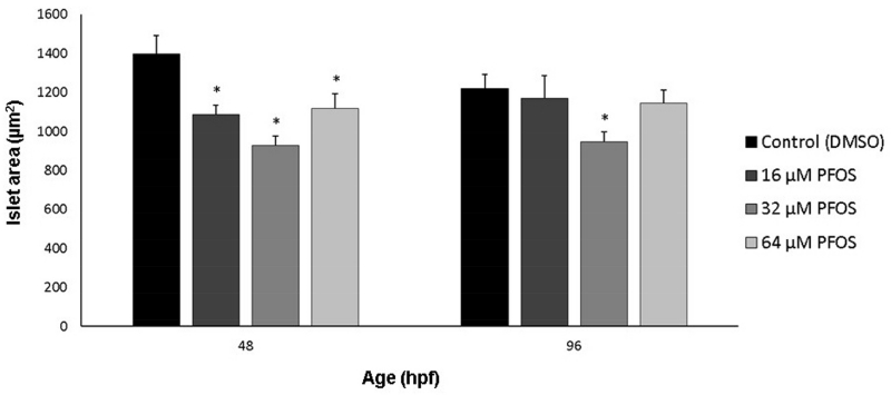
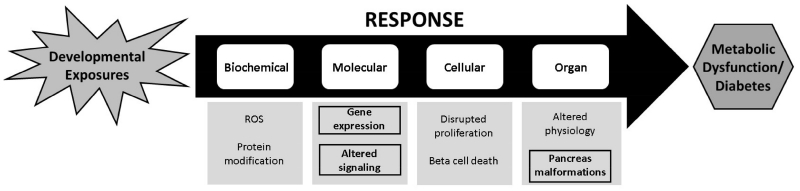
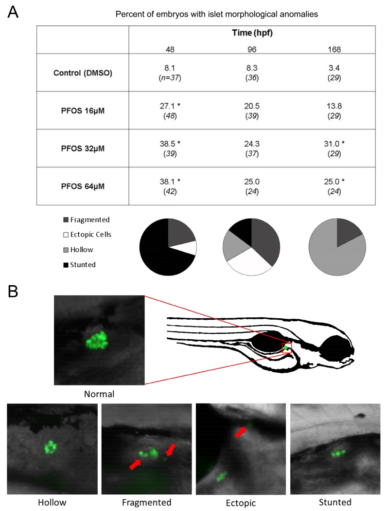
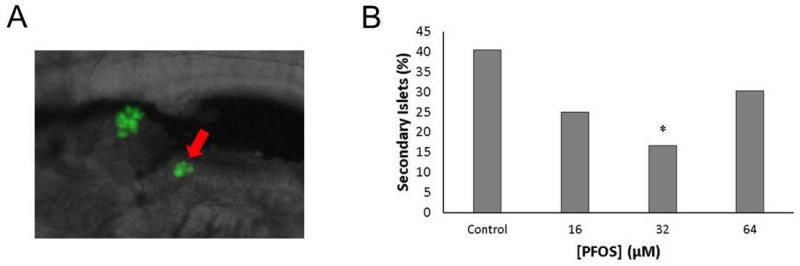
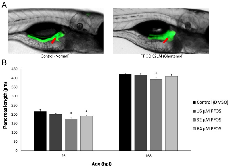
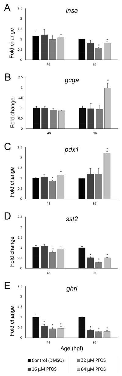
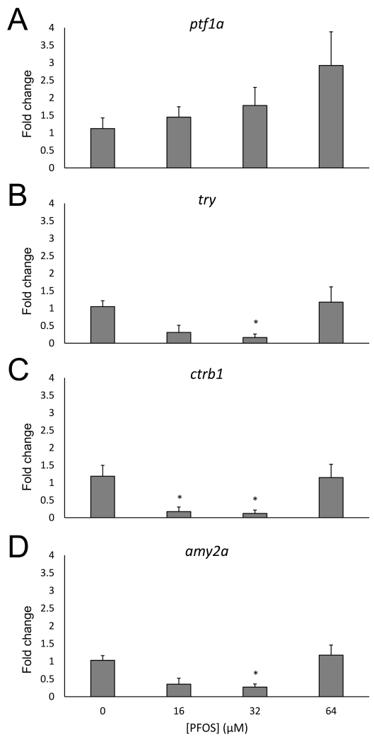
Perfluorooctanesulfonic acid (PFOS) is a ubiquitous environmental contaminant, previously utilized as a non-stick application for consumer products and firefighting foam. It can cross the placenta, and has been repeatedly associated with increased risk for diabetes in epidemiological studies. Here, we sought to establish the hazard posed by embryonic PFOS exposures on the developing pancreas in a model vertebrate embryo, and develop criteria for an adverse outcome pathway (AOP) framework to study the developmental origins of metabolic dysfunction. Zebrafish (Danio rerio) embryos were exposed to 16, 32, or 64 μM PFOS beginning at the mid-blastula transition. We assessed embryo health, size, and islet morphology in Tg(insulin-GFP) embryos at 48, 96 and 168 hpf, and pancreas length in Tg(ptf1a-GFP) embryos at 96 and 168 hpf. QPCR was used to measure gene expression of endocrine and exocrine hormones, digestive peptides, and transcription factors to determine whether these could be used as a predictive measure in an AOP. Embryos exposed to PFOS showed anomalous islet morphology and decreased islet size and pancreas length in a U-shaped dose-response curve, which resemble congenital defects associated with increased risk for diabetes in humans. Expression of genes encoding islet hormones and exocrine digestive peptides followed a similar pattern, as did total larval growth. Our results demonstrate that embryonic PFOS exposures can disrupt pancreatic organogenesis in ways that mimic human congenital defects known to predispose individuals to diabetes; however, future study of the association between these defects and metabolic dysfunction are needed to establish an improved AOP framework.

Keywords: Embryo; Exocrine pancreas; Insulin; Islets; Pancreas development; β cells.


Figures

Fig 1
Fig 1
PFOS decreases islet area at 48 and 96 hpf. Islet area was measured in Tg(insulin-GFP) embryos using EVOS software. Islet area was decreased along a U-shaped curve. Asterisks (*) indicate a difference between designated treatment group and the controls (p<0.05); n=30-45 embryos at 48 hpf; n=20-25 eleutheroembryos at 96 hpf
Fig 2
Fig 2
PFOS exposure increases the frequency of anomalous pancreas morphologies during development. (A) Islet morphology was examined in Tg(insulin-GFP) transgenic fish at 48, 96, and 168 hpf after subchronic PFOS exposure beginning at 3 hpf. Islets were screened for fragmentation, hollowness, and severely stunted growth (shown in B at 20× magnification). Numbers presented are the percent of embryos/larvae with variant islets. Italicized numbers are the number of embryos/larvae sampled, cumulative across several study replicates. Fewer than 5% of embryos and larvae were severely deformed at the time of sampling, and were excluded from pancreas imaging. The distribution of islet morphologies are shown in pie charts under each respective time point, indicating a difference in the types of variants observed throughout development. No significant temporal differences were observed. The position of the islet within the zebrafish is shown (B, left). Asterisks (*) indicate a difference between designated treatment group and the controls (p<0.05); n=30-45 embryos at 48 hpf; n=20-25 eleutheroembryos at 96 hpf; n=24-29 larvae at 168 hpf
Fig 3
Fig 3
PFOS exposure delays formation of secondary islets. (A) Secondary islets are characterized by one or more beta cells developing after the primary islet (arrow), typically after 120 hpf. (B) The number of secondary islets at 7 dpf was quantified in Tg(insulin-GFP) larvae. Incidence of islet defects was 19/47 (40%) in controls, 9/36 (25%) in the 16 μM group, 6/36 (17%) in the 32 μM group, and 13/43 (30%) in the 64 μM group. Bars represent the percent of larvae with secondary islets. Asterisks (*) indicate a difference between designated treatment group and the controls (p<0.05); n=36-47 larvae per group.
Fig 4
Fig 4
PFOS exposure decreases exocrine pancreas length at 96 and 168 hpf. (A) Pancreas length was measured in Tg(ptf1a-GFP) transgenic fish, shown at 168 hpf. Pancreas length was measured by quantifying the distance from the center of the islet (arrow) to the posterior tail of the pancreas. A control pancreas of normal length is shown at left, and a PFOS-exposed and shortened pancreas is shown at right. (B) Pancreas length is significantly decreased in fish exposed to 32 and 64 μM PFOS at 96 hpf, and to 32 μM PFOS at 168 hpf. Asterisks (*) indicate a difference between designated treatment group and the controls (p<0.05); n=22-28 larvae
Fig 5
Fig 5
Embryonic PFOS exposure alters pancreas endocrine gene expression. RNA was isolated from embryos collected at 48 and 96 hpf, following subchronic PFOS exposure since 3 hpf. Expression of insa (A), gcga (B), pdx1 (C), sst2 (D), and ghrl (E) was analyzed using qPCR and the ΔΔCT method. Bars represent the average fold change (relative to beta actin; shown on y-axis) and the control group, and stars represent a PFOS-associated statistically significant change of expression from the control group. Age of the embryos and eleutheroembryos is shown on the x-axis in hpf. Asterisks (*) indicate a difference between designated treatment group and the controls (p<0.05); n=7-9 samples of 9 pooled embryos at 48 hpf; n=4-5 samples of 5 pooled eleutheroembryos at 96 hpf
Fig 6
Fig 6
Embryonic PFOS exposure alters pancreas exocrine gene expression. RNA was isolated from 96 hpf following subchronic PFOS exposure since 3 hpf. Expression of ptf1a (A), try (B), ctrb1 (C), and amy2a (D) was analyzed using qPCR and the ΔΔCT method. Bars represent the average fold change (relative to beta actin; shown on y-axis) and the control group, and stars represent a PFOS-associated statistically significant change of expression from the control group. Asterisks (*) indicate a difference between designated treatment group and the controls (p<0.05); n=4-5 samples of 5 pooled eleutheroembryos at 96 hpf
Fig 7
Fig 7
This study helps to expand an AOP framework for the developmental origins of metabolic dysfunction and diabetes. Findings of this study (highlighted in black boxes) provide new criteria for use in an AOP framework for the association between developmental exposures and metabolic dysfunction. This framework (flowing from left to right) has guided the identification of several key biochemical, molecular, cellular, and organ changes that lead to these disorders; however, the effects of exposures such as PFOS on pancreas structure had not been studied. In the future, we seek to elucidate a mechanism by which these exposures may cause dysmorphogenesis of the endocrine and exocrine pancreas, and further how these structural anomalies are associated with the development of metabolic dysfunction later in the lifecourse.

References

    1. Agabi JO, Akhigbe AO. Comparative sonographic evaluation of the anteroposterior dimensions of the pancreas in diabetics and nondiabetics. Niger J Clin Pract. 2016;19:175–181. - PubMed
    1. Akirav E, Kushner JA, Herold KC. β-Cell Mass and Type 1 Diabetes: Going, Going, Gone? Diabetes. 2008;57:2883–2888. - PMC - PubMed
    1. Aldinger KA, Elsen GE. Ptf1a Is a Molecular Determinant for Both Glutamatergic and GABAergic Neurons in the Hindbrain. The Journal of neuroscience. 2008;28:338–339. - PMC - PubMed
    1. Balakrishnan V, Narayanan VA, Siyad I, Radhakrishnan L, Nair P. Agenesis of the dorsal pancreas with chronic calcific pancreatitis. case report, review of the literature and genetic basis. JOP. 2006;7:651–659. - PubMed
    1. Bosco D, Armanet M, Morel P, Niclauss N, Sgroi A, Muller YD, Giovannoni L, Parnaud G, Berney T. Unique Arrangement of α- and β-Cells in Human Islets of Langerhans. Diabetes. 2010;59:1202–1210. - PMC - PubMed

MeSH terms

LinkOut - more resources