

Body Diffusion Weighted Imaging Using Non-CPMG Fast Spin Echo
- PMID: 27810802
- PMCID: PMC5492898
- DOI: 10.1109/TMI.2016.2622238
Body Diffusion Weighted Imaging Using Non-CPMG Fast Spin Echo
Abstract
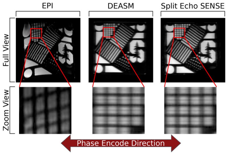
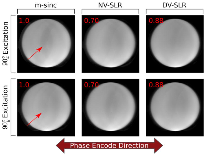
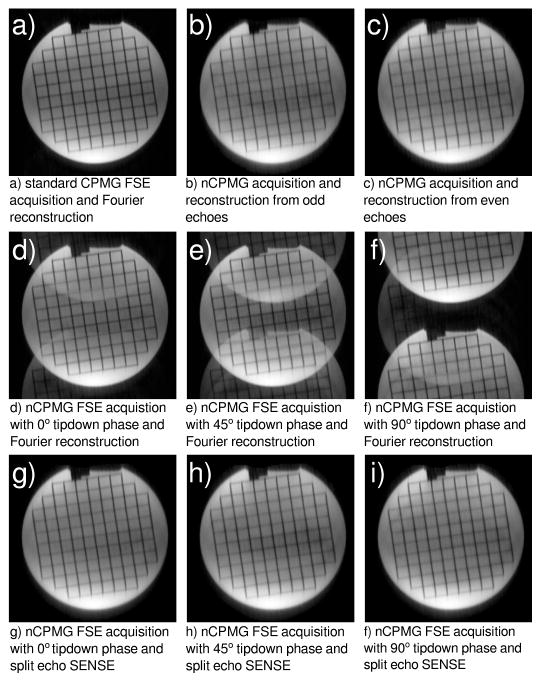
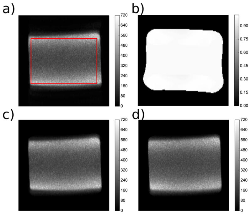
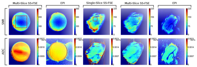
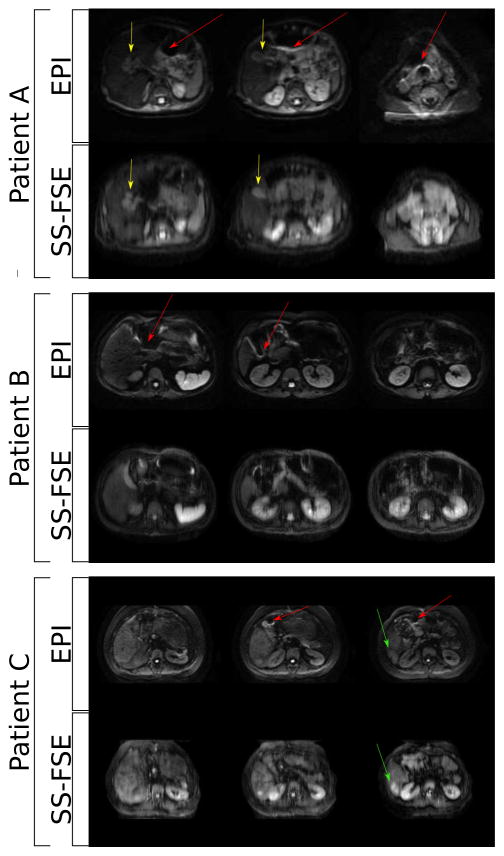
SS-FSE is a fast technique that does not suffer from off-resonance distortions to the degree that EPI does. Unlike EPI, SS-FSE is ill-suited to diffusion weighted imaging (DWI) due to the Carr-Purcell-Meiboom-Geill (CPMG) condition. Non-CPMG phase cycling does accommodate SS-FSE and DWI but places constraints on reconstruction, which are resolved here through parallel imaging. Additionally, improved echo stability can be achieved by using short duration and highly selective DIVERSE radiofrequency pulses. Here, signal-to-noise ratio (SNR) comparisons between EPI and nCPMG SS-FSE acquisitions and reconstruction techniques give similar values. Diffusion imaging with nCPMG SS-FSE gives similar SNR to an EPI acquisition, though apparent diffusion coefficient values are higher than seen with EPI. In vivo images have good image quality with little distortion. This method has the ability to capture distortion-free DWI images near areas of significant off-resonance as well as preserve adequate SNR. Parallel imaging and DIVERSE refocusing RF pulses allow shorter ETL compared to previous implementations and thus reduces phase encode direction blur and SAR accumulation.
Figures
Similar articles
-
Slice profile effects on nCPMG SS-FSE.Magn Reson Med. 2018 Jan;79(1):430-438. doi: 10.1002/mrm.26694. Epub 2017 Mar 31. Magn Reson Med. 2018. PMID: 28370409 Free PMC article.
-
Body diffusion-weighted imaging using magnetization prepared single-shot fast spin echo and extended parallel imaging signal averaging.Magn Reson Med. 2018 Jun;79(6):3032-3044. doi: 10.1002/mrm.26971. Epub 2017 Oct 17. Magn Reson Med. 2018. PMID: 29044721 Free PMC article.
-
Diffusion-weighted imaging of the spine with a non-carr-purcell-meiboom-gill single-shot fast spin-echo sequence: initial experience.AJNR Am J Neuroradiol. 2007 Mar;28(3):575-80. AJNR Am J Neuroradiol. 2007. PMID: 17353340 Free PMC article. Clinical Trial.
-
A joint linear reconstruction for multishot diffusion weighted non-Carr-Purcell-Meiboom-Gill fast spin echo with full signal.Magn Reson Med. 2022 Nov;88(5):2139-2156. doi: 10.1002/mrm.29393. Epub 2022 Jul 30. Magn Reson Med. 2022. PMID: 35906924 Free PMC article.
-
Diffusion-weighted imaging of the appendicular skeleton with a non-Carr-Purcell-Meiboom-Gill single-shot fast spin-echo sequence.AJR Am J Roentgenol. 2007 Dec;189(6):1494-501. doi: 10.2214/AJR.07.2512. AJR Am J Roentgenol. 2007. PMID: 18029891
Cited by
-
Isotropic resolution diffusion tensor imaging of lumbosacral and sciatic nerves using a phase-corrected diffusion-prepared 3D turbo spin echo.Magn Reson Med. 2018 Aug;80(2):609-618. doi: 10.1002/mrm.27072. Epub 2018 Jan 29. Magn Reson Med. 2018. PMID: 29380414 Free PMC article.
-
Slice profile effects on nCPMG SS-FSE.Magn Reson Med. 2018 Jan;79(1):430-438. doi: 10.1002/mrm.26694. Epub 2017 Mar 31. Magn Reson Med. 2018. PMID: 28370409 Free PMC article.
-
Body diffusion-weighted imaging using magnetization prepared single-shot fast spin echo and extended parallel imaging signal averaging.Magn Reson Med. 2018 Jun;79(6):3032-3044. doi: 10.1002/mrm.26971. Epub 2017 Oct 17. Magn Reson Med. 2018. PMID: 29044721 Free PMC article.
-
Robust Self-Calibrating nCPMG Acquisition: Application to Body Diffusion-Weighted Imaging.IEEE Trans Med Imaging. 2018 Jan;37(1):200-209. doi: 10.1109/TMI.2017.2741421. Epub 2017 Aug 17. IEEE Trans Med Imaging. 2018. PMID: 28829307 Free PMC article.
-
Volumetric and multispectral DWI near metallic implants using a non-linear phase Carr-Purcell-Meiboom-Gill diffusion preparation.Magn Reson Med. 2022 Jun;87(6):2650-2666. doi: 10.1002/mrm.29153. Epub 2022 Jan 11. Magn Reson Med. 2022. PMID: 35014729 Free PMC article.
References
-
- Harry VN, Deans H, Ramage E, Parkin DE, Gilbert FJ. Magnetic resonance imaging in gynecological oncology. International Journal of Gynecological Cancer. 2009;19(2):186–193. - PubMed
-
- Shinagare AB, Ip IK, Raja AS, Sahni VA, Banks P, Khorasani R. Use of CT and MRI in emergency department patients with acute pancreatitis. Abdominal Imaging. 2015;40(2):272–277. - PubMed
-
- Le Bihan D, Breton E, et al. Imagerie de diffusion in-vivo par resonance magnetique nucleaire. Comptes-Rendus de l’Académie des Sciences. 1985;93(5):27–34.
-
- Eastwood JD, Lev MH, Wintermark M, Fitzek C, Barboriak DP, Delong DM, Lee TY, Azhari T, Herzau M, Chilukuri VR, et al. Correlation of early dynamic CT perfusion imaging with whole-brain MR diffusion and perfusion imaging in acute hemispheric stroke. American Journal of Neuroradiology. 2003;24(9):1869–1875. - PMC - PubMed
MeSH terms
Grants and funding
LinkOut - more resources
Full Text Sources
Other Literature Sources
Miscellaneous
