. 2016 Dec 23;291(52):26794-26805.
doi: 10.1074/jbc.M116.736991. Epub 2016 Nov 3.

Endoplasmic Reticulum Stress-induced Hepatocellular Death Pathways Mediate Liver Injury and Fibrosis via Stimulator of Interferon Genes

Affiliations

Endoplasmic Reticulum Stress-induced Hepatocellular Death Pathways Mediate Liver Injury and Fibrosis via Stimulator of Interferon Genes

Arvin Iracheta-Vellve et al. J Biol Chem. .

Abstract

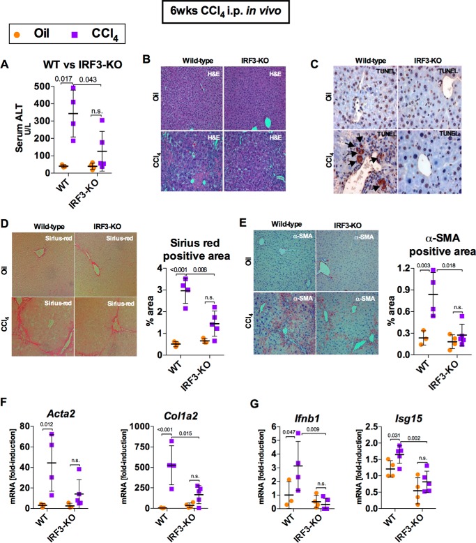
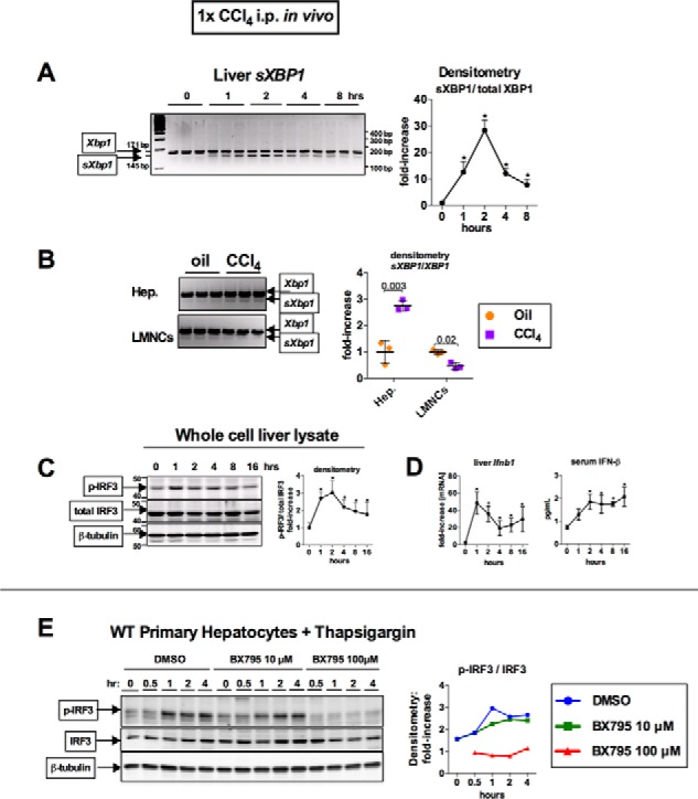
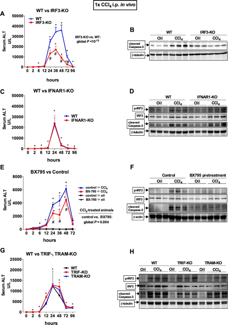
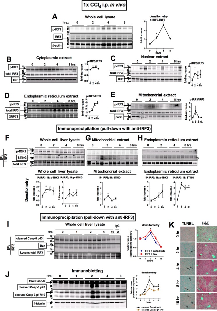
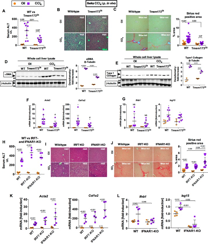
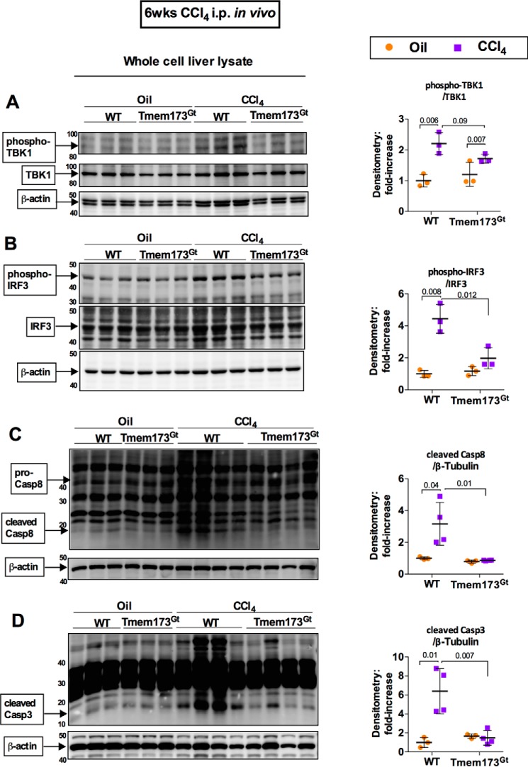
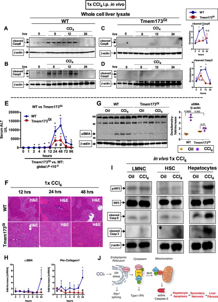
Fibrosis, driven by inflammation, marks the transition from benign to progressive stages of chronic liver diseases. Although inflammation promotes fibrogenesis, it is not known whether other events, such as hepatocyte death, are required for the development of fibrosis. Interferon regulatory factor 3 (IRF3) regulates hepatocyte apoptosis and production of type I IFNs. In the liver, IRF3 is activated via Toll-like receptor 4 (TLR4) signaling or the endoplasmic reticulum (ER) adapter, stimulator of interferon genes (STING). We hypothesized that IRF3-mediated hepatocyte death is an independent determinant of chemically induced liver fibrogenesis. To test this, we performed acute or chronic CCl4 administration to WT and IRF3-, Toll/Interleukin-1R (TIR) domain-containing adapter-inducing interferon-β (TRIF)-, TRIF-related adaptor molecule (TRAM)-, and STING-deficient mice. We report that acute CCl4 administration to WT mice resulted in early ER stress, activation of IRF3, and type I IFNs, followed by hepatocyte apoptosis and liver injury, accompanied by liver fibrosis upon repeated administration of CCl4 Deficiency of IRF3 or STING prevented hepatocyte death and fibrosis both in acute or chronic CCl4 In contrast, mice deficient in type I IFN receptors or in TLR4 signaling adaptors, TRAM or TRIF, upstream of IRF3, were not protected from hepatocyte death and/or fibrosis, suggesting that the pro-apoptotic role of IRF3 is independent of TLR signaling in fibrosis. Hepatocyte death is required for liver fibrosis with causal involvement of STING and IRF3. Thus, our results identify that IRF3, by its association with STING in the presence of ER stress, couples hepatocyte apoptosis with liver fibrosis and indicate that innate immune signaling regulates outcomes of liver fibrosis via modulation of hepatocyte death in the liver.

Keywords: apoptosis; endoplasmic reticulum stress (ER stress); fibrosis; interferon regulatory factor (IRF); liver injury.


Figures

FIGURE 1.
FIGURE 1.
IRF3 deficiency attenuates chronic CCl4-mediated liver injury and fibrosis. WT or IRF3-KO mice injected with oil or CCl4 for 6 weeks and sacrificed 48 h after final injection. A and B, liver injury was assessed by serum ALT (A) and H&E staining (B). C, liver apoptosis was assessed via TUNEL staining (black arrows) on paraffin-embedded liver sections. D and E, liver fibrosis was evaluated by Sirius Red staining (D) and α-SMA immunohistochemistry (E), and quantified using ImageJ. F and G, mRNA expression in liver for Acta2 and Col1a2 (F) and Ifnb1 and Isg15 (G) were measured by RT-PCR to assess liver fibrosis and IRF3 activation, respectively. n = 6–7 mice (CCl4-treated, per genotype); 3–4 mice (oil-treated, per genotype).
FIGURE 2.
FIGURE 2.
Acute CCl4 induces ER stress and TBK1-mediated phosphorylation of IRF3 in hepatocytes. WT mice received a single injection of corn oil or CCl4. A, at the indicated time points, mice were sacrificed; splicing of Xbp1 mRNA (sXBP1) in the liver, indicating ER stress, was evaluated by PCR; and products were separated on 3% agarose gel. B, 2 h after CCl4 administration, hepatocytes (Hep.), or LMNCs from WT mice were isolated and analyzed for sXBP1 by PCR. C and D, at the indicated time points, phosphorylation of IRF3 was assessed by immunoblotting in liver (C), whereas Ifnb1 mRNA expression in liver and as serum IFN-β were measured by RT-PCR and ELISA, respectively (D). n = 4 mice per time point. E, primary hepatocytes isolated from WT mice were treated with 1 μm thapsigargin and were pretreated with DMSO or BX795 (10 or 100 μm), and analyzed for phospho-IRF3.
FIGURE 3.
FIGURE 3.
Acute CCl4-induced liver injury is ameliorated by the deficiency of IRF3 or inhibition of TBK1, independent of TRIM or TRAM, type I IFN signaling. A and B, WT or IRF3-KO mice received a single injection of CCl4. Serum ALT levels were assessed (A), and apoptosis was assessed by measuring caspase-3 cleavage in the liver by immunoblot (B). C and D, WT or IFNAR1-KO mice received a single injection of CCl4. Serum ALT levels were assessed (C), and IRF3 activation and apoptosis were assessed by probing for phosphor-IRF3 and caspase-3 cleavage in the liver by immunoblot (D). E and F, WT were pretreated with DMSO or BX795, a TBK1 inhibitor, 2 h before receiving a single injection of corn oil (vehicle) or CCl4. Serum ALT levels were assessed (E), and IRF3 activation and apoptosis were assessed by probing for phosphor-IRF3 and caspase-3 cleavage in the liver by immunoblot (F). G and H, WT or TRIF- or TRAM-KO mice received a single injection of CCl4. G and H, serum ALT levels were assessed (G), and IRF3 activation and apoptosis were assessed by probing for phosphor-IRF3 and caspase-3 cleavage in the liver by immunoblot (H). The mice were bled serially at the indicated time points. n = 8–9 mice (CCl4-treated, per genotype); 3–4 mice (oil-treated, per genotype) (A, C, and G). n = 3–5 mice (CCl4-treated, pretreated with BX795); 3 mice (oil-treated, pretreated with DMSO) (E).
FIGURE 4.
FIGURE 4.
Acute CCl4 induces early association of IRF3 with STING in the ER and association of IRF3 with Bax in the mitochondria in the liver. WT mice received a single injection of CCl4. A–E, at the indicated time points, the mice were sacrificed, and phosphorylation of IRF3 was assessed by immunoblotting in liver whole cell lysate (A) or cytoplasmic (B), nuclear (C), ER (D), and mitochondrial (E) extracts. F–H, using immunoprecipitation with anti-total IRF3 antibody for protein pulldown, we evaluated association between total IRF3 and phosphorylated TBK1 or STING in the whole cell (F), mitochondrial (G), and ER (H) extracts. Similarly, we evaluated association between total IRF3 and cleaved caspase-8 p43 fragment or BAX in liver whole cell lysate (I), with representative IgG control shown (2 h after injection). J, at the indicated time points, cleavage of caspase-8 and caspase-3 was assessed by immunoblotting in liver. K, liver injury and apoptosis was assessed by H&E liver histology and TUNEL staining on paraffin-embedded liver sections. n = 4 mice per time point. IP, immunoprecipitation.
FIGURE 5.
FIGURE 5.
Deficiency in STING, but not in type I IFN signaling, attenuates liver fibrosis. WT or STING-deficient mice (Tmem173Gt) were injected with oil or CCl4 for 6 weeks and sacrificed 48 h after final injection. A and B, liver injury was assessed by serum ALT (A) and H&E staining (B). C, liver fibrosis was evaluated by measuring Sirius Red staining, and the percent-positive area stained was quantified using ImageJ. D and E, liver fibrosis was also evaluated by immunoblot for α-SMA (D) or type 1 collagen (E). F and G, mRNA expression in liver for Acta2 and Col1a2 (F) and Ifnb1 and Isg15 (G) was measured by PCR to assess liver fibrosis and IRF3 activation, respectively. WT or IRF7- or IFNAR1-KO mice injected with oil or CCl4 for 6 weeks and sacrificed 48 h after final injection. H and I, liver injury was assessed by serum ALT (H) and H&E staining (I). J, liver fibrosis was evaluated by Sirius Red staining and quantified using ImageJ. K and L, mRNA expression in liver for Acta2 and Col1a2 (K) and Ifnb1 and Isg15 (L) were measured by RT-PCR. n = 8–9 mice (CCl4-treated, per genotype); 3–4 mice (oil-treated, per genotype).
FIGURE 6.
FIGURE 6.
STING mediates pro-apoptotic activation of IRF3 in a chronic CCl4-induced model of fibrosis. WT or STING-deficient mice (Tmem173Gt) were injected with oil or CCl4 for 6 weeks and sacrificed. Whole cell liver lysates were probed for phosphorylated TBK1 (A), phosphorylated IRF3 (B), cleaved caspase-8 (C), and cleaved caspase-3 (D) by immunoblot. n = 8–9 mice (CCl4-treated, per genotype); 3–4 mice (oil-treated, per genotype).
FIGURE 7.
FIGURE 7.
Early pro-apoptotic activation of IRF3 by CCl4 is hepatocyte-specific and mediated by STING. WT or STING-deficient mice (Tmem173Gt) received a single injection of CCl4 or oil (baseline) and sacrificed at indicated time points. A–D, whole cell liver lysates were probed for cleaved caspase-8 (A and B) and cleaved caspase-3 (C and D) by immunoblot. E and F, liver injury was assessed by serum ALT (E) and H&E staining at the indicated time points (F). G and H, deposition of α-SMA 24 h after injection was evaluated by immunoblots from whole cell liver lysate (G), whereas mRNA expression in liver for Acta2 and Col1a2 (H) was assessed by PCR. n = 5–8 mice per time point, per genotype (A–H). I, WT mice received a single injection of CCl4 or oil (baseline) and sacrificed 9 h later. LMNCs, HSCs, or hepatocytes were isolated from livers and probed for phosphorylated IRF3, total IRF3, cleaved caspase-8, and cleaved caspase-3 by immunoblot. n = 3 per condition (LMNCs and HSCs were pooled from 3 mice); n = 1 (hepatocytes isolated from single mouse per condition); the experiment was repeated three times. J, schematic of pro-apoptotic STING and IRF3 activation from CCl4 administration in mice. In CCl4-treated hepatocytes, ER stress results in phosphorylation of TBK1 via STING, followed by phosphorylation of IRF3. IRF3 associates with BAX in the mitochondria through its BH3-only domain, leading to pro-apoptotic caspase-3 activation and hepatocyte apoptosis. After chronic CCl4 administration, hepatocyte apoptosis is associated with secondary necrosis, which results in liver fibrosis.

References

    1. Seki E., De Minicis S., Osterreicher C. H., Kluwe J., Osawa Y., Brenner D. A., and Schwabe R. F. (2007) TLR4 enhances TGF-β signaling and hepatic fibrosis. Nat. Med. 13, 1324–1332 - PubMed
    1. Rivera C. A., Bradford B. U., Hunt K. J., Adachi Y., Schrum L. W., Koop D. R., Burchardt E. R., Rippe R. A., and Thurman R. G. (2001) Attenuation of CCl4-induced hepatic fibrosis by GdCl3 treatment or dietary glycine. Am. J. Physiol. Gastrointest. Liver Physiol. 281, G200–G207 - PubMed
    1. Malhi H., and Gores G. J. (2008) Cellular and molecular mechanisms of liver injury. Gastroenterology 134, 1641–1654 - PMC - PubMed
    1. Danial N. N., and Korsmeyer S. J. (2004) Cell death: critical control points. Cell 116, 205–219 - PubMed
    1. Jiang X., and Wang X. (2004) Cytochrome c-mediated apoptosis. Annu. Rev. Biochem. 73, 87–106 - PubMed

MeSH terms