. 2016 Nov 14;213(12):2671-2689.
doi: 10.1084/jem.20160041. Epub 2016 Oct 24.

--LUBAC deficiency perturbs TLR3 signaling to cause immunodeficiency and autoinflammation

Affiliations

--LUBAC deficiency perturbs TLR3 signaling to cause immunodeficiency and autoinflammation

Julia Zinngrebe et al. J Exp Med. .

Abstract

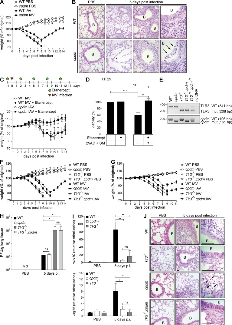
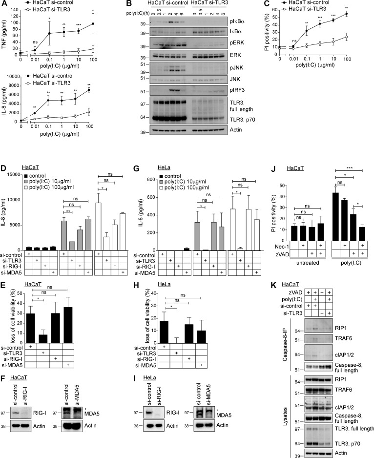
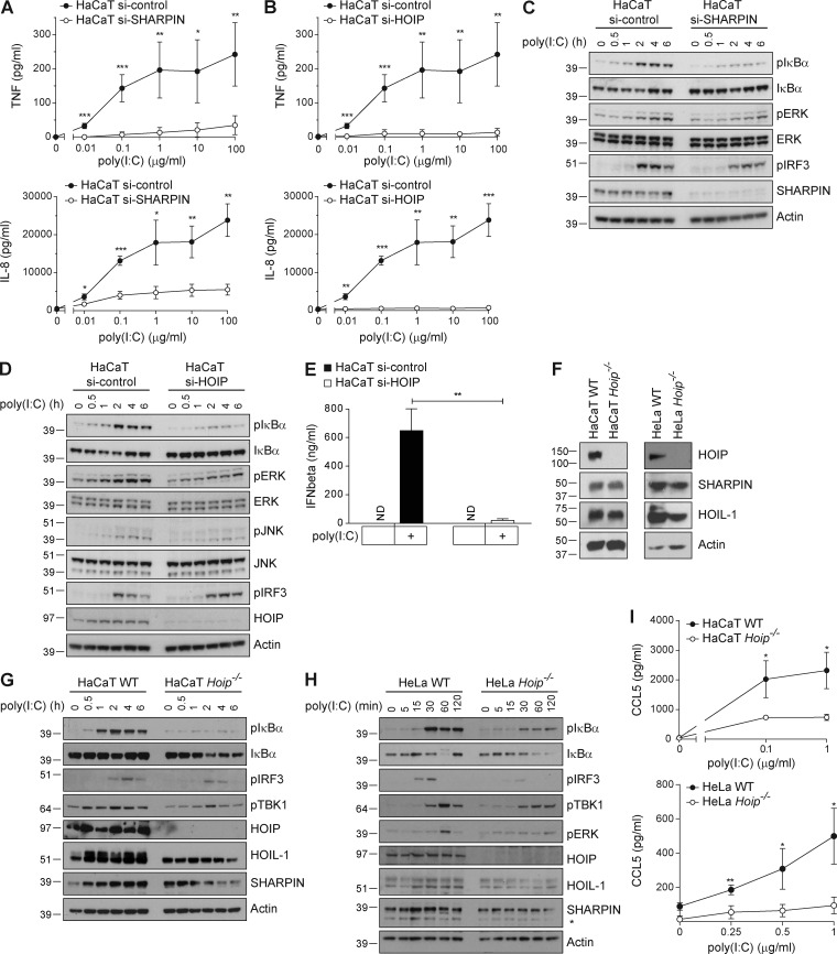
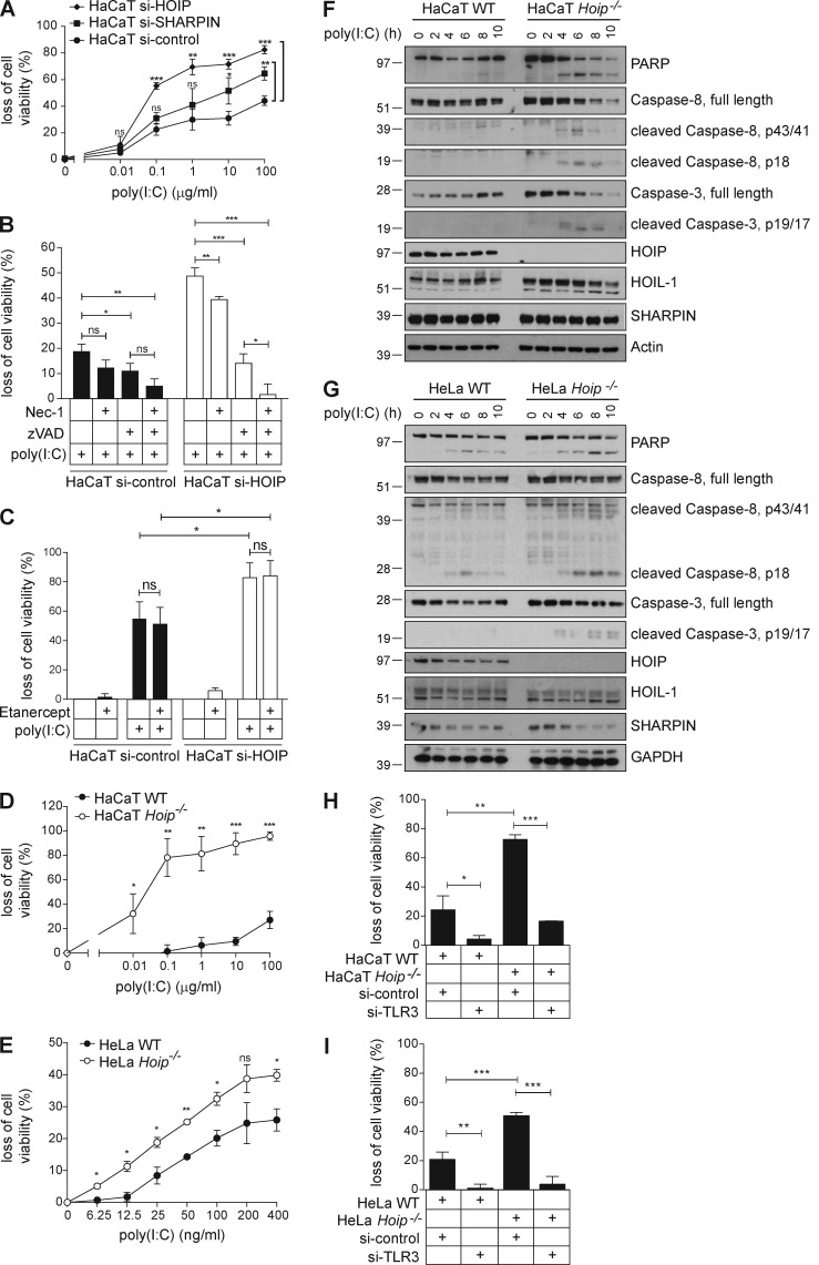
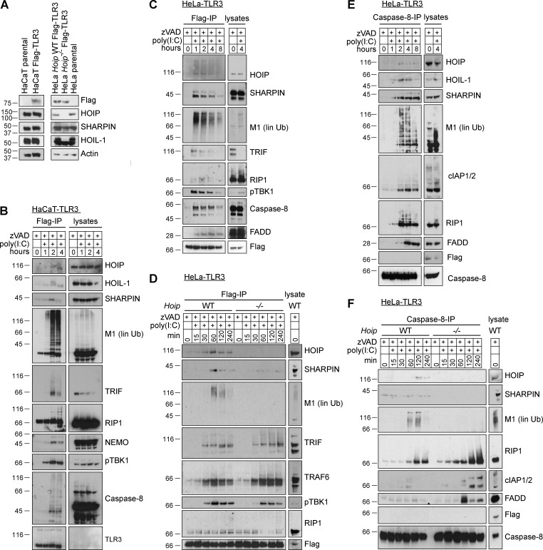
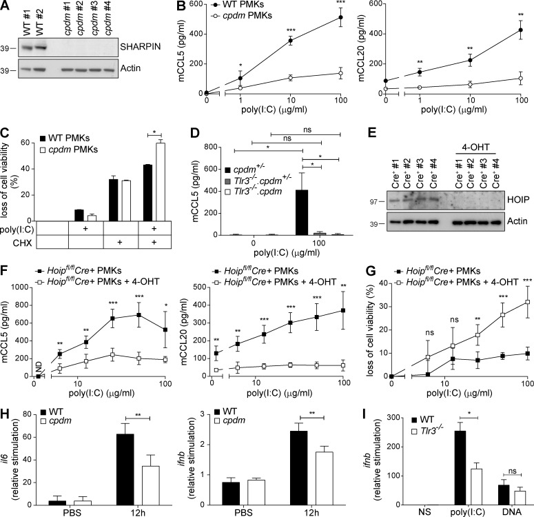
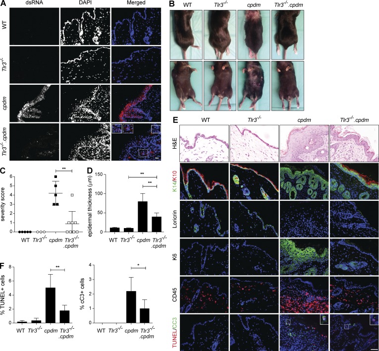
The linear ubiquitin chain assembly complex (LUBAC), consisting of SHANK-associated RH-domain-interacting protein (SHARPIN), heme-oxidized IRP2 ubiquitin ligase-1 (HOIL-1), and HOIL-1-interacting protein (HOIP), is a critical regulator of inflammation and immunity. This is highlighted by the fact that patients with perturbed linear ubiquitination caused by mutations in the Hoip or Hoil-1 genes, resulting in knockouts of these proteins, may simultaneously suffer from immunodeficiency and autoinflammation. TLR3 plays a crucial, albeit controversial, role in viral infection and tissue damage. We identify a pivotal role of LUBAC in TLR3 signaling and discover a functional interaction between LUBAC components and TLR3 as crucial for immunity to influenza A virus infection. On the biochemical level, we identify LUBAC components as interacting with the TLR3-signaling complex (SC), thereby enabling TLR3-mediated gene activation. Absence of LUBAC components increases formation of a previously unrecognized TLR3-induced death-inducing SC, leading to enhanced cell death. Intriguingly, excessive TLR3-mediated cell death, induced by double-stranded RNA present in the skin of SHARPIN-deficient chronic proliferative dermatitis mice (cpdm), is a major contributor to their autoinflammatory skin phenotype, as genetic coablation of Tlr3 substantially ameliorated cpdm dermatitis. Thus, LUBAC components control TLR3-mediated innate immunity, thereby preventing development of immunodeficiency and autoinflammation.


Figures

Figure 1.
Figure 1.
Adequate host response against IAV infection requires SHARPIN and TLR3. (A) Weight of cpdm mice and WT littermate controls (n ≥ 6 mice per group) infected i.n. with 103 PFU IAV was monitored for 14 consecutive days after infection. (B) Representative H&E stainings of lungs of cpdm mice and WT littermate controls noninfected (PBS) or infected i.n. with IAV are shown (lungs of n ≥ 3 mice per genotype and group were analyzed). Alveolar damage and dead/dying cells are indicated by asterisk and arrows, respectively. Bars, 100 µm. B, bronchiole. (C) Treatment schedule. cpdm mice and WT littermate controls (n ≥ 9 mice per group) were injected i.p. with Etanercept or Pentaglobin as control at a dose of 0.5 mg/mouse 1 d before infection with 50 PFU IAV, and then every third day. Weight was monitored for 13 consecutive days after infection. (D) HT29 cells were incubated with 50 µg/ml Etanercept before stimulation with 100 nM SM and 20 µM zVAD. Cell viability was assessed after 48 h. Data are presented as means ± SD of three independent experiments performed in triplicates. (E) Representative PCR genotyping of experimental mice is shown. (F and G) Weight of WT, cpdm, Tlr3−/−, and Tlr3−/−.cpdm mice (n ≥ 5 mice per group) infected i.n. with 300 PFU IAV (F) or 100 PFU IAV (G) was monitored for 11 consecutive days after infection. (H and I) IAV lung titers (H) and induction of cxcl10 and isg15 by RT-PCR (I) were determined at day 5 after infection (lungs of n ≥ 3 mice per genotype and group were analyzed). (J) Representative H&E stainings of lungs of mice of indicated genotypes (n ≥ 3 per genotype and group were analyzed) at day 5 after infection or noninfection (PBS) are shown. Dead/dying cells are indicated by arrows. Bars, 100 µm. B, bronchiole. Values are plotted as means ± SEM in A, C, F, and G and as means ± SD in D, H, and I. n.d., nondetectable; p.i., post infection. *, P < 0.05; **, P < 0.01; ***, P < 0.001, unpaired Student’s t test.
Figure 2.
Figure 2.
TLR3 presence is required to mediate poly(I:C)-induced signaling. (A) HaCaT cells transfected with control or TLR3 RNAi were stimulated with poly(I:C) as indicated and concentrations of TNF and IL-8 were determined by ELISA. (B) HaCaT cells transfected with control or TLR3 RNAi were stimulated with 5 µg/ml poly(I:C) as indicated and subjected to analysis by immunoblotting. (C) HaCaT cells transfected with control or TLR3 RNAi were stimulated with poly(I:C) as indicated and analyzed for propidium iodide positivity by FACS after 24 h. (D–I) HaCaT cells (D–F) or HeLa cells (G–I) transfected with control siRNA or siRNA targeting TLR3, RIG-I, or MDA5 were stimulated with 10 or 100 µg/ml poly(I:C). After 24 h, IL-8 secretion was determined by ELISA (D and G) and loss of cell viability was assessed in parallel (E and H). Knockdown efficiency of MDA5 and RIG-I was determined by immunoblotting (F and I). (J) HaCaT cells were preincubated for 1 h with 10 µM 7-Cl-O-Nec-1 (Nec-1), 20 µM zVAD (zVAD), or a combination thereof before stimulation with 50 µg/ml poly(I:C). Propidium iodide positivity was determined by FACS 24 h later. (K) HaCaT cells transfected with control or TLR3 siRNA were stimulated with 20 µg/ml poly(I:C) in the presence of 20 µM zVAD. Caspase-8 was immunoprecipitated and coimmunoprecipitated proteins were analyzed by Western blot. All values are means ± SD of at least three independent experiments performed in triplicates. *, P < 0.05; **, P < 0.01; ***, P < 0.001, unpaired Student’s t test. Western blots are representative of at least two independent experiments. Nonspecific bands are indicated by asterisk.
Figure 3.
Figure 3.
LUBAC components are required for TLR3-mediated gene activation. (A and B) HaCaT cells transfected with control or siRNA targeting SHARPIN (A) or HOIP (B) were stimulated with poly(I:C) as indicated. Cell supernatants were collected after 24 h of stimulation and concentration of TNF and IL-8 was determined by ELISA. (C and D) HaCaT cells transfected with control or RNAi targeting SHARPIN (C) or HOIP (D) were stimulated with 5 µg/ml poly(I:C) as indicated and subjected to analysis by immunoblotting. (E) HaCaT cells transfected with nontargeting or HOIP RNAi were stimulated with 10 µg/ml poly(I:C). Cell supernatants were collected after 4 h of stimulation and analyzed for concentration of IFN-β by ELISA. (F) HaCaT Hoip WT and Hoip−/− cells and HeLa Hoip WT and Hoip−/− cells were generated, and the absence of HOIP protein was confirmed by Western blotting. Protein levels of SHARPIN and HOIL-1 were determined in parallel. Actin served as loading control. (G and H) HaCaT Hoip WT and Hoip−/− cells (G) or HeLa Hoip WT and Hoip−/− cells (H) were stimulated with 5 µg/ml poly(I:C) as indicated and subjected to analysis by immunoblotting. (I) HaCaT and HeLa Hoip WT and Hoip−/− cells were stimulated with poly(I:C) as indicated. Cell supernatants were collected 24 h later and analyzed for concentration of CCL5 by ELISA. All values are means ± SD of at least three independent experiments performed in triplicates. *, P < 0.05; **, P < 0.01; ***, P < 0.001, unpaired Student’s t test. Western blots are representative of at least two independent experiments. Nonspecific bands are indicated by asterisk.
Figure 4.
Figure 4.
LUBAC components limit TLR3-induced cell death. (A) HaCaT cells transfected with control, SHARPIN, or HOIP siRNA were stimulated with poly(I:C) as indicated and loss of cell viability was determined after 24 h. (B) HaCaT cells transfected with control or HOIP siRNA were incubated with 10 µM 7-Cl-O-Nec-1 (Nec-1), 20 µM zVAD (zVAD), or a combination thereof 1 h before stimulation with 1 µg/ml poly(I:C). Loss of cell viability was assessed after 24 h. (C) HaCaT cells transfected with control or HOIP siRNA were incubated in the presence or absence of 50 µg/ml Etanercept 2 h before stimulation with 100 µg/ml poly(I:C). Loss of cell viability was determined after 24 h. (D and E) HaCaT Hoip WT and Hoip−/− cells (D) and HeLa Hoip WT and Hoip−/− cells (E) were stimulated with poly(I:C) as indicated. Loss of cell viability was assessed after 24 h. (F and G) HaCaT Hoip WT and Hoip−/− cells (F) or HeLa Hoip WT and Hoip−/− cells (G) were stimulated with 5 µg/ml poly(I:C) as indicated and subjected to further analysis by immunoblotting. (H) HaCaT Hoip WT and Hoip−/− cells were transfected with control RNAi or RNAi targeting TLR3. Cells were stimulated with 1 µg/ml and loss of cell viability was assessed after 24 h. (I) HeLa Hoip WT and Hoip−/− cells were transfected with control or TLR3 siRNA. Cells were stimulated with 100 µg/ml and loss of cell viability was assessed after 24 h. All values are presented as means ± SD of at least three independent experiments performed in triplicates. *, P < 0.05; **, P < 0.01; ***, P < 0.001, unpaired Student’s t test. Images are representative of at least two independent experiments.
Figure 5.
Figure 5.
LUBAC components form part of the TLR3-SC and prevent TLR3-induced DISC formation. (A) HaCaT parental and Flag-tagged TLR3-expressing cells and HeLa parental and Flag-tagged TLR3-expressing HeLa Hoip WT and Hoip−/− cells were lysed, and levels of TLR3 (by staining for Flag), HOIP, HOIL-1, and SHARPIN were determined by immunoblotting. Actin served as loading control. (B) HaCaT-TLR3 cells were stimulated with 20 µg/ml poly(I:C) in the presence of 20 µM zVAD as indicated. Cells were subsequently lysed and TLR3 was immunoprecipitated via Flag and coimmunoprecipitated proteins were analyzed by immunoblotting. Levels of Flag-tagged TLR3 were determined by staining for TLR3. (C) HeLa cells stably expressing Flag-TLR3 were incubated in the presence of 20 µM zVAD before stimulation with 20 µg/ml poly(I:C) as indicated. TLR3 was immunoprecipitated via its Flag-tag and analysis of coimmunoprecipitated proteins by immunoblotting was performed as indicated. (D) HeLa Hoip WT and Hoip−/− cells stably expressing Flag-TLR3 were stimulated with 20 µg/ml poly(I:C) in the presence of 20 µM zVAD and TLR3 was immunoprecipitated via Flag and coimmunoprecipitated proteins were analyzed by immunoblotting. (E) Caspase-8-IP was performed after lysates were depleted of the TLR3-SC by Flag-IP (C), and coimmunoprecipitated proteins were analyzed by immunoblotting. (F) Caspase-8-IP was performed after lysates were depleted of the TLR3-SC by Flag-IP (shown in D) and coimmunoprecipitated proteins were subjected to analysis by immunoblotting. Images are representative of at least three independent experiments. M1, linear ubiquitin linkages.
Figure 6.
Figure 6.
LUBAC components regulate TLR3 signaling induced by poly(I:C) ex vivo and in vivo. (A) SHARPIN protein expression of PMKs from WT mice (n = 2) and cpdm (n = 4) littermates was analyzed by Western blot. Actin served as loading control. (B) PMKs isolated from newborn WT (n = 2) and cpdm (n = 4) littermates were stimulated by poly(I:C) as indicated. Concentrations of murine CCL5 (mCCL5) and murine CCL20 (mCCL20) in the supernatants were determined by ELISA after 24 h of stimulation. One representative of two independent experiments performed in triplicates is shown. (C) PMKs from WT mice (n = 2) and cpdm littermates (n = 2) were isolated at the age of 4 wk and cells were stimulated with 50 µg/ml poly(I:C) in absence versus presence of 250 ng/ml CHX. Loss of cell viability was determined after 48 h of stimulation. One representative of two independent experiments performed in triplicates is shown. (D) PMKs from cpdm+/− (n = 3), Tlr3−/−.cpdm+/− (n = 3), and Tlr3−/−.cpdm−/− (n = 3) littermates isolated at the age of 4 wk were stimulated with 100 µg/ml poly(I:C) and concentration of mCCL5 was determined in the supernatant after 24 h by ELISA. (E) HOIP protein expression of PMKs isolated from one litter of newborn Cre+ Hoipfl/fl mice (n = 4) cultured in absence versus presence of 1 µM 4-OHT for at least 72 h was analyzed by Western blot. Actin served as loading control. (F) PMKs with 4-OHT–inducible deletion of HOIP from Cre+ Hoipfl/fl newborn littermates (n = 4) were stimulated with poly(I:C) as indicated. Concentrations of mCCL5 and mCCL20 were determined by ELISA after 24 h of stimulation. One representative of two independent experiments performed in triplicates is shown. (G) Loss of cell viability was determined in parallel. (H) Naked poly(I:C) was injected into the ear pinnae of WT and cpdm littermates (n = 5) and induction of il6 and ifnb mRNA was assessed by RT-PCR. (I) Naked poly(I:C) was injected into the ear pinnae of WT and Tlr3−/− littermates (n = 5) and ifnb mRNA level was assessed by RT-PCR. Values are plotted as means ± SD for the indicated number of mice. *, P < 0.05; **, P < 0.01; ***, P < 0.001, unpaired Student’s t test. Images are representative of at least two independent experiments.
Figure 7.
Figure 7.
TLR3 deficiency ameliorates cpdm dermatitis. (A) Representative images of dsRNA staining (J2) in the skin of 8-wk-old WT, Tlr3−/−, cpdm, and Tlr3−/−.cpdm littermates are shown (two independent experiments, n = 3 per genotype). (B) Representative pictures of 8-wk-old littermates of indicated genotypes are shown. (C) Severity scoring of dermatitis was assessed at the age of 8 wk. Dots represent scoring of individual mice ± SD. (D) Quantification of epidermal thickness of mice of indicated genotype (n = 3 per genotype) is shown. (E) Representative H&E, keratin 14/10, loricrin, keratin 6, CD45, and TUNEL/cleaved Caspase-3 stainings of skin sections of 8-wk-old WT, Tlr3−/−, cpdm, and Tlr3−/−.cpdm littermates are shown (three independent experiments, n ≥ 3 per genotype). Bar, 50 µm. (F) Quantification of TUNEL-positive cells and cleaved Caspase-3-positive cells in the skin sections of indicated genotypes is shown (n ≥ 3 per genotype). *, P < 0.05, **, P < 0.01, unpaired Student’s t test.

References

    1. Akira S., Uematsu S., and Takeuchi O.. 2006. Pathogen recognition and innate immunity. Cell. 124:783–801. 10.1016/j.cell.2006.02.015 - DOI - PubMed
    1. Alexopoulou L., Holt A.C., Medzhitov R., and Flavell R.A.. 2001. Recognition of double-stranded RNA and activation of NF-kappaB by Toll-like receptor 3. Nature. 413:732–738. 10.1038/35099560 - DOI - PubMed
    1. Andersen L.L., Mørk N., Reinert L.S., Kofod-Olsen E., Narita R., Jørgensen S.E., Skipper K.A., Höning K., Gad H.H., Østergaard L., et al. . 2015. Functional IRF3 deficiency in a patient with herpes simplex encephalitis. J. Exp. Med. 212:1371–1379. 10.1084/jem.20142274 - DOI - PMC - PubMed
    1. Bernard J.J., Cowing-Zitron C., Nakatsuji T., Muehleisen B., Muto J., Borkowski A.W., Martinez L., Greidinger E.L., Yu B.D., and Gallo R.L.. 2012. Ultraviolet radiation damages self noncoding RNA and is detected by TLR3. Nat. Med. 18:1286–1290. 10.1038/nm.2861 - DOI - PMC - PubMed
    1. Boisson B., Laplantine E., Prando C., Giliani S., Israelsson E., Xu Z., Abhyankar A., Israël L., Trevejo-Nunez G., Bogunovic D., et al. . 2012. Immunodeficiency, autoinflammation and amylopectinosis in humans with inherited HOIL-1 and LUBAC deficiency. Nat. Immunol. 13:1178–1186. 10.1038/ni.2457 - DOI - PMC - PubMed

Publication types

MeSH terms