. 2017 Mar;102(3):466-475.
doi: 10.3324/haematol.2016.153312. Epub 2016 Nov 3.

Small-molecule factor D inhibitors selectively block the alternative pathway of complement in paroxysmal nocturnal hemoglobinuria and atypical hemolytic uremic syndrome

Affiliations

Small-molecule factor D inhibitors selectively block the alternative pathway of complement in paroxysmal nocturnal hemoglobinuria and atypical hemolytic uremic syndrome

Xuan Yuan et al. Haematologica. 2017 Mar.

Abstract

Paroxysmal nocturnal hemoglobinuria and atypical hemolytic uremic syndrome are diseases of excess activation of the alternative pathway of complement that are treated with eculizumab, a humanized monoclonal antibody against the terminal complement component C5. Eculizumab must be administered intravenously, and moreover some patients with paroxysmal nocturnal hemoglobinuria on eculizumab have symptomatic extravascular hemolysis, indicating an unmet need for additional therapeutic approaches. We report the activity of two novel small-molecule inhibitors of the alternative pathway component Factor D using in vitro correlates of both paroxysmal nocturnal hemoglobinuria and atypical hemolytic uremic syndrome. Both compounds bind human Factor D with high affinity and effectively inhibit its proteolytic activity against purified Factor B in complex with C3b. When tested using the traditional Ham test with cells from paroxysmal nocturnal hemoglobinuria patients, the Factor D inhibitors significantly reduced complement-mediated hemolysis at concentrations as low as 0.01 μM. Additionally the compound ACH-4471 significantly decreased C3 fragment deposition on paroxysmal nocturnal hemoglobinuria erythrocytes, indicating a reduced potential relative to eculizumab for extravascular hemolysis. Using the recently described modified Ham test with serum from patients with atypical hemolytic uremic syndrome, the compounds reduced the alternative pathway-mediated killing of PIGA-null reagent cells, thus establishing their potential utility for this disease of alternative pathway of complement dysregulation and validating the modified Ham test as a system for pre-clinical drug development for atypical hemolytic uremic syndrome. Finally, ACH-4471 blocked alternative pathway activity when administered orally to cynomolgus monkeys. In conclusion, the small-molecule Factor D inhibitors show potential as oral therapeutics for human diseases driven by the alternative pathway of complement, including paroxysmal nocturnal hemoglobinuria and atypical hemolytic uremic syndrome.


Figures

Figure 1.
Figure 1.
Inhibition of Factor D proteolytic activity. (A) Depiction of Factor D inhibitors in the alternative complement pathway (APC). Factor D (fD) participates in C3 convertase generation by cleavage of Factor B (fB) at two steps in the APC cascade: generation of the initial C3 convertase [C3(H2O)Bb] following spontaneous APC activation (tickover); and the production of surface-bound C3 convertase (C3bBb) which mediates dramatic amplification of the initial activation (amplification loop) and the consequent opsonization of target surfaces by C3b, formation of the terminal complement complex (aka, membrane attack complex, MAC), and release of the anaphylatoxins C3a and C5a. An erythrocyte is shown to depict the membrane-bound events. Additional regulatory proteins not shown here can promote (properdin) or attenuate (Factor H, Factor I, multiple membrane-bound proteins) APC activity. (B) Factor D proteolytic activity against Factor B in complex with C3b. Two stained gels from a single representative experiment show dose-dependent inhibition by compounds of Factor B (fB) cleavage to its products Ba and Bb. Control reactions included one omitting C3b and fB (labeled “fD”) and one omitting fD (labeled “No fD”). (C) Dose-response curves and IC50 values from the representative experiment of (B). Average IC50 values ± standard deviation for ACH-3856 and ACH-4471 were 0.0058 ± 0.0005 μM and 0.015 ± 0.003 μM in 4 independent experiments.
Figure 2.
Figure 2.
Factor D inhibitors block hemolysis of erythrocytes from paroxymal nocturnal hemoglobinuria (PNH) patients. (A) Hemolysis of erythrocytes obtained from the 3 PNH patients in 20% acidified normal human serum (Ham test). The control samples “Buffer”, “Serum + EDTA”, and “H2O”, and the patient-specific samples without inhibition, “Serum”, were as defined in the Methods section. (B) Inhibition by ACH-4471 (○) and ACH-3856 (□) of hemolysis reactions using erythrocytes from the 3 PNH patients. Each inhibitor concentration was tested in duplicate with mean±standard deviation (SD) shown. (C) IC50 and IC90 values of ACH-4471 and ACH-3856 from the hemolysis reactions in (B).
Figure 3.
Figure 3.
Factor D inhibitor ACH-4471 blocks C3 fragment deposition on erythrocytes from a paroxymal nocturnal hemoglobinuria (PNH) patient. (A) Representative flow cytometric scattergrams of erythrocytes from PNH Patient 2 treated with acidified C5-depleted serum (ACH-4471-treated or EDTA-treated). C3 fragment deposition was assessed using anti-human C3c antibody (x-axis). PNH mutant erythrocytes were identified by the absence of cell-surface CD59 expression (y-axis). Respective gating thresholds are indicated by vertical and horizontal lines. Note, a fraction of CD59-negative cells (approx. 10%) were positive when stained with anti-C3d antibody despite staining negative for anti-C3b and anti-C3c antibodies when tested in the presence of EDTA to prevent in vitro complement activation; this result indicates that some CD59-negative cells had been coated with C3 fragments but were transformed into the terminal opsonin C3dg in vivo before harvesting, a process known to be rather rapid (below and Online Supplementary Figure S3). (B) ACH-4471 dose-response curve from the experiment in (A); IC50 and IC90 values were 0.031 μM and 0.089 μM, respectively.
Figure 4.
Figure 4.
Factor D inhibitors block cell killing by sera from atypical hemolytic uremic syndrome (aHUS) patients. Significant killing of PIGA-null reagent cells by the 5 aHUS patients’ sera in the modified Ham test is shown (0.000 μM ACH-3856, left; 0.000 μM ACH-4471, right). The addition of Factor D inhibitors caused a dose-dependent reduction of cell killing at the indicated concentrations (ACH-3856, left; ACH-4471, right). Activity was also abrogated by heat inactivation of serum complement (data not shown). Horizontal dashed line indicates the 20% threshold that is considered indicative of cell killing due to alternative pathway of complement (APC) dysregulation in aHUS serum. The 5 patient samples are shown for each condition with mean ± standard deviation.
Figure 5.
Figure 5.
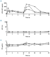
Factor D inhibitor inhibits alternative pathway of complement (APC) activity following oral dosing in cynomolgus monkeys. (A) ACH-4471 concentrations at the indicated time points in plasma samples collected from 3 cynomolgus monkeys following oral dosing with ACH-4471 at 200 mg/kg at 0 and 12 hours (arrows). Key pharmacokinetic parameters were calculated as follows: maximum concentration (Cmax), 4020±1480 ng/mL; time at Cmax (Tmax), 15.3±1.2 hours (3.3±1.2 hours following the second dose), and 30-hour exposure level (AUC0-30), 48,300±19,100 ng/mL/h. Parameters were calculated as mean ± standard deviation (SD) from the 3 animals. (B) APC activity assessed by Wieslab assay in serum samples collected at the indicated time points. Activity in each sample was normalized to the activity in the same animal at 0 h (pre-dosing). Mean±SD are shown from duplicate assay values. (C) Serum FD concentrations at the indicated time points. Circle, square, and triangle shapes; animals 1, 2, and 3, respectively.

References

    1. Varela JC, Tomlinson S. Complement: An Overview for the Clinician. Hematol Oncol Clin North Am. 2015;29(3):409–427. - PMC - PubMed
    1. Walport MJ. Complement. First of two parts. N Engl J Med. 2001;344(14):1058–1066. - PubMed
    1. Brodsky RA. Paroxysmal nocturnal hemoglobinuria. Blood. 2014;124(18):2804–2811. - PMC - PubMed
    1. Cataland SR, Wu HM. Diagnosis and management of complement mediated thrombotic microangiopathies. Blood Rev. 2014; 28(2):67–74. - PubMed
    1. Brodsky RA. Complement in hemolytic anemia. Blood. 2015;126(22):2459–2465. - PubMed

Publication types

MeSH terms