. 2016 Oct 17;213(11):2269-2279.
doi: 10.1084/jem.20151975. Epub 2016 Oct 10.

M-CSF improves protection against bacterial and fungal infections after hematopoietic stem/progenitor cell transplantation

Affiliations

M-CSF improves protection against bacterial and fungal infections after hematopoietic stem/progenitor cell transplantation

Prashanth K Kandalla et al. J Exp Med. .

Abstract

Myeloablative treatment preceding hematopoietic stem cell (HSC) and progenitor cell (HS/PC) transplantation results in severe myeloid cytopenia and susceptibility to infections in the lag period before hematopoietic recovery. We have previously shown that macrophage colony-stimulating factor (CSF-1; M-CSF) directly instructed myeloid commitment in HSCs. In this study, we tested whether this effect had therapeutic benefit in improving protection against pathogens after HS/PC transplantation. M-CSF treatment resulted in an increased production of mature myeloid donor cells and an increased survival of recipient mice infected with lethal doses of clinically relevant opportunistic pathogens, namely the bacteria Pseudomonas aeruginosa and the fungus Aspergillus fumigatus M-CSF treatment during engraftment or after infection efficiently protected from these pathogens as early as 3 days after transplantation and was effective as a single dose. It was more efficient than granulocyte CSF (G-CSF), a common treatment of severe neutropenia, which showed no protective effect under the tested conditions. M-CSF treatment showed no adverse effect on long-term lineage contribution or stem cell activity and, unlike G-CSF, did not impede recovery of HS/PCs, thrombocyte numbers, or glucose metabolism. These results encourage potential clinical applications of M-CSF to prevent severe infections after HS/PC transplantation.


Figures

Figure 1.
Figure 1.
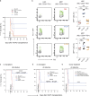
M-CSF protects against P. aeruginosa infection by inducing increased myelopoiesis of transplanted HS/PCs. (a) Lethally irradiated CD45.2 mice were transplanted with 2,500 CD45.1 HS/PCs, treated with three doses of control PBS or M-CSF during transplantation (−1 h, 5 h, and 18 h), challenged after 1 wk by intraperitoneal injection of 500 CFU of P. aeruginosa, and analyzed for bacterial load and myeloid donor cells 1 d after infection or for survival. (b and c) Survival of mice after HS/PC transplantation and bacterial infection (arrow) for PBS control (n = 13) or mice treated with three doses of 10 µg of baculovirus-expressed rmM-CSF (n = 8; b) or three doses of 10 µg rhM-CSF (n = 10; c). Transplanted, not infected (black line; n = 4) and irradiated (XR), untransplanted and uninfected control mice (dashed line; n = 6) are shown. P < 0.001 (rmM-CSF) and P < 0.05 (rhM-CSF) by a Mantel-Cox test. (d) Bacterial load in indicated organs 18 h after infection of transplanted mice treated with rhM-CSF or control PBS. (e and f) Median of donor monocytes (CD11b+, Ly6C+) and monocyte-derived macrophages (CD11b+, Ly6C+, F4/80int) in the liver or granulocytes (Ly6G+, CD11b+) and mononuclear phagocytes (Ly6G, CD11b+) in the spleen of PBS control or M-CSF–treated recipient mice 1 d after infection (e) or 9 d after transplantation of uninfected mice (f). Gating strategy is shown in Fig. S1. All data are representative of at least two independent experiments. (d–f) P-values were obtained by Mann-Whitney U tests. **, P < 0.01; ***, P < 0.001.
Figure 2.
Figure 2.
M-CSF protects against A. fumigatus infection after HS/PC transplantation. (a) Mice were transplanted and treated with M-CSF as described in Fig. 1 a, infected after 1 wk by intranasal instillation of A. fumigatus (4–6 × 10⁶ CFU), and analyzed for fungal load after 2 d or for survival. (b and c) Survival of mice after HS/PC transplantation and fungal infection (arrow) for PBS control (n = 10) or mice treated with three doses of 10 µg rmM-CSF (n = 10; b) or three doses of 10 µg rhM-CSF (n = 10; c). Transplantation (n = 4) and irradiation (XR) controls (n = 4) are shown. P < 0.001 (rmM-CSF) and P < 0.01 (rhM-CSF) by Mantel-Cox tests. (d) Fungal load in indicated organs 48 h after infection of transplanted mice treated with rhM-CSF (n = 4) or control PBS (n = 4). Photos of cultures from two representative mice are shown. All data are representative of at least two independent experiments.
Figure 3.
Figure 3.
Specific M-CSF effect on early HSC commitment correlates with better antibacterial protection than G-CSF during HS/PC transplantation. (a) Mice were transplanted, treated, and infected as described in Fig. 1 a. Survival of mice after transplantation and bacterial infection (arrow) for PBS control (n = 10) or mice treated with rmM-CSF (n = 10), rhM-CSF (n = 11), or rhG-CSF (n = 10) is shown. P < 0.05 (rmM-CSF or rhM-CSF) by a Mantel-Cox test. XR, irradiation. (b) Representative FACS profiles and median of GFP expression in HSCs (KSL CD34 Flt3) and multipotent progenitors (MPP; KSL CD34+ Flt3+) of PU.1-GFP reporter mice 20 h after control PBS, rhM-CSF, or rhG-CSF injection. **, P < 0.01 by a Mann-Whitney U test. (c and d) Survival of nontransplanted lethally irradiated mice treated with three doses of control PBS (n = 6) or 10 µg rmM-CSF (n = 6) after irradiation (6 h, 12 h, and 24 h) and challenged after 8 (c) or 3 (d) d (arrow) by i.p. injection of 500 CFU of P. aeruginosa. Control mice were treated identically but transplanted with 2,500 HS/PCs (n = 6). ****, P < 0.0001 (rmM-CSF + HS/PC) by a Mantel-Cox test. (e) Survival of mice transplanted with donor HS/PCs from mice treated with three doses (−5 h, −3 h, and −1 h) of 10 µg rmM-CSF (n = 10) or PBS (n = 8) and infected after 3 d (arrow) with 500 CFU of P. aeruginosa. Control recipient mice were treated with three doses of 10 µg rmM-CSF (n = 8) during transplantation. ***, P < 0.001 (rmM-CSF–treated donor HS/PCs) by a Mantel-Cox test. Transplantation (n = 4) and irradiation controls (n = 4) are shown in all survival analyses. All data are representative of two independent experiments.
Figure 4.
Figure 4.
M-CSF provides antibacterial protection of HS/PC transplant recipients for early infection and single-dose or postinfection cytokine treatment. (a) Mice were transplanted and treated with PBS, M-CSF, or G-CSF as described in Fig. 1 a but infected after 3 d. Survival of mice after transplantation and bacterial infection (arrow) for PBS control (n = 10) or mice treated with three doses of 10 µg rmM-CSF (n = 10) or rhG-CSF (n = 10) is shown. **, P < 0.01 (rmM-CSF) by Mantel-Cox test. XR, irradiation. (b and c) Confocal images with quantification of bacterial colonies (b) or donor cells (c) in spleens 24 h after early infection on day 3 of transplanted mice treated with rmM-CSF or control PBS. ****, P < 0.0001 by a Mann-Whitney U test. Bars, 50 µm. Arrowheads indicate the maximum colony size in the M-CSF–treated group. (d) Mice were transplanted and infected as described in Fig. 1 a but treated with a single dose of control PBS, M-CSF, or G-CSF during transplantation (5 h). Survival of mice after transplantation and bacterial infection (arrow) for PBS control (n = 12) or mice treated with a single dose of 10 µg rmM-CSF (n = 12) or rhG-CSF (n = 12) is shown. **, P < 0.01 by a Mantel-Cox test. (e) Mice were transplanted and infected as described in Fig. 1 a but treated with three doses (1 h, 3 h, and 5 h) of 10 µg rmM-CSF or PBS after infection. Survival of mice after transplantation and bacterial infection (arrow) for PBS control (n = 10) or rmM-CSF (n = 12) is shown. *, P < 0.05 by a Mantel-Cox test. Transplantation (n = 4) and irradiation controls (n = 4) are shown in all survival analyses. All data are representative of two independent experiments.
Figure 5.
Figure 5.
M-CSF treatment does not compromise HSC self-renewal, multilineage differentiation, or stem cell activity. (a) Mice were transplanted with actin-GFP HS/PCs and treated with rmM-CSF, rhG-CSF, or PBS as described in Fig. 1 a and analyzed after 17 wk for donor cell contribution in blood and bone marrow. (b and c) Percentage of GFP+ donor cell contribution to CD11b+ myeloid, CD19+ B cells, or CD3e+ T cells in blood of reconstituted mice (b) or expressed as a ratio of CD11b+ myeloid cells to CD19+ lymphoid cells (c). (d) Percentage of GFP+ donor cell contribution and percentage of total bone marrow cells for c-kit+ progenitors and HS/PCs (KSL). (e) Percentage of GFP+ donor cell contribution to indicated stem and progenitor cell populations. (f) Percentage of GFP+ donor cell contribution to myeloid and lymphoid cells in blood 7 wk after secondary transplantation. Error bars represent the median with range. *, P < 0.05; **, P < 0.01 by Mann-Whitney U tests. All data are representative of two independent experiments.
Figure 6.
Figure 6.
M-CSF treatment does not affect platelet recovery or blood glucose levels. (a–c) Mice were transplanted with actin-GFP HS/PCs and treated with rmM-CSF, rhG-CSF, or PBS as described in Fig. 1 a and analyzed for donor side scatter (SSC)lo forward scatter (FSC)lo platelet contribution in the blood shown as time course of median absolute numbers (b) or quantification at indicated time points (c). Non-irradiated (noXR) nontransplanted controls were used as a reference. For 1 wk, * = 0.011 (>0.01 and <0.06). For 7 wk, * = 0.06. (d) Time course of weight and blood glucose levels of mice transplanted and treated with rmM-CSF (n = 6) or PBS (n = 6) as indicated in Fig. 1 a. Error bars represent the median and error with interquartile range. **, P < 0.01; ***, P < 0.001 by Mann-Whitney U tests. Data are representative of two independent experiments.

References

    1. Baldridge M.T., King K.Y., and Goodell M.A.. 2011. Inflammatory signals regulate hematopoietic stem cells. Trends Immunol. 32:57–65. 10.1016/j.it.2010.12.003 - DOI - PMC - PubMed
    1. BitMansour A., Burns S.M., Traver D., Akashi K., Contag C.H., Weissman I.L., and Brown J.M.. 2002. Myeloid progenitors protect against invasive aspergillosis and Pseudomonas aeruginosa infection following hematopoietic stem cell transplantation. Blood. 100:4660–4667. 10.1182/blood-2002-05-1552 - DOI - PubMed
    1. BitMansour A., Cao T.M., Chao S., Shashidhar S., and Brown J.M.. 2005. Single infusion of myeloid progenitors reduces death from Aspergillus fumigatus following chemotherapy-induced neutropenia. Blood. 105:3535–3537. 10.1182/blood-2004-07-2676 - DOI - PMC - PubMed
    1. Bodine D.M., Seidel N.E., and Orlic D.. 1996. Bone marrow collected 14 days after in vivo administration of granulocyte colony-stimulating factor and stem cell factor to mice has 10-fold more repopulating ability than untreated bone marrow. Blood. 88:89–97. - PubMed
    1. Bryder D., Rossi D.J., and Weissman I.L.. 2006. Hematopoietic stem cells: the paradigmatic tissue-specific stem cell. Am. J. Pathol. 169:338–346. 10.2353/ajpath.2006.060312 - DOI - PMC - PubMed

MeSH terms

Substances