. 2016 Nov 4:6:36498.
doi: 10.1038/srep36498.

On-chip, multisite extracellular and intracellular recordings from primary cultured skeletal myotubes

Affiliations

On-chip, multisite extracellular and intracellular recordings from primary cultured skeletal myotubes

Noha Rabieh et al. Sci Rep. .

Abstract

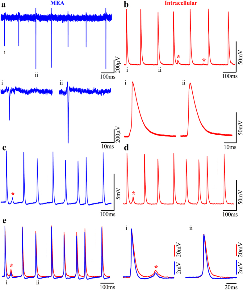
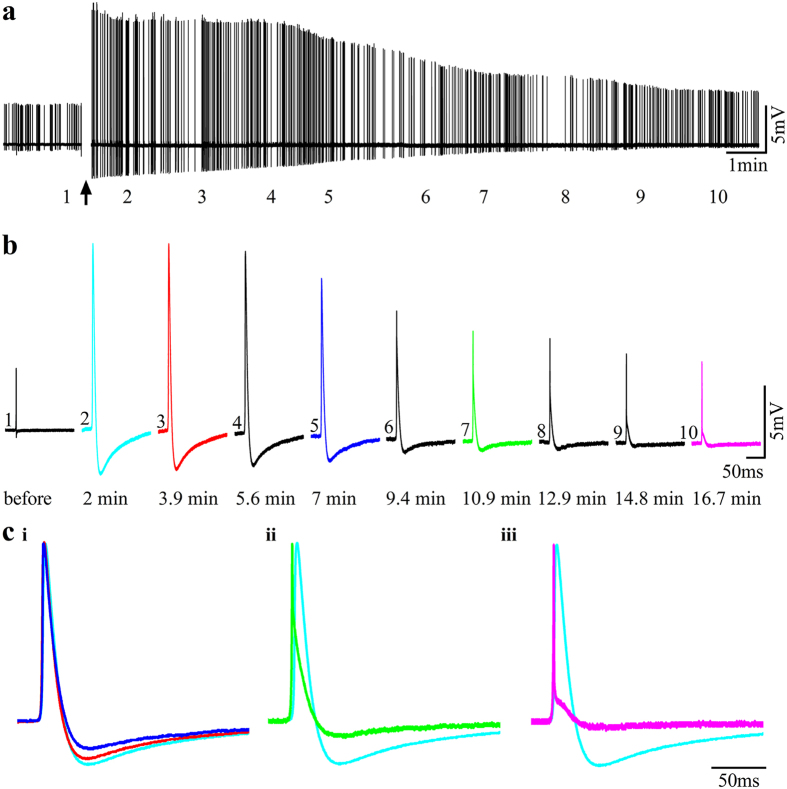
In contrast to the extensive use of microelectrode array (MEA) technology in electrophysiological studies of cultured neurons and cardiac muscles, the vast field of skeletal muscle research has yet to adopt the technology. Here we demonstrate an empowering MEA technology for high quality, multisite, long-term electrophysiological recordings from cultured skeletal myotubes. Individual rat skeletal myotubes cultured on micrometer sized gold mushroom-shaped microelectrode (gMμE) based MEA tightly engulf the gMμEs, forming a high seal resistance between the myotubes and the gMμEs. As a consequence, spontaneous action potentials generated by the contracting myotubes are recorded as extracellular field potentials with amplitudes of up to 10 mV for over 14 days. Application of a 10 ms, 0.5-0.9 V voltage pulse through the gMμEs electroporated the myotube membrane, and transiently converted the extracellular to intracellular recording mode for 10-30 min. In a fraction of the cultures stable attenuated intracellular recordings were spontaneously produced. In these cases or after electroporation, subthreshold spontaneous potentials were also recorded. The introduction of the gMμE-MEA as a simple-to-use, high-quality electrophysiological tool together with the progress made in the use of cultured human myotubes opens up new venues for basic and clinical skeletal muscle research, preclinical drug screening, and personalized medicine.


Figures

Figure 1
Figure 1. Transmission electron micrographs of 10 day old cultured myotubes.
(a) Low magnification of a long multinucleated myotube (3 nuclei –N) with a distinct region showing sarcomere structures (S). The sarcomeres are enlarged in (b,c). (di and dii) a cultured myotube “resting” on top of gMμPs (black arrowheads) does not adhere to the culture substrate that is spattered with residual small gold particles (empty arrow heads). (e) Example of the engulfment of gMμPs by cultured myotubes. (ei) low magnification of a long myotube engulfing three gMμPs. The translucent areas around the gMμPs were formed by the electron beam of the microscope during the observation. (eii and eiii), examples of tight engulfment of gMμPs by myotubes which also adhere to the gold substrate (black).
Figure 2
Figure 2. Skeletal myotubes cultured on polydopamine and matrigel functionalized gMμE-MEA.
(a) 1DIV and (b) 4 DIV after the final plating cycle.
Figure 3
Figure 3. Raw recordings of spontaneous field potentials by gMμE-MEA from cultured myotube 4 DIV.
Each box show FPs recordings by a single gMμE. Biphasic field potentials are depicted in green (a) and enlarged in (b, electrode 77). FPs dominated by a negative component are labeled in blue, and enlarged in (c, electrode 83) and FPs dominated by a positive component are labeled red, and enlarged in (d, electrode 34). Electrodes that peaked up FPs <0.1 mV are marked in black. Note that a fraction of the gMμE record both negative and biphasic FPs and others both negative and positive FPs. Whereas the FPs waveform shape (c) suggests that the majority of the individual gMμE pick up the activity from a single myotube, it is interesting to note that the FP amplitudes recorded by individual gMμE were not constant.
Figure 4
Figure 4. Concomitant bursts of field potentials recorded from a population of myotube 7 DIV after the final replating.
(a) Raw FP recordings from 26 gMμE reveal that gMμE spaced over the entire recording surface area (total recording surface of 0.8 × 0.8 mm) fire in apparent synchrony. Enlargement of the FPs marked by a box in (a) shows that the FPs recorded by the different gMμE have different waveform shapes (b,c) and are not generated at the same time (c). The FPs propagates at an estimated rate of 150–200 cm/s. This may represent the conduction velocity of action potentials along large myotubes or that a fraction of the 7 DIV myotubes were electrically coupled.
Figure 5
Figure 5. Comparison of intracellular recorded potentials to IN-CELL recordings from cultured myotubes.
Concomitant spontaneous extracellular FPs recordings by a gMμE (a) and intracellular recordings by sharp electrodes (b) from 3 DIV myotubes after the final replating. The recordings in (a,b) revealed identical firing patterns and similar qualitative alterations in the amplitudes of the recorded action potentials. The field potentials labeled i, and ii, and action potentials i and ii are shown in the lower traces with an expanded time scale. (c) Electroporation of the myotube changed the gMμE mode of recording from extracellular to the IN-CELL mode. Note that although the recorded amplitude of the spikes is about an order of magnitude lower than that of the intracellular electrode, the shape of the recorded potentials are identical. Also, note that in (c,d) both electrodes recorded subthreshold potentials (red asterisk) in between the spikes. (e) Merge traces of c (blue) and d (red), i and ii are traces with an expanded time scale.
Figure 6
Figure 6. Accessing IN-CELL recording by membrane electroporation and its reversal by membrane repair (3DIV after the final replating).
(a1) Before electroporation, a gMμE recorded an extracellular positive FP (a1 and b1) characterized by 3.6 mV and a short duration of 0.25 ms (50% height). After the delivery of an electroporating pulse, the extracellular FP transformed into a 12 mV, 4.9 ms. IN-CELL recorded potential. The amplitude of the action potential gradually diminished over a period of approximately 30 minutes (a,b), resuming the shape of an extracellular field potential (ci–iii). Super-positioning of the first IN-CELL recorded potential (light blue) on normalized potentials recorded at different points in time after the electroporation (color coded as in b) revealed that aside from a gradual reduction of the spike amplitude shown in (b) the duration of the potential was gradually and significantly reduced as the recording configuration changed from IN-CELL to extracellular (c).
Figure 7
Figure 7. Spontaneous subthreshold potentials are generated by the spread of current generated among electrically coupled myotubes.
Intracellular recordings from spontaneously firing myotubes (3 DIV after the final plating cycle) cultured in the absence of neurons were made by a sharp intracellular microelectrode. The electrode was used for both current injection and voltage recordings. (a) hyperpolarizing square current pulse injunction. Traces of spontaneous firing (b–e) during hyperpolarization of the myotube from which the recordings were made reduced the frequency of the subthreshold potentials. In (b) spontaneous spikes and subthreshold potentials (red asterisks) are shown whereas in (c–e) the recordings were trimmed along the dashed line shown in (b). On the other hand depolarization (f) increased the frequency of the subthreshold potentials (g–j red asterisk). In (g) spontaneous spikes and subthreshold potentials (red asterisks) are shown whereas in (h–j) the recordings were trimmed along the dashed line as shown in (g).
Figure 8
Figure 8. Simulation of action potential amplitudes and their electrotonic spread among coupled myotubes.
(aii) An 80 mV action potential generated by myotube spike current pulse injection (2.22 nA) into an isolated model myotube (as shown in ai). (bii) When the same current pulse (2.22 nA) is injected into the same myotube but after its coupling to a second myotube (as shown in bi) the voltage amplitude is reduced to 70 mV. Concomitantly (cii), an attenuated potential-an electrotonic excitatory potential is recorded in the second coupled myotube (red asterisk). Action potential and voltage spread recorded “intracellularly” from myotube 1 when an action potential current is injected first into myotube 2 and then into myotube 1 (ci). The time interval between the two current injections is reduced from −60 to 0 ms (in 10 ms steps) and then increased to + 60 ms. As the decremental potential (red asterisk) and the action potential summate, the peak amplitude of the action potential is increased and reaches a maximum at an interval of −10. (dii) The attenuated potentials as “recorded” by a gMuE (di). Note that although the shapes of the simulated potentials are similar, the interface with a gMμE simulation circuit leads to attenuation of the potentials’ amplitudes.

References

    1. Verschuuren J., Strijbos E. & Vincent A. Neuromuscular junction disorders. Handbook of clinical neurology 133, 447–466, doi: 10.1016/B978-0-444-63432-0.00024-4 (2016). - DOI - PubMed
    1. Liang C. L. & Han S. Neuromuscular junction disorders. PM R 5, S81–S88, doi: 10.1016/j.pmrj.2013.03.016 (2013). - DOI - PubMed
    1. Gilhus N. E. et al.. Myasthenia gravis - autoantibody characteristics and their implications for therapy. Nature reviews. Neurology 12, 259–268, doi: 10.1038/nrneurol.2016.44 (2016). - DOI - PubMed
    1. Picher-Martel V., Valdmanis P. N., Gould P. V., Julien J. P. & Dupre N. From animal models to human disease: a genetic approach for personalized medicine in ALS. Acta neuropathologica communications 4, 70, doi: 10.1186/s40478-016-0340-5 (2016). - DOI - PMC - PubMed
    1. Fuller H. R., Gillingwater T. H. & Wishart T. M. Commonality amid diversity: Multi-study proteomic identification of conserved disease mechanisms in spinal muscular atrophy. Neuromuscular disorders: NMD, doi: 10.1016/j.nmd.2016.06.004 (2016). - DOI - PubMed

Publication types