Comparative Study
. 2017 Jan 1;102(1):200-209.
doi: 10.1210/jc.2016-3001.

Increased Proliferation of the Pancreatic Duct Gland Compartment in Type 1 Diabetes

Affiliations
Comparative Study

Increased Proliferation of the Pancreatic Duct Gland Compartment in Type 1 Diabetes

Abu Saleh Md Moin et al. J Clin Endocrinol Metab. .

Abstract

Context: Pancreatic duct glands (PDGs) have been proposed as a source of regeneration in response to exocrine pancreas injury, and thus may serve as an organ stem cell niche. There is evidence to suggest ongoing β-cell formation in longstanding type 1 diabetes (T1D), but the source is unknown.

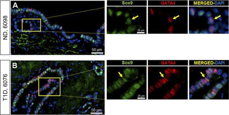
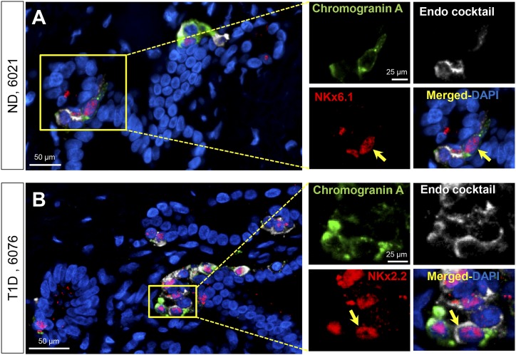
Objective: To investigate the PDG compartment of the pancreas in humans with T1D for evidence of an active regenerative signature (presence of progenitor cells and increased proliferation) and, in particular, as a potential source of β-cells.

Design, setting, and participants: Pancreases from 46 brain dead organ donors (22 with T1D, 24 nondiabetic controls) were investigated for activation (increased proliferation) and markers of pancreatic exocrine and endocrine progenitors.

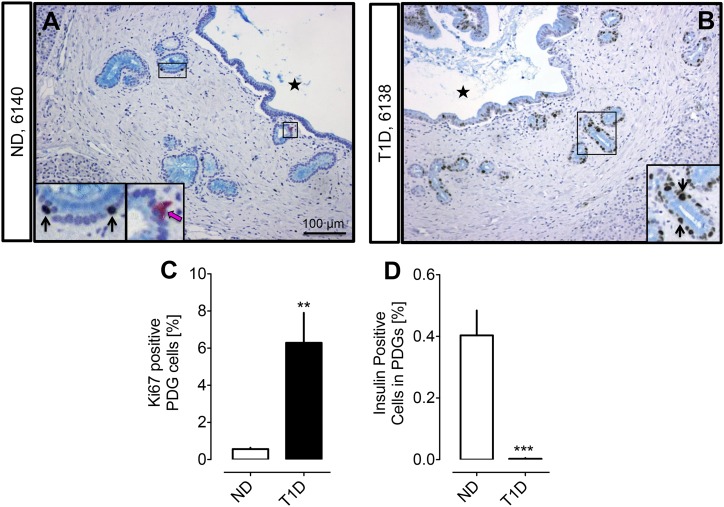
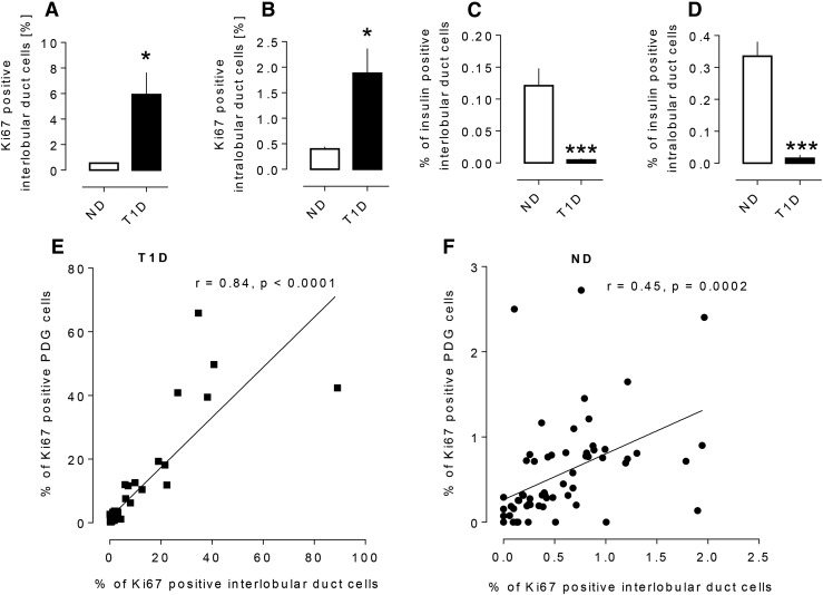
Results: PDG cell replication was increased in T1D (6.3% ± 1.6% vs 0.6% ± 0.1%, P < 0.001, T1D vs nondiabetic), most prominently in association with pancreatic inflammation. There were increased progenitor-like cells in PDGs of T1D, but predominantly with an exocrine fate.

Conclusion: The PDG compartment is activated in T1D consistent with a response to ongoing inflammation, and via resulting ductal hyperplasia may contribute to local obstructive pancreatitis and eventual pancreatic atrophy characteristic of T1D. However, there is no evidence of effective endocrine cell formation from PDGs.


Figures

Figure 1.
Figure 1.
Examples of PDG compartments in ND (A) and subjects with T1D (B). Pancreas sections were stained by immunohistochemistry for insulin (pink), Ki67 (brown), and alcian blue with hemotoxylin counterstain. Insets, magnified areas of the low-power images marked by black rectangle. Black arrows indicate Ki67-positive nuclei in PDG cells. Pink arrows indicate insulin-positive PDG cells. Black stars indicate the interlobular duct lumen. (C) The frequency of replicating cells in the epithelium of PDGs is higher in T1D compared with ND (6.3% ± 1.6% vs 0.6% ± 0.1%, T1D vs ND, **P < 0.001). (D) The frequency of insulin-positive cells in the epithelium of PDGs is higher in ND compared with T1D (0.003% ± 0.002% vs 0.40% ± 0.08%, T1D vs ND, ***P < 0.0001). White bars = ND, black bars = T1D. Scale bar, 100 μm.
Figure 2.
Figure 2.
The frequency of replicating cells in the epithelium of interlobular duct cells (5.9 ± 1.7 vs 0.6 ± 0.1% Ki67-positive cells, T1D vs ND, *P < 0.01) (A) and intralobular duct cells (1.9 ± 0.5 vs 0.4 ± 0.0% Ki67-positive cells, *P < 0.01, T1D vs ND) (B) in T1D and ND subjects. The frequency of insulin-positive cells in the epithelium of interlobular ducts (0.005 ± 0.002 vs 0.12 ± 0.03% insulin-positive cells T1D vs ND, ***P < 0.0001) (C) and intralobular ducts (0.02 ± 0.01 vs 0.34 ± 0.05% insulin-positive cells, T1D vs ND, ***P < 0.0001) (D) in T1D and ND subjects. The relationship between frequency of Ki67 positivity in interlobular duct cells and in PDG cells from subjects with T1D (E) and ND subjects (F). The percentage of Ki67-positive cells in the PDGs (the number of Ki67+ PDG cells/total number of PDG cells) and the percentage of Ki67-positive cells in the interlobular pancreatic ducts (the number of Ki67+ interlobular duct cells/total number of interlobular duct cells) show a positive correlation in both the T1D donors (r = 0.84, P < 0.0001) and the ND donors (r = 0.45, P = 0.0002). Data were present as mean ± SEM, n = 22 donors for T1D, the data points representing 22 sections of head of pancreas, 21 sections of body of pancreas, and 22 sections of tail of pancreas; n = 24 donors for ND, the data points representing 24 sections of head of pancreas, 16 sections of body of pancreas, and 24 sections of tail of pancreas. White bars, ND, black bars, T1D.
Figure 3.
Figure 3.
Examples of PDG cells expressing Sox9-GATA4 in pancreas from an adult ND (A) and in an adult T1D donor (B). Individual layers stained for Sox9 (green) and GATA4 (red) are shown along with merged (with 4′,6-diamidino-2-phenylindole) images. Insets, higher magnification of individual layers as marked by the yellow square in the low-power images. Yellow arrows indicate a PDG cell costaining for Sox9 and GATA4. Scale bars, 50 µm (for low-power images) and 25 µm (for insets).
Figure 4.
Figure 4.
Examples of PDG cells expressing chromogranin A, NKx6.1, and endocrine cocktail (insulin, glucagon, somatostatin, pancreatic polypeptide, and ghrelin) in pancreas from an adult ND donor (A) and chromogranin A, NKx2.2, and endocrine cocktail in an adult T1D donor (B). Individual layers stained for NKx6.1/NKx2.2 (red), endocrine cocktail (insulin, glucagon, somatostatin, pancreatic polypeptide, and ghrelin) (white), and chromogranin A (green) are shown along with the merged (with 4′,6-diamidino-2-phenylindole) image. Insets, higher magnification of individual layers as marked by the yellow square in the low-power images. Yellow arrows indicate a PDG cell costaining for Nkx6.1, chromogranin A, and a cocktail of all the endocrine hormones (A) or Nkx2.2, chromogranin A, and a cocktail of all the endocrine hormones (B). Scale bars, 50 µm (for low-power images) and 25 µm (for insets).

References

    1. Atkinson MA, von Herrath M, Powers AC, Clare-Salzler M. Current concepts on the pathogenesis of type 1 diabetes: considerations for attempts to prevent and reverse the disease. Diabetes Care. 2015;38(6):979–988. - PMC - PubMed
    1. Klöppel G, Löhr M, Habich K, Oberholzer M, Heitz PU. Islet pathology and the pathogenesis of type 1 and type 2 diabetes mellitus revisited. Surv Synth Pathol Res. 1985;4(2):110–125. - PubMed
    1. Meier JJ, Bhushan A, Butler AE, Rizza RA, Butler PC. Sustained beta cell apoptosis in patients with long-standing type 1 diabetes: indirect evidence for islet regeneration? Diabetologia. 2005;48(11):2221–2228. - PubMed
    1. Gepts W. Pathologic anatomy of the pancreas in juvenile diabetes mellitus. Diabetes. 1965;14(10):619–633. - PubMed
    1. Sjöberg S, Gunnarsson R, Gjötterberg M, Lefvert AK, Persson A, Ostman J. Residual insulin production, glycaemic control and prevalence of microvascular lesions and polyneuropathy in long-term type 1 (insulin-dependent) diabetes mellitus. Diabetologia. 1987;30(4):208–213. - PubMed

Publication types

MeSH terms