

Review
doi: 10.1016/j.stem.2016.10.015.
Neural Subtype Specification from Human Pluripotent Stem Cells
Affiliations
- PMID: 27814479
- PMCID: PMC5127287
- DOI: 10.1016/j.stem.2016.10.015
Item in Clipboard
Review
Neural Subtype Specification from Human Pluripotent Stem Cells
Cell Stem Cell.
.
Abstract
Human pluripotent stem cells (hPSCs) provide a model to study early neural development, model pathological processes, and develop therapeutics. The generation of functionally specialized neural subtypes from hPSCs relies on fundamental developmental principles learned from animal studies. Manipulation of these principles enables production of highly enriched neural types with functional attributes that resemble those in the brain. Further development to promote faster maturation or aging as well as circuit integration will help realize the potential of hPSC-derived neural cells in disease modeling and cell therapy.
Copyright © 2016 Elsevier Inc. All rights reserved.
Figures
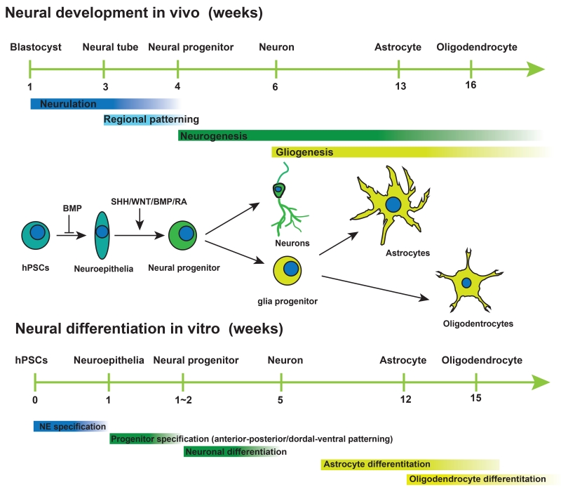
Cartoon illustrating the major developmental events in vivo and neural differentiation process in vitro and the morphogens that govern the neural induction, patterning of neural progenitors and neuronal and glial differentiation of the progenitors.
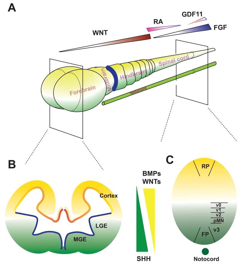
A. A-P patterning is under the regulation of several morphogens during development. The gradient of WNTs dictates the regionalization of the forebrain, mid-hindbrain, and anterior spinal cord whereas gradients of RA and FGFs govern the spinal cord segmentation. B and C, D-V patterning in the forebrain (B) and spinal cord (C) is set by the dorsally derived morphogens WNTs and BMPs (yellow color) and the (notocord) ventrally derived SHH (green color).
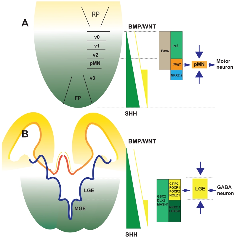
A. To enrich the neural progenitors from the pMN domain in the spinal cord, a high concentration of SHH or Purmorphamine (SHH signalling agonist) is employed to induce the OLIG2+ and the more ventral NKX2.2+ progenitors. Since the more ventral NKX2.2+ cells give rise to interneurons rather than motor neurons, CHIR (activate the WNT pathway) is used to antagonize the effect of SHH in induction of NKX2.2 but not OLIG2, leading to the enrichment of the OLIG2+ progenitors. Such a strategy enables generation of highly enriched population of spinal motor neurons. B. To enrich LGE progenitors and striatal GABA neurons, forebrain NE are ventralized to the LGE and MGE domains. At the same time, the most ventral MGE domain is antagonized by activin (on the BMP pathway). Together, the opposing morphogens restrict the cells to the LGE domain, thus producing enriched striatal GABA neurons.
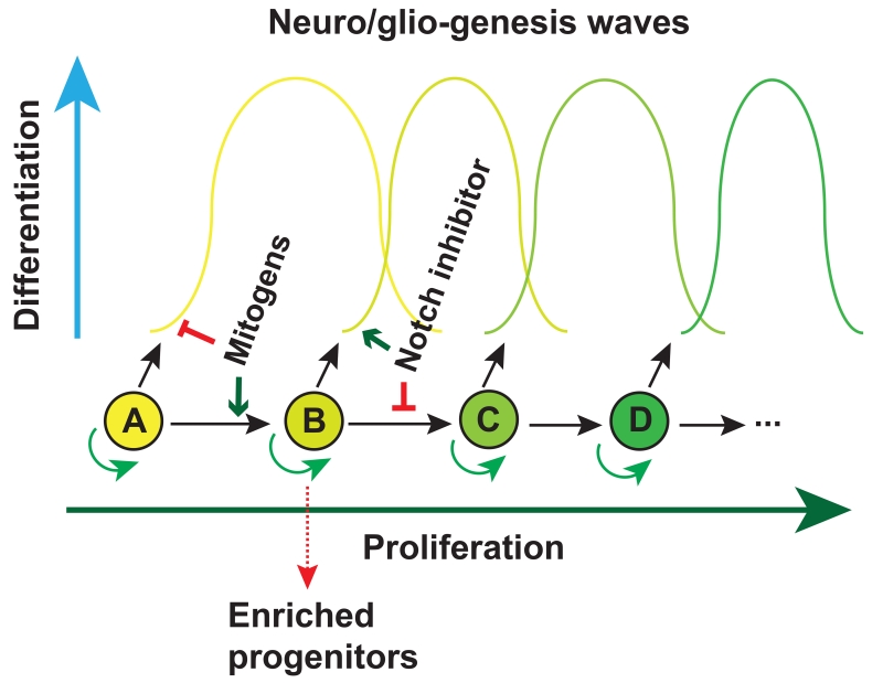
Neural progenitors such as radial glia or retinal progenitors give rise to multiple progenies (waves) over development. If the progenitor cell is kept in cell cycle (to prevent differentiation) it shifts to the next progenitor pool. If the progenitor is forced to exit cell cycle (e.g., by γ-secretase inhibitors (also known as Notch inhibitors)), no subsequent progenitor pools are available. Together, a particular neuronal subtype is enriched.
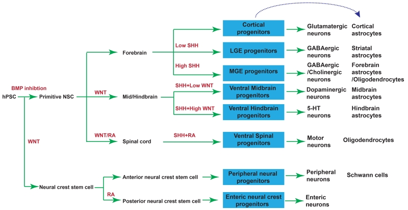
Neuronal and glial subtypes that have been successfully differentiated from hPSCs through NE induction, regional patterning, and neural differentiation. Red indicates morphogens applied in each step. Blue boxes indicate subtype progenitors.
References
-
- Bardy C, van den Hurk M, Eames T, Marchand C, Hernandez RV, Kellogg M, Gorris M, Galet B, Palomares V, Brown J, et al. Neuronal medium that supports basic synaptic functions and activity of human neurons in vitro. Proceedings of the National Academy of Sciences of the United States of America. 2015;112:E2725–2734. - PMC - PubMed
-
- Borghese L, Dolezalova D, Opitz T, Haupt S, Leinhaas A, Steinfarz B, Koch P, Edenhofer F, Hampl A, Brustle O. Inhibition of notch signaling in human embryonic stem cell-derived neural stem cells delays G1/S phase transition and accelerates neuronal differentiation in vitro and in vivo. Stem Cells. 2010;28:955–964. - PubMed
Publication types
MeSH terms
Grants and funding
LinkOut - more resources
Full Text Sources
Other Literature Sources
