. 2016 Dec 15;5(12):1834-1843.
doi: 10.1242/bio.012021.

BMP4 acts as a dorsal telencephalic morphogen in a mouse embryonic stem cell culture system

Affiliations

BMP4 acts as a dorsal telencephalic morphogen in a mouse embryonic stem cell culture system

Momoko Watanabe et al. Biol Open. .

Abstract

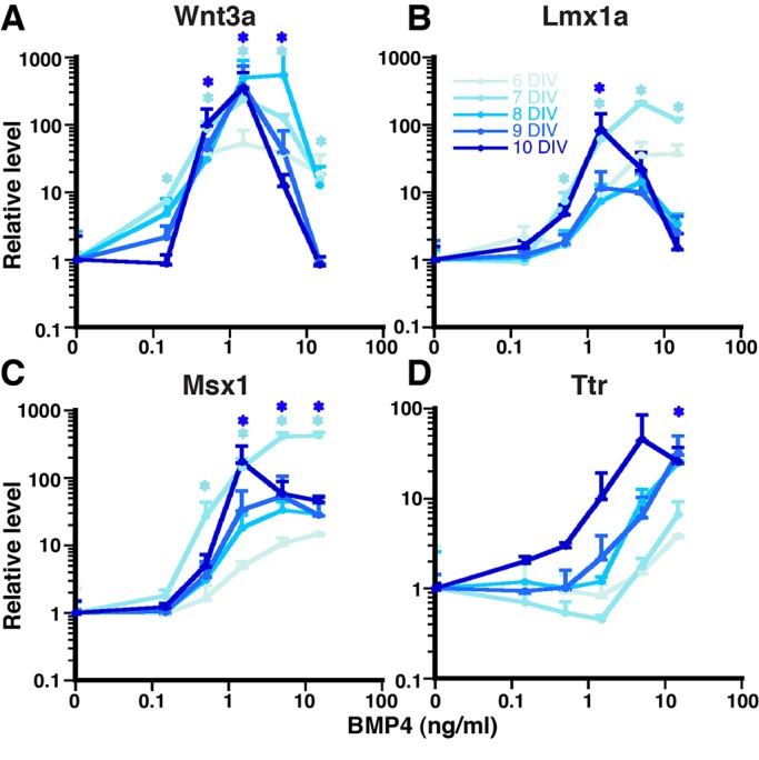
The concept of a morphogen - a molecule that specifies two or more cell fates in a concentration-dependent manner - is paradigmatic in developmental biology. Much remains unknown, however, about the existence of morphogens in the developing vertebrate central nervous system (CNS), including the mouse dorsal telencephalic midline (DTM). Bone morphogenetic proteins (BMPs) are candidate DTM morphogens, and our previous work demonstrated BMP4 sufficiency to induce one DTM cell fate - that of choroid plexus epithelial cells (CPECs) - in a mouse embryonic stem cell (mESC) culture system. Here we used BMP4 in a modified mESC culture system to derive a second DTM fate, the cortical hem (CH). CH and CPEC markers were induced by BMP4 in a concentration-dependent manner consistent with in vivo development. BMP4 concentrations that led to CH fate also promoted markers for Cajal-Retzius neurons, which are known CH derivatives. Interestingly, single BMP4 administrations also sufficed for appropriate temporal regulation of CH, CPEC, and cortical genes, with initially broad and overlapping dose-response profiles that sharpened over time. BMP4 concentrations that yielded CH- or CPEC-enriched populations also had different steady-state levels of phospho-SMAD1/5/8, suggesting that differences in BMP signaling intensity underlie DTM fate choice. Surprisingly, inactivation of the cortical selector gene Lhx2 did not affect DTM expression levels, dose-response profiles, or timing in response to BMP4, although neural progenitor genes were downregulated. These data indicate that BMP4 can act as a classic morphogen to orchestrate both spatial and temporal aspects of DTM fate acquisition, and can do so in the absence of Lhx2.

Keywords: Choroid plexus epithelial cell; Cortical hem; ES cell.


Conflict of interest statement

The authors declare no competing or financial interests.

Figures

Fig. 1.
Fig. 1.
BMP4 dose-dependency to drive CH and CPEC fates. (A) Schematic of the monolayer (M) system to induce CH and CPEC fates from mESCs and the chart for DTM expression profile. (B) RT-qPCR of dissociated 5-day M2 and M1 aggregates with a single application of BMP4 at 10 DIV. Three BMP4-dose ranges are found to preferentially regulate cortex, CH, and CPECs markers. (C-J) Immunocytochemical analysis of dissociated 5-day M2 aggregates treated with BMP4 for another 2 DIV (C-F) or 5 DIV (G-J). Lmx1 (green) and Msx1 (blue) are initially highly upregulated with 1.5 ng/ml and 15 ng/ml BMP4 at 7 DIV, and restricted to 1.5 ng/ml BMP4 culture at 10 DIV. Ttr::RFP takes time to upregulate mainly in 15 ng/ml BMP4 culture and few in 1.5 ng/ml BMP4 culture. Corresponding fields for Hoechst staining (white) are shown in C′-J′ for cell density comparison. Blue arrowheads: Msx1 single positive; white arrowheads: Msx1/Lmx1 double positive; yellow arrowheads: Msx1/Lmx1/Ttr triple positive; pink arrowheads: Ttr single or Ttr/Msx1 double positive. Scale bar: 100 μm. (K) Quantification of DTM markers represented in C-J. Immuno-positive cells/total cells (Hoechst). Because C-J are magnified selected fields, lower magnification pictures are provided in Fig. S2. (L) Enriched Cajal-Retzius (CR) markers from CH-abundant cultures. M2 dissociated 5-day aggregates treated with 1.5 ng/ml (RT-qPCR, normalized to no BMP4 control at each time point) demonstrated increased levels of CR markers, p73 and Reelin, in a temporally sequential manner. For K and L, data are presented as mean±s.e.m.; *P<0.05 compared to no BMP culture.
Fig. 2.
Fig. 2.
Temporal regulation of DTM markers by BMP4. (A-D) RT-qPCR of dissociated 5-day M2 aggregates treated with BMP4 (0.15-15 ng/ml) for another 1-5 DIV. (A,B) CH markers, Wnt3a and Lmx1a, are increased quickly at moderate-high BMP4 concentration (0.15-15 ng/ml). However, CH markers are maintained only at moderate BMP4 concentration (0.5-1.5 ng/ml for Wnt3a, 1.5-5 ng/ml for Lmx1a), while they are downregulated at high (>5 ng/ml) BMP4 concentration. (C) Msx1 is a direct target of BMP signaling and activated at moderate-high BMP4 concentration (0.5-15 ng/ml) at early to late time points. (D) Ttr is a mature CPEC marker and activated only at high BMP4 concentration (>5 ng/ml) at later time points (after 8 DIV), consistent with Ttr being a mature late-onset CPEC marker in vivo. Similar results are obtained, using the M1 mESC line (Fig. S4). Data are presented as mean±s.e.m.; *P<0.05 compared to no BMP culture.
Fig. 3.
Fig. 3.
BMP4 mediated-neural progenitor marker regulation in a dose-dependent fashion. (A-J) 5-day SFEBq aggregates are dissociated and plated in monolayer with varying BMP4 (0.15-15 ng/ml for M2 and 0.15-150 ng/ml for M1) for another 1-5 DIV (6 to 10 DIV total). Usually, at higher BMP4 concentration, all neural progenitor expression is suppressed. Selectively, some neural progenitor markers are slightly upregulated in a temporal manner at lower concentration of BMP4. Data are presented as mean±s.e.m.; *P<0.05 compared to no BMP culture.
Fig. 4.
Fig. 4.
No facilitation of BMP4-mediated DTM upregulation by Lhx2 inactivation. (A) Experimental design. All SFEBq aggregates are treated with (dotted lines) or without (solid lines) 4HT (1 μM) at 4 DIV. After 24 h, Lhx2 is inactivated to about 95% (Fig. S5). 5-day SFEBq aggregates are then dissociated to single cells in monolayer with fresh media containing varying BMP4 concentrations (0.15-150 ng/ml) for another 2 DIV (B) or 5 DIV (C) for RT-qPCR analyses. All data points are normalized to monolayer cultures without BMP4 and 4HT. No significant gene expression change was detected, indicating that Lhx2 absence does not facilitate BMP4-mediated DTM induction. Data are presented as mean±s.e.m.; *P<0.05 compared to no BMP culture.
Fig. 5.
Fig. 5.
Downregulation of other neural progenitor markers by inactivation of Lhx2. 1 μM of 4HT or vehicle is applied to 4-day SFEBq aggregates and 5-day aggregates are dissociated with varying BMP4 concentrations (0.15-150 ng/ml). Gene expression is analyzed by RT-qPCR 2 DIV (A-E) or 5 DIV (F-J) later. At no-moderate BMP concentration (around 0-1.5 ng/ml), inactivation of Lhx2 reduces all neural progenitor markers, consistent with Lhx2 being an early hierarchy of telencephalic development. BMP4-mediated suppression of neural progenitor markers is not influenced by Lhx2 inactivation except Pax6 that is genetically linked with Lhx2. This indicates that Lhx2 does not directly interact with BMP signaling. Data are presented as mean±sem; *P<0.05 compared to no 4HT culture.

References

    1. Ashe H. L. and Briscoe J. (2006). The interpretation of morphogen gradients. Development 133, 385-394. 10.1242/dev.02238 - DOI - PubMed
    1. Cheng X., Hsu C.-m., Currle D. S., Hu J. S., Barkovich A. J. and Monuki E. S. (2006). Central roles of the roof plate in telencephalic development and holoprosencephaly. J. Neurosci. 26, 7640-7649. 10.1523/JNEUROSCI.0714-06.2006 - DOI - PMC - PubMed
    1. Chizhikov V. V., Lindgren A. G., Mishima Y., Roberts R. W., Aldinger K. A., Miesegaes G. R., Currle D. S., Monuki E. S. and Millen K. J. (2010). Lmx1a regulates fates and location of cells originating from the cerebellar rhombic lip and telencephalic cortical hem. Proc. Natl. Acad. Sci. USA 107, 10725-10730. 10.1073/pnas.0910786107 - DOI - PMC - PubMed
    1. Cornell R. A. and Ohlen T. V. (2000). Vnd/nkx, ind/gsh, and msh/msx: conserved regulators of dorsoventral neural patterning? Curr. Opin. Neurobiol. 10, 63-71. 10.1016/S0959-4388(99)00049-5 - DOI - PubMed
    1. Currle D. S., Cheng X., Hsu C.-m. and Monuki E. S. (2005). Direct and indirect roles of CNS dorsal midline cells in choroid plexus epithelia formation. Development 132, 3549-3559. 10.1242/dev.01915 - DOI - PubMed