

S-Ketamine Rapidly Reverses Synaptic and Vascular Deficits of Hippocampus in Genetic Animal Model of Depression
- PMID: 27815416
- PMCID: PMC5408982
- DOI: 10.1093/ijnp/pyw098
S-Ketamine Rapidly Reverses Synaptic and Vascular Deficits of Hippocampus in Genetic Animal Model of Depression
Abstract
Background: The neurovascular plasticity of hippocampus is an important theory underlying major depression. Ketamine as a novel glutamatergic antidepressant drug can induce a rapid antidepressant effect within hours. In a mechanistic proof of this concept, we examined whether ketamine leads to an increase in synaptogenesis and vascularization within 24 hours after a single injection in a genetic rat model of depression.
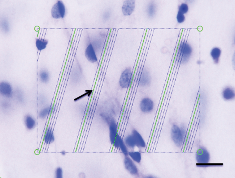
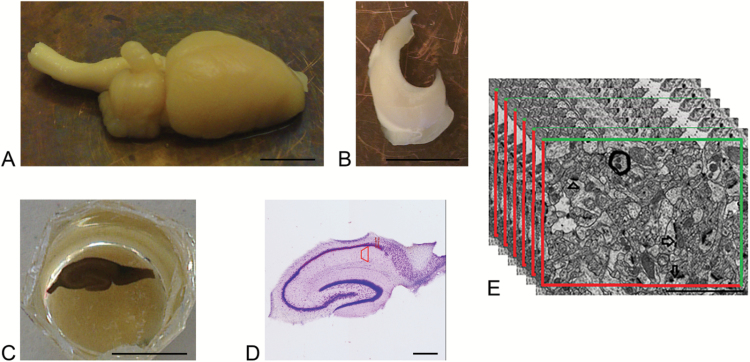
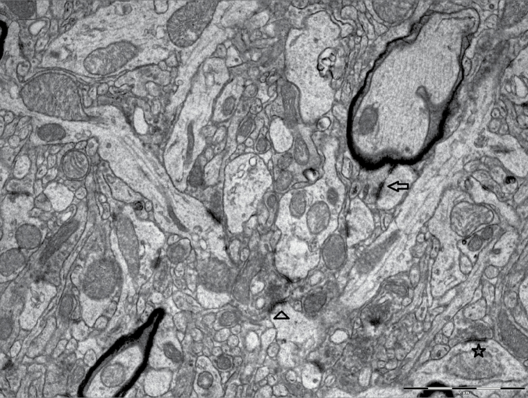
Methods: Flinders Sensitive Line and Flinders Resistant Line rats were given a single intraperitoneal injection of ketamine (15 mg/kg) or saline. One day later, their behavior was evaluated by a modified forced swim test. Microvessel length was evaluated with global spatial sampling and optical microscopy, whereas the number of asymmetric synapses was quantified through serial section electron microscopy by using physical disector method in the CA1.stratum radiatum area of hippocampus.
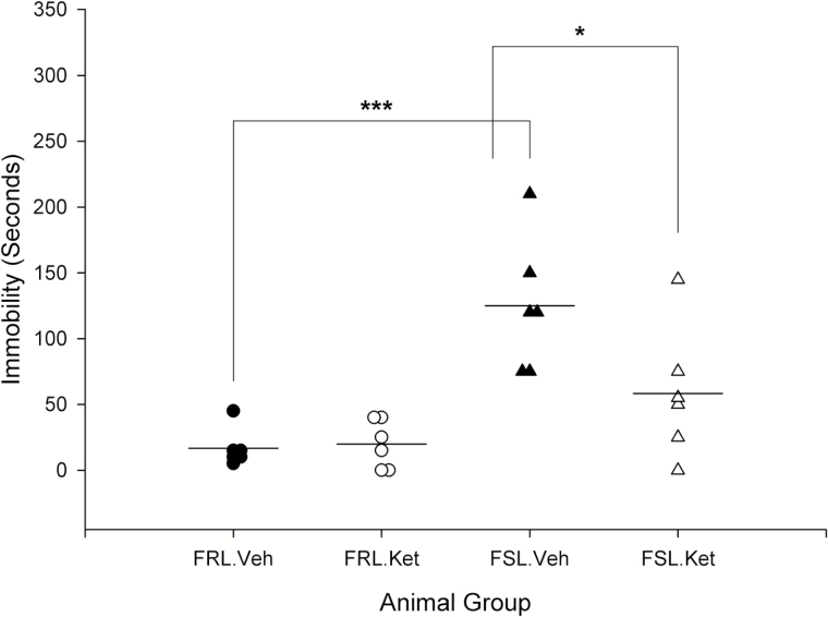
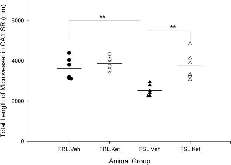
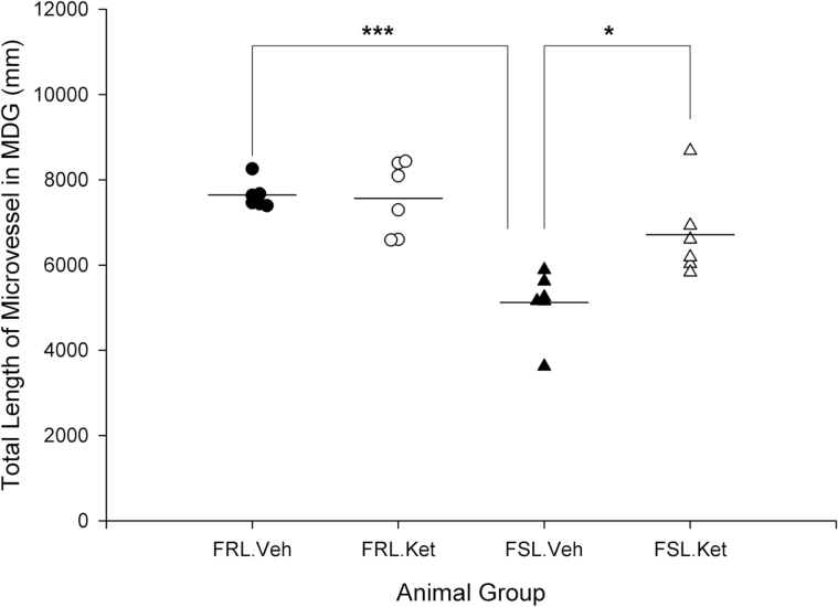
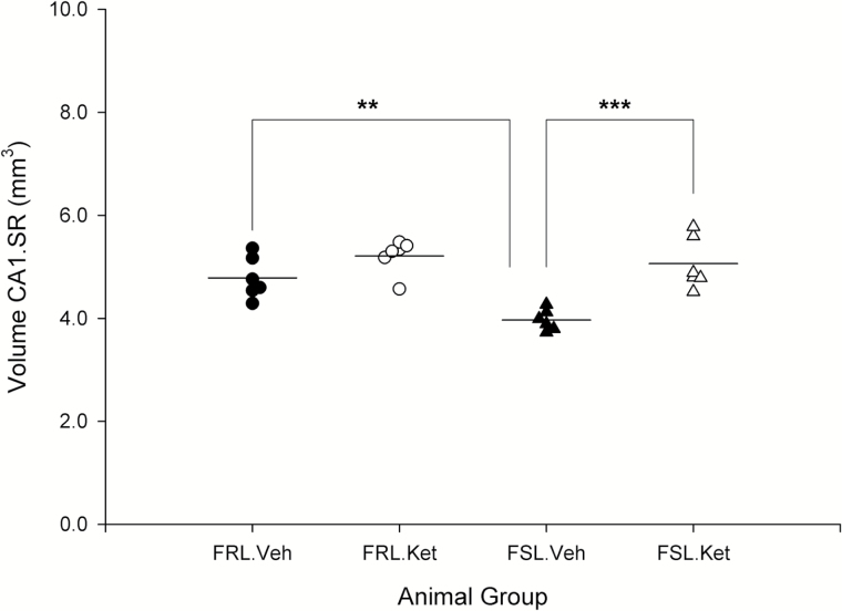
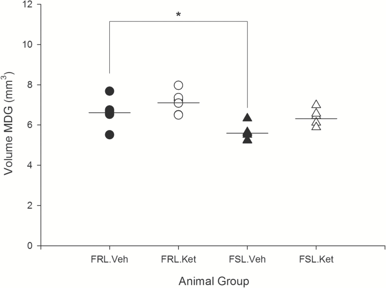
Results: The immobility time in the forced swim test among Flinders Sensitive Line rats with ketamine treatment was significantly lower compared with Flinders Sensitive Line rats without treatment. The number of nonperforated and perforated synapses was significantly higher in the Flinders Sensitive Line-ketamine vs the Flinders Sensitive Line-vehicle group; however, ketamine did not induce a significant increase in the number of shaft synapses. Additionally, total length of microvessels was significantly increased 1 day after ketamine treatment in Flinders Sensitive Line rats in the hippocampal subregions, including the CA1.stratum radiatum.
Conclusion: Our findings indicate that hippocampal vascularization and synaptogenesis is co-regulated rapidly after ketamine, and microvascular elongation may be a supportive factor for synaptic plasticity and neuronal activity. These findings go hand-in-hand with the behavioral observations, where ketamine acts as a potent antidepressant.
Keywords: antidepressant; hippocampus; ketamine; synaptic plasticity; vascularization.
© The Author 2016. Published by Oxford University Press on behalf of CINP.
Figures
References
-
- Alves GS, Carvalho AF, Sudo FK, Oertel-Knochel V, Knochel C, de Carvalho Lde A, Laks J, Engelhardt E, Pantel J. (2014) Structural neuroimaging findings in major depressive disorder throughout aging: a critical systematic review of prospective studies. CNS Neurol Disord Drug Targets 13:1846–1859. - PubMed
-
- Bonhoeffer T, Yuste R. (2002) Spine motility. Phenomenology, mechanisms, and function. Neuron 35:1019–1027. - PubMed
-
- Boyer PA, Skolnick P, Fossom LH. (1998) Chronic administration of imipramine and citalopram alters the expression of NMDA receptor subunit mRNAs in mouse brain. A quantitative in situ hybridization study. J Mol Neurosci 10:219–233. - PubMed
-
- Camus V, Kraehenbuhl H, Preisig M, Bula CJ, Waeber G. (2004) Geriatric depression and vascular diseases: what are the links? J Affect Disord 81:1–16. - PubMed
Publication types
MeSH terms
Substances
LinkOut - more resources
Full Text Sources
Other Literature Sources
Medical
Miscellaneous
