. 2017 Aug;37(8):2857-2869.
doi: 10.1177/0271678X16676287. Epub 2016 Jan 1.

Altered hippocampal arteriole structure and function in a rat model of preeclampsia: Potential role in impaired seizure-induced hyperemia

Affiliations

Altered hippocampal arteriole structure and function in a rat model of preeclampsia: Potential role in impaired seizure-induced hyperemia

Abbie C Johnson et al. J Cereb Blood Flow Metab. 2017 Aug.

Abstract

We investigated the effect of experimental preeclampsia on hyperemia during seizure in the hippocampus and vascular function and structure of hippocampal arterioles using Sprague Dawley rats (n = 14/group) that were nonpregnant, pregnant (d20), or had experimental preeclampsia (induced by a high cholesterol diet d7-20). Hyperemia was measured via hydrogen clearance basally and during pentylenetetrazol-induced seizure (40-130 mg/kg i.v.). Reactivity of isolated and pressurized hippocampal arterioles to KCl, nitric oxide synthase inhibition with NG-nitro-L-arginine methyl ester and the nitric oxide donor sodium nitroprusside were investigated. Capillary density was quantified via immunohistochemistry. Cerebral blood flow increased during seizure vs. baseline in pregnant (118 ± 14 vs. 87 ± 9 mL/100 g/min; p < 0.05) and nonpregnant rats (106 ± 9 vs. 82 ± 9 mL/100 g/min; p < 0.05) but was unchanged in preeclamptic rats (79 ± 16 vs. 91 ± 4 mL/100 g/min; p > 0.05), suggesting impaired seizure-induced hyperemia in preeclampsia. Hippocampal arterioles from preeclamptic rats had less basal tone, and dilated less to 15 mM KCl (9 ± 8%) vs. pregnant (61 ± 27%) and nonpregnant rats (20 ± 11%). L-NAME had no effect on hippocampal arterioles in any group, but dilation to sodium nitroprusside was similar. Structurally, hippocampal arterioles from preeclamptic rats underwent inward hypotrophic remodeling and capillary rarefaction. Impaired seizure-induced hyperemia, vascular dysfunction, and limited vasodilatory reserve of hippocampal arterioles could potentiate hippocampal injury in preeclampsia especially during eclampsia.

Keywords: Cerebral blood flow; cerebral vascular function; hippocampus; preeclampsia/eclampsia; seizure.


Figures

Figure 1.
Figure 1.
Anatomic location and photomicrograph of hippocampal arterioles (HippAs). (a) Illustration of the hippocampal vasculature showing the branches of the longitudinal hippocampal artery (Long hipp a.) that penetrate into the hippocampal cleft. Internal transverse hippocampal arteries (Int trans hipp a.) were dissected to access the hippocampal arterioles (boxed inset) supplying the CA3 region of the dorsal hippocampus. Reprinted from P. Coyle, Vascular Patterns of the Rat Hippocampal Formation, Exp Neurol, 1976; 52(3): 450, 1976, with permission from Elsevier. (b) Wide field image (20×) of an Int trans hipp a. from a nonpregnant rat secured to a glass cannula with a HippA branching off at a 90° angle. HippA was pressurized to 60 mmHg. The region of the HippA where lumen diameter and wall thickness were measured is delineated by the boxed inset.
Figure 2.
Figure 2.
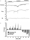
Changes in cerebral blood flow (CBF) in the CA3 region of the hippocampus in response to seizure in nonpregnant (Nonpreg), pregnant (Preg), and preeclamptic (PE) rats. (a) Representative hydrogen (H2) desaturation curves under baseline conditions and during seizure in a Nonpreg rat from which CBF was determined using the half-life of desaturation (t1/2). (b) Graph showing baseline CBF and CBF during seizure in Nonpreg, Preg, and PE rats. CBF significantly increased during seizure compared with baseline in Nonpreg and Preg rats, but was unchanged during seizure in PE rats. (c) Percent change in CBF during seizure was positive and similar between Nonpreg and Preg rats; however, the change in CBF was negative in PE rats. *p < 0.05 vs. baseline by paired t-test; ^p < 0.05 by one-way ANOVA with post-hoc Bonferroni test.
Figure 3.
Figure 3.
Myogenic reactivity of hippocampal arterioles (HippAs) from nonpregnant (Nonpreg), pregnant (Preg), and preeclamptic (PE) rats. Pressure-diameter curves of lumen diameters in both the active (circles) and passive (fully relaxed; squares) states of HippAs from Nonpreg (a), Preg (b), and PE (c) rats. Active diameters of HippAs from all groups were smaller than passive diameters. However, the difference between active and passive diameters was considerably less in PE rats. *p < 0.05, **p < 0.01 vs. passive lumen diameter by t-test.
Figure 4.
Figure 4.
Reactivity of hippocampal arterioles (HippAs) to elevated extracellular KCl. (a) Representative inner diameter traces from nonpregnant (Nonpreg; top panel), pregnant (Preg; middle panel), and preeclamptic (PE; bottom panel) rats in response to increased extracellular KCl concentrations from 5 to 10 mM. Diameters increased with increasing KCl concentration in HippAs from Nonpreg and Preg rats, but remained unchanged in the HippA from the PE rat. (b) Graph showing the percent change in vessel diameter from baseline of HippAs from Nonpreg, Preg, and PE rats in response to elevated extracellular KCl. HippAs dilated in a dose dependent manner to elevations in KCl up to 15 mM. There was a minimal dilation of HippAs from PE rats. At KCl concentrations greater than 20 mM, arterioles constricted in a dose-dependent manner with the greatest constriction occurring in HippAs from PE rats at 40 and 60 mM KCl. ^p < 0.05 vs. Preg; *p < 0.05 vs. Nonpreg and Preg by one-way ANOVA with a post-hoc Bonferroni test.
Figure 5.
Figure 5.
Reactivity of hippocampal arterioles (HippAs) to nitric oxide (NO). (a) Lumen diameters of HippAs from nonpregnant (Nonpreg), pregnant (Preg), and preeclamptic (PE) rats at baseline and after 20-min incubation with the NOS inhibitor L-NAME (10−3 M). Diameters remained unchanged after NOS inhibition in all groups. (b) Reactivity of HippAs to the NO-donor SNP. All HippAs dilated in a dose-dependent manner.
Figure 6.
Figure 6.
Structural and biomechanical properties of hippocampal arterioles (HippAs). (a) Inner diameters of fully relaxed HippAs from Nonpregnant (Nonpreg), pregnant (Preg), and preeclamptic (PE) rats. Diameters were smaller in HippAs from PE rats compared with Nonpreg rats. (b) Outer diameters of fully relaxed HippAs were smaller in PE rats than Nonpreg rats. (c) Wall thickness was decreased in HippAs from both PE and Preg rats. (d) Stress–strain curves of HippAs show a leftward shift in the stress–strain curve of HippAs from PE rats, with an intermediary left shift in HippAs from Preg rats. *p < 0.05, **p < 0.01 vs. Nonpreg; ^^p < 0.01 vs. Nonpreg and Preg by one-way ANOVA with a post-hoc Bonferroni test.
Figure 7.
Figure 7.
Capillary density in the hippocampus of nonpregnant (Nonpreg), pregnant (Preg), and preeclamptic (PE) rats. (a) Representative photomicrographs of collagen IV+ vessels within the CA3 region of the hippocampus of a Nonpreg (left), Preg (middle), and PE (right) rat. (b) Graph showing capillary density in the CA3 region of the hippocampus was decreased in PE rats compared with Nonpreg rats. Capillary density was similar between groups in the dentate gyrus (c) and CA1 region (d). *p < 0.05 vs. Nonpreg by one-way ANOVA with a post-hoc Bonferroni test.

References

    1. Lindheimer MD, Taylor RN, Roberts JM, et al. Introduction, history, controversies, and definitions. In: Taylor RN, Roberts JM, Cunningham FG, et al. (eds). Chesley's hypertensive disorders in pregnancy, 4th ed Boston: Academic Press/Elsevier, 2014.
    1. Roberts JM, Redman CW. Pre-eclampsia: more than pregnancy-induced hypertension. Lancet 1993; 341: 1447–1451. - PubMed
    1. MacKay AP, Berg CJ, Atrash HK. Pregnancy-related mortality from preeclampsia and eclampsia. Obstet Gynecol 2001; 97: 533–538. - PubMed
    1. Zeeman G, Cipolla M, Cunningham G. Cerebrovascular (patho) physiology in preeclampsia/eclampsia. In: Lindhiemer M, Roberts J, Cunningham G. (eds). Chesley's hypertensive disorders in pregnancy, San Diego, CA: Elsevier, 2009, pp. 227–248.
    1. Shah AK, Rajamani K, Whitty JE. Eclampsia: a neurological perspective. J Neurol Sci 2008; 271: 158–167. - PubMed

LinkOut - more resources