. 2016 Dec 16;291(51):26320-26331.
doi: 10.1074/jbc.M116.734012. Epub 2016 Nov 4.

A Hidden Active Site in the Potential Drug Target Mycobacterium tuberculosis dUTPase Is Accessible through Small Amplitude Protein Conformational Changes

Affiliations

A Hidden Active Site in the Potential Drug Target Mycobacterium tuberculosis dUTPase Is Accessible through Small Amplitude Protein Conformational Changes

Anna Lopata et al. J Biol Chem. .

Abstract

dUTPases catalyze the hydrolysis of dUTP into dUMP and pyrophosphate to maintain the proper nucleotide pool for DNA metabolism. Recent evidence suggests that dUTPases may also represent a selective drug target in mycobacteria because of the crucial role of these enzymes in maintaining DNA integrity. Nucleotide-hydrolyzing enzymes typically harbor a buried ligand-binding pocket at interdomain or intersubunit clefts, facilitating proper solvent shielding for the catalyzed reaction. The mechanism by which substrate binds this hidden pocket and product is released in dUTPases is unresolved because of conflicting crystallographic and spectroscopic data. We sought to resolve this conflict by using a combination of random acceleration molecular dynamics (RAMD) methodology and structural and biochemical methods to study the dUTPase from Mycobacterium tuberculosis In particular, the RAMD approach used in this study provided invaluable insights into the nucleotide dissociation process that reconciles all previous experimental observations. Specifically, our data suggest that nucleotide binding takes place as a small stretch of amino acids transiently slides away and partially uncovers the active site. The in silico data further revealed a new dUTPase conformation on the pathway to a relatively open active site. To probe this model, we developed the Trp21 reporter and collected crystallographic, spectroscopic, and kinetic data that confirmed the interaction of Trp21 with the active site shielding C-terminal arm, suggesting that the RAMD method is effective. In summary, our computational simulations and spectroscopic results support the idea that small loop movements in dUTPase allow the shuttlingof the nucleotides between the binding pocket and the solvent.

Keywords: Mycobacterium tuberculosis; X-ray crystallography; dUTPase; molecular dynamics; molecular modeling; nucleotide; pre-steady-state kinetics; solvent accessibility; substrate binding.


Figures

FIGURE 1.
FIGURE 1.
Three-dimensional structure and conserved motifs of the M. tuberculosis dUTPase. A, three-dimensional structure of the M. tuberculosis dUTPase enzyme complexed with dUPNPP (PDB code 2PY4 (33)). The subunits are shown as white, cyan, and blue ribbons, whereas the fifth conserved motif located on the C-terminal arm is highlighted as thick yellow ribbon in all subunits. B, three-dimensional structure of the active site of M. tuberculosis dUTPase enzyme complexed with dUPNPP (PDB code 2PY4 (33)). The protein backbone is shown as white ribbon, and the conserved motifs are highlighted with stick representation of different colors (violet, green, orange, blue, and yellow for first, second, third, fourth, and fifth conserved motif, respectively). dUTP is visualized by atomic colored stick representation. Mg2+ ion, the water molecules of the Mg2+ ion coordination sphere, and the catalytic water molecule are shown as green and red spheres, respectively. C, amino acid sequence of the M. tuberculosis dUTPase. The conserved motifs are color-coded according to B.
FIGURE 2.
FIGURE 2.
A representative of the 17 unproductive RAMD runs. The enzyme can be seen with gray surface representation. For clarity, the C-terminal arm is omitted from this picture. dUTP is visualized as atomic coloring stick model, and Mg2+ is an orange sphere. The path of the center of mass of the dUTP-Mg2+ complex is visualized as a blue, white, and red line.
FIGURE 3.
FIGURE 3.
Exit routes of five representative simulations from the five major ligand unbinding pathways. A, the enzyme is shown with light blue ribbon model, and the C-terminal arm is highlighted as flat yellow ribbon. The curves, representing the five major pathways, are the sphere models of the center of mass of the dUTP-Mg2+ complex during the course of simulations. B, the same structures upon rotating the protein by 90°.
FIGURE 4.
FIGURE 4.
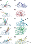
Initial structures and exit routes of the dUTP-Mg2+ complex of all simulations (panels A1–E1) and end point protein structures of representative runs (panels A2–E2) from the five major pathways (A–E). The enzyme is shown as light blue ribbon model, and the C-terminal arm is highlighted as flat ribbon. The curves representing the runs are the sphere models of the center of the dUTP-Mg2+ complex during the course of simulations. Arrows indicate the notable conformational changes between the start and end point structures.
FIGURE 5.
FIGURE 5.
Schematic representation of the dUTP-Mg2+ complex. The coordination sphere of the Mg2+ ion is constituted from the phosphate chain of dUTP and three water molecules.
FIGURE 6.
FIGURE 6.
Maximal RMSD values of single amino acids in the C-terminal arm during the five representative RAMD runs.
FIGURE 7.
FIGURE 7.
Snapshots from pathways A and E showing the His21–Arg140 interaction. The initial enzyme structure is shown as light blue ribbon model, whereas the actual structure during the course of the simulation is shown as a blue and yellow ribbon model for pathways A (panels A1–A3) and E (panels B1–B3), respectively. The C-terminal arm is highlighted as thick ribbon in all cases. The dUTP-Mg2+ complex and the His21 and Arg140 residues are visualized as stick models with atomic coloring, and the hydrogen atoms are omitted for clarity.
FIGURE 8.
FIGURE 8.
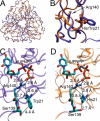
Crystal structure of the H21W mutant. A, superimposition of the WT M. tuberculosis dUTPase (PDB code 2PY4 (33), orange) and the H21W mutant (PDB code 4GCY, violet). The proteins are visualized as colored ribbon model, and dUPNPP can be seen as atomic colored sticks. The global structures of the two proteins are highly similar, which is supported by the 0.2 Å RMSD of the protein backbone atoms. B, enlarged view of A, where the side chains of the mutated residues are shown as stick models. The indole ring of the Trp residue and the imidazole ring of the His residue are in the same position. C and D, possible interacting points of the Trp21 residue (C) or the His21 residue (D) are Ser139 and Arg140 from the crystal structure of the H21W mutant (C) or the WT M. tuberculosis dUTPase (D). Arg140 is coordinating the γ-phosphate oxygen of the substrate. The protein backbone is shown as violet (C) or orange (D) ribbon model, and the residues and the non-hydrolyzable substrate analogue dUPNPP can be seen as atomic colored sticks.
FIGURE 9.
FIGURE 9.
Fast kinetics of the H21W and H145W mutants. The latter was described earlier to display WT behavior (35). A, the fluorescence time courses of the H21W mutant hydrolyzing sub- and superstoichiometric amounts of substrate show similar progression to those reported for the H145W mutant. 3 μm enzyme was mixed with various concentrations of dUTP (post-mix concentrations). Triple exponential function was fitted to the single turnover curve obtained for the lowest, substoichiometric dUTP concentration which yielded 3.1 s−1 for the third phase (k1,obs = 69 s−1, k2,obs = 3.6 s−1). In earlier works, this rate constant was identified as the single turnover rate constant (kSTO) (12). B and C, dUTP binding to the H21W (B) or to the H145W (C) mutant. 1 μm enzyme was mixed with buffer or with various concentrations of dUTP in pseudo first order conditions. Double exponential function was fitted to the data. D, concentration dependence of the observed catalytic rate constants of the fast phase of dUTP binding (kobs,1). E, concentration dependence of the observed catalytic rate constants of the slow phase of dUTP binding (kobs,2). This phase seems to be independent of the substrate concentration in agreement with previous data.
FIGURE 10.
FIGURE 10.
Fluorescence and circular dichroism studies of ligand binding to the H145W and H21W mutants. A, fluorescence spectra of the H21W mutant enzyme with and without nucleoside mono-, di-, and triphosphate ligands. B, fluorescence intensities of the H145W (49) and H21W mutant enzymes with saturating ligand concentrations. C, fluorescence titration of H145W and H21W with the dUPNPP substrate analogue; quadratic fit to the data yielded dissociation constants (Kd,dUPNPP) shown in Table 3. D, circular dichroism titration of H145W and H21W with dUDP; quadratic fit to the data yielded dissociation constants (Kd,dUDP) shown in Table 3.
FIGURE 11.
FIGURE 11.
Thermal mobility of the Arg140 residue in various ligand bound complexes. The enzyme is shown as thick transparent ribbon, whereas the ligand and Arg140 are visualized as sticks. Coloring represents the relative crystallographic B factor for each atom (blue, less mobility; red, more mobility). A, the structure of M. tuberculosis dUTPase complexed with dUPNPP (PDB code 1SIX (10)). B, the structure of M. tuberculosis dUTPase complexed with dUDP (PDB code 1SLH (10)). Note that Arg140 is in different conformation and that Arg140 has much more mobility compared with the dUPNPP-complexed structure.
FIGURE 12.
FIGURE 12.
Solvent accessibility of Trp21 with and without ligand bound to the protein. Acrylamide titration curves are shown. The modified Stern-Volmer equation was fitted to the data yielding quenching constants 18.8, 6.4, 7.1, 6.7, and 5.3 m−1 for NATA, apo H21W, dUPNPP-bound H21W, apo active site Trp, and dUPNPP-bound active site Trp, respectively.

References

    1. Lee J. Y., and Yang W. (2006) UvrD helicase unwinds DNA one base pair at a time by a two-part power stroke. Cell 127, 1349–1360 - PMC - PubMed
    1. Gai D., Zhao R., Li D., Finkielstein C. V., and Chen X. S. (2004) Mechanisms of conformational change for a replicative hexameric helicase of SV40 large tumor antigen. Cell 119, 47–60 - PubMed
    1. Velankar S. S., Soultanas P., Dillingham M. S., Subramanya H. S., and Wigley D. B. (1999) Crystal structures of complexes of PcrA DNA helicase with a DNA substrate indicate an inchworm mechanism. Cell 97, 75–84 - PubMed
    1. Sablin E. P., and Fletterick R. J. (2001) Nucleotide switches in molecular motors: structural analysis of kinesins and myosins. Curr. Opin Struct. Biol. 11, 716–724 - PubMed
    1. Shuman S., and Lima C. D. (2004) The polynucleotide ligase and RNA capping enzyme superfamily of covalent nucleotidyltransferases. Curr. Opin Struct. Biol. 14, 757–764 - PubMed

MeSH terms