Comparative Study
. 2016 Dec 23;291(52):26698-26706.
doi: 10.1074/jbc.M116.760892. Epub 2016 Nov 4.

Substrate Trapping in Crystals of the Thiolase OleA Identifies Three Channels That Enable Long Chain Olefin Biosynthesis

Affiliations
Comparative Study

Substrate Trapping in Crystals of the Thiolase OleA Identifies Three Channels That Enable Long Chain Olefin Biosynthesis

Brandon R Goblirsch et al. J Biol Chem. .

Abstract

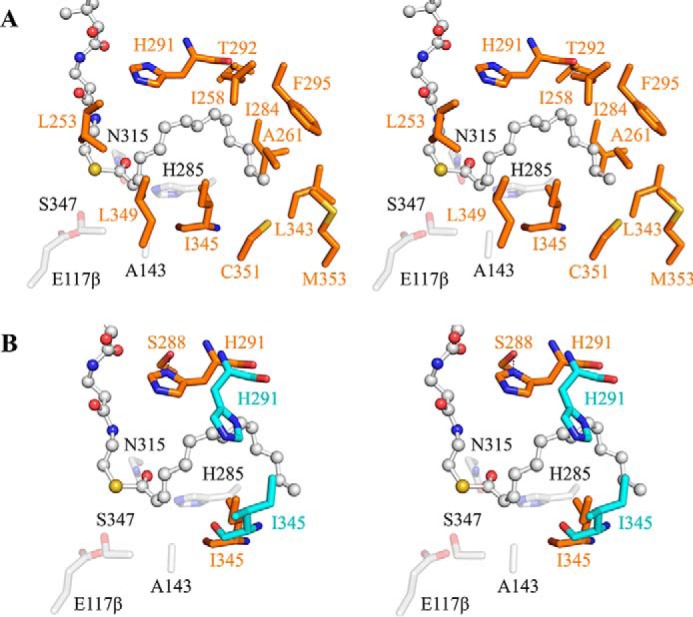
Phylogenetically diverse microbes that produce long chain, olefinic hydrocarbons have received much attention as possible sources of renewable energy biocatalysts. One enzyme that is critical for this process is OleA, a thiolase superfamily enzyme that condenses two fatty acyl-CoA substrates to produce a β-ketoacid product and initiates the biosynthesis of long chain olefins in bacteria. Thiolases typically utilize a ping-pong mechanism centered on an active site cysteine residue. Reaction with the first substrate produces a covalent cysteine-thioester tethered acyl group that is transferred to the second substrate through formation of a carbon-carbon bond. Although the basics of thiolase chemistry are precedented, the mechanism by which OleA accommodates two substrates with extended carbon chains and a coenzyme moiety-unusual for a thiolase-are unknown. Gaining insights into this process could enable manipulation of the system for large scale olefin production with hydrocarbon chains lengths equivalent to those of fossil fuels. In this study, mutagenesis of the active site cysteine in Xanthomonas campestris OleA (Cys143) enabled trapping of two catalytically relevant species in crystals. In the resulting structures, long chain alkyl groups (C12 and C14) and phosphopantetheinate define three substrate channels in a T-shaped configuration, explaining how OleA coordinates its two substrates and product. The C143A OleA co-crystal structure possesses a single bound acyl-CoA representing the Michaelis complex with the first substrate, whereas the C143S co-crystal structure contains both acyl-CoA and fatty acid, defining how a second substrate binds to the acyl-enzyme intermediate. An active site glutamate (Gluβ117) is positioned to deprotonate bound acyl-CoA and initiate carbon-carbon bond formation.

Keywords: OleA; X-ray crystallography; bacteria; biosynthesis; fatty acid; long-chain olefins; mutant; thiolase.


Figures

FIGURE 1.
FIGURE 1.
OleA-catalyzed reactions with acyl-CoA substrates. A, condensation of acyl-CoA substrates to produce β-ketoacid product. B, direct hydrolysis of acyl-CoA substrate.
FIGURE 2.
FIGURE 2.
Substrate binding channels in OleA C143 mutants. A, C143A OleA bound with myristoyl-CoA (ball and stick). B, C143S OleA bound with myristic acid and myristoyl-CoA. C, C143S OleA bound with lauric acid and lauroyl-CoA. The single monomer of OleA is shown in gray cartoon with alkyl channels A (orange cartoon) and B (green cartoon) color-coded for clarity. The mutated residue is shown as a stick model (gray carbon). Bound substrates are represented as ball and stick models with blue mesh illustrating the simulated annealing ligand omit FoFc electron density maps contoured at 2.5 σ.
FIGURE 3.
FIGURE 3.
A, overlay of C143A and C143S co-crystal structures. Bound ligands (ball and stick) are color-coded by structure: C143A-myristoyl-CoA (gray), C143S-myristoyl-CoA (magenta), and C143S-lauroyl-CoA (brown). Sulfur atoms are colored yellow for orientation. B, OleA WT monomer highlighting the three-channel nexus at the active site. Active site residues are shown as sticks (gray carbon). Alkyl channels A and B colored as in Fig. 2.
FIGURE 4.
FIGURE 4.
Stereoviews (cross-eyed) of the C143A and C143S myristoyl-CoA co-crystal active sites. A, acyl-CoA bound in the OleA pantetheinate channel. Pantetheinate channel residues forming hydrogen bonds with the CoA moiety are shown as sticks (gray carbon). The guanidium group of residue Arg195 additionally pi-stacks with the adenine ring of coenzyme A. B, C143A bound with myristoyl-CoA (ball and stick). Active site residues are shown as sticks (gray carbon). C, C143S bound with myristic acid and myristoyl-CoA (ball and stick) occupying alkyl channels A and B, respectively. Active site residues are shown as sticks (pink carbon). D, overlay of C143A and C143S co-crystal structures. Color coding for active site residues are conserved as in B and C. Bound myristoyl-CoA (gray, C143A co-crystal; pink, C143S co-crystal) and myristic acid (pink, C143S co-crystal) are explicitly labeled. Note significant movement of the side chains of Gluβ117 and Ser347 occurs to accommodate the alkyl chain in channel B. In all panels, hydrogen bond contacts are represented by dashed lines, and the ordered waters are shown as red spheres.
FIGURE 5.
FIGURE 5.
Stereoviews (cross-eyed) of OleA alkyl channel A. A, C143A bound with myristoyl-CoA (ball and stick). Residue side chains forming the channel are indicated by sticks (orange carbon and label). B, overlay of unbound C143A OleA (blue carbon) with C143A co-crystallized with myristoyl-CoA (orange carbon). Residues His291 and Ile345 undergo significant conformational changes to bind myristoyl-CoA. Active site residues are shown as faded sticks (gray carbon). Hydrogen bond contacts are represented by dashed lines.
FIGURE 6.
FIGURE 6.
Stereoview (cross-eyed) of OleA alkyl channel B. C143S OleA bound with myristoyl-CoA (ball and stick) within alkyl channel B. Residue side chains forming the channel are indicated by sticks (green carbon). Active site residues are shown as faded sticks (gray carbon).
FIGURE 7.
FIGURE 7.
Substrate coordination and proposed reaction scheme of OleA. The active site of a single monomer is depicted with three substrate channels labeled. Alkyl channel A (orange) and alkyl channel B (green) are color-coded to be consistent with all previous figures. Coenzyme A is represented by a gray oval connected to the phosphopantetheine arm depicted as a wavy line terminating in the reactive thiol group. A, resting state of OleA. B, binding of the first acyl-CoA substrate within alkyl channel A prior to transesterfication. The variable length of the alkyl group is signified by n repeating units. C, binding of the second acyl-CoA substrate within alkyl channel B prior to C–C bond formation. Note a base, putatively assigned as Gluβ117, required to preempt C–C bond formation by proton abstraction. D, nucleophilic attack by the α-carbanion of the second acyl-CoA on the thioester of the enzyme-acyl intermediate to form a C–C bond. E, bound β-ketoacyl-CoA prior to hydrolysis. The release of free coenzyme A and β-ketoacid product complete OleA turnover back to the resting state.
FIGURE 8.
FIGURE 8.
Top-down view of the OleA dimer shown as schematic with each monomer colored coded as tan or gray. The loop (residues 238–248) that lies over alkyl channel B is explicitly shown as green schematic. The image orientation is ∼90° rotation about an axis horizontally in the plane of the paper from the orientation in Fig. 2.

Similar articles

Cited by

References

    1. Wackett L. P., and Wilmot C. M. (2015) Hydrocarbon biosynthesis in microorganisms. In Direct Microbial Conversion of Biomass to Advanced Biofuels (Himmel M. E., ed) pp. 442–470, Elsevier, Amsterdam, The Netherlands
    1. Sukovich D. J., Seffernick J. L., Richman J. E., Hunt K. A., Gralnick J. A., and Wackett L. P. (2010) Structure, function, and insights into the biosynthesis of a head-to-head hydrocarbon in Shewanella oneidensis strain MR-1. Appl. Environ. Microbiol. 76, 3842–3849 - PMC - PubMed
    1. Sukovich D. J., Seffernick J. L., Richman J. E., Gralnick J. A., and Wackett L. P. (2010) Widespread head-to-head hydrocarbon biosynthesis in bacteria and role of OleA. Appl. Environ. Microbiol. 76, 3850–3862 - PMC - PubMed
    1. Beller H. R., Goh E. B., and Keasling J. D. (2010) Genes involved in long-chain alkene biosynthesis in Micrococcus luteus. Appl. Environ. Microbiol. 76, 1212–1223 - PMC - PubMed
    1. Friedman L., and Rude M. (November 13, 2008) U. S. Patent WO 2008/147781 A2

Publication types

MeSH terms

LinkOut - more resources