. 2016 Dec 23;291(52):26636-26646.
doi: 10.1074/jbc.M116.758680. Epub 2016 Nov 4.

The Arrhythmogenic Calmodulin Mutation D129G Dysregulates Cell Growth, Calmodulin-dependent Kinase II Activity, and Cardiac Function in Zebrafish

Affiliations

The Arrhythmogenic Calmodulin Mutation D129G Dysregulates Cell Growth, Calmodulin-dependent Kinase II Activity, and Cardiac Function in Zebrafish

Martin W Berchtold et al. J Biol Chem. .

Abstract

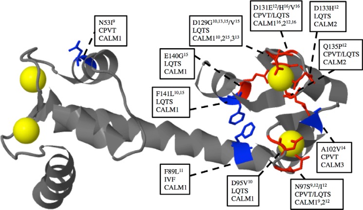
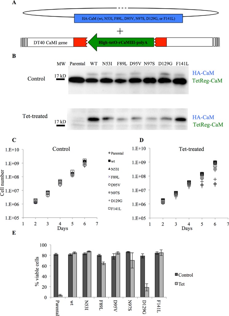
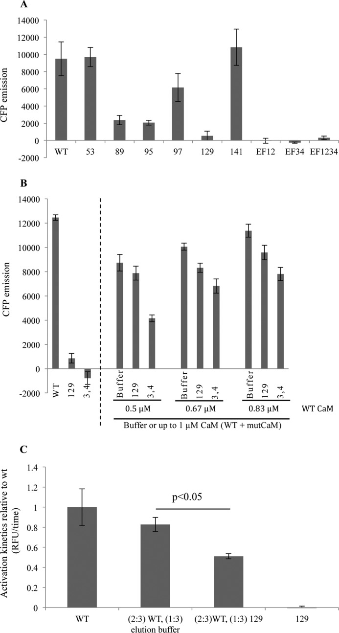
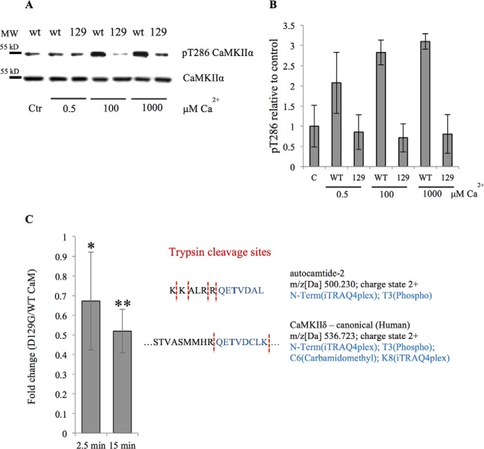
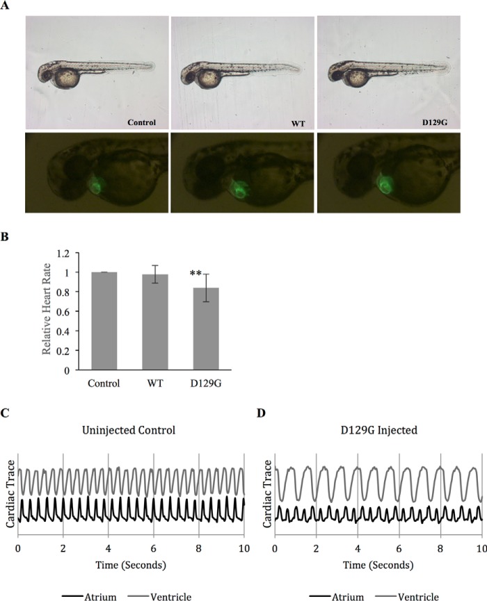
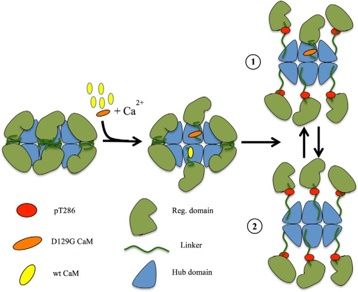
Calmodulin (CaM) is a Ca2+ binding protein modulating multiple targets, several of which are associated with cardiac pathophysiology. Recently, CaM mutations were linked to heart arrhythmia. CaM is crucial for cell growth and viability, yet the effect of the arrhythmogenic CaM mutations on cell viability, as well as heart rhythm, remains unknown, and only a few targets with relevance for heart physiology have been analyzed for their response to mutant CaM. We show that the arrhythmia-associated CaM mutants support growth and viability of DT40 cells in the absence of WT CaM except for the long QT syndrome mutant CaM D129G. Of the six CaM mutants tested (N53I, F89L, D95V, N97S, D129G, and F141L), three showed a decreased activation of Ca2+/CaM-dependent kinase II, most prominently the D129G CaM mutation, which was incapable of stimulating Thr286 autophosphorylation. Furthermore, the CaM D129G mutation led to bradycardia in zebrafish and an arrhythmic phenotype in a subset of the analyzed zebrafish.

Keywords: Ca2+/calmodulin-dependent protein kinase II (CaMKII); DT40; calcium; calmodulin (CaM); catecholaminergic polymorphic ventricular tachycardia (CPVT); cell signaling; heart failure.


Figures

FIGURE 1.
FIGURE 1.
Representation of pathogenic CaM variants associated with CPVT, LQTS, or iodiopathic ventricular fibrillation (IVF). The backbone is shown in gray except for the mutated residues where the side chains in stick representation have been included and color-coded either blue or red (red indicates the residue is directly involved in Ca2+ coordination). Ca2+ is shown in yellow space fill presentation. Text boxes show the amino acid conversion, as well as the arrhythmia associated with the mutation and in which of the three calmodulin genes (CALM) the mutation has been identified and references to the discoveries. The CaM model is based on the 1.7 Å structure of Ca2+ bound WT CaM (Protein Data Bank code 1CLL) modified from Ref. .
FIGURE 2.
FIGURE 2.
The CaM mutant D129G is not able to replace WT CaM in DT40 cells. The CaM1 gene of the chicken DT40 cells was silenced by disrupting exon 3 (red) by insertion of a tetracyclin-regulatable rat CaM cDNA (green) and the other allele by insertion of a selection marker (23). In this cell line, both alleles of the CaM 2 gene had previously been disrupted (24) rendering the tetracyclin-regulatable rCaM (TetReg-CaM) as the only CaM expressed. Stable transfection of HA-tagged versions of CaM (blue) followed by down-regulation of rCaM enables exchange of WT for HA-tagged WT and mutant CaM. B, Western blot analysis of 25 μg of total protein/lane from DT40 clones showing levels of TetReg-CaM and HA-tagged CaM non-treated (control, upper panel incubated with anti-CaM antibody) and 4 days of tet treatment showing almost complete exchange of WT for mutated HA-tagged CaM (lower panel incubated with both anti-CaM and anti-HA antibodies). Because the CaM antibody has different affinities for the mutant versions of CaM, the signals shown for HA-CaM do not represent the precise amounts of these proteins. C and D, growth curves of cells expressing HA-tagged CaM WT and mutants grown in the absence (C) or presence (D) of tetracyclin (tet). Error bars are S.E. from five independent repetitions. E, bar diagram showing % viable cells at 120-h tet treatment for all clones compared with the control without tet. Error bars are S.E. from five independent repetitions.
FIGURE 3.
FIGURE 3.
CaM D129G activates CaMKII only to a minor degree and decreases WT CaM-mediated activation. A, recombinant heart disease-related CaMs with the indicated amino acids mutated or WT CaM were incubated with HeLa cell lysate containing the Camui CaMKII sensor, and peak unquenching of mCerulean was measured in the presence of 2.5 μm [Ca2+]. CaM with disrupted Ca2+ binding at EF hands 1 and 2 (EF12), 3 and 4 (EF34), or 1–4 (EF1234) were used as negative controls. B, bar diagram shows the effect of CFP emission on adding 1 μm WT CaM, CaM 129, or EF34 alone (left-hand side), or mixing WT CaM with the mutant CaM proteins (right-hand side). To test different ratios of WT:mutCaM, the mutCaM was added to give a final total CaM concentration (WT + mutCaM) of 1 μm. Addition of buffer served as a control for the effect of reducing WT CaM. C, activation kinetics of CaMKII in the presence of D129G CaM. p < 0.05 as calculated by paired two-tailed analysis. Error bars represent S.E. from three independent experiments.
FIGURE 4.
FIGURE 4.
Lack of CaMKII Thr286 phosphorylation in the presence of CaM D129G and reduced phosphorylation of a downstream target peptide. Purified CaMKII was stimulated with either WT CaM or D129G CaM under various [Ca2+]. A, representative Western blot analysis of phosphorylation analyzed with an anti-Thr(P)286/287 specific antibody. Ctr indicates control without added Ca2+. B, quantification of Western blot analysis showing Thr(P)286 CamKII to total CaMKII ratios relative to non-Ca2+-stimulated control from three independent experiments. Error bars represents S.D. C, quantitative mass spectrometry analysis of the autocamtide 2 target tryptic peptide (QET(phosp)VDAL, highlighted in blue) following recombinant CaMKIIδ stimulation with either WT or D129G CaM. Measurements were performed in triplicate at 2.5 and 15 min after the reaction was initiated. *, p < 0.05; **, p < 0.005 calculated using either homoscedastic or heteroscedastic two-tailed t test, depending on the statistical value of the F-test (heteroscedastic if p < 0.05). Proteins with t test p values smaller than 0.05 were considered as significantly altered between the two tested conditions. The values are given as fold change of the D129G versus WT CaM. Detected CaMKIIδ tryptic Thr286 phosphopeptide is shown for comparison.
FIGURE 5.
FIGURE 5.
Impact of CaM D129G on the heart rate and conduction of the developing heart in zebrafish. A, representative images of zebrafish injected with RNA coding for either WT CaM or the mutant D129G, as well as non-injected control (upper panel). The lower panel shows GFP expression in the zebrafish hearts. B, bar diagram showing relative heartbeat normalized to non-injected control embryos. Heartbeat measurements were based on fluorescent intensities from cardiac contractions in non-injected control embryos, WT CaM-injected embryos, and D129G-injected embryos. D129G-injected embryos exhibit a heart rate of 83% compared with non-injected control and CaM-WT-injected embryos. ***, p < 0.001. C, contractions in the atrium and ventricle of a non-injected animal. D, a representative trace from a D129G-injected embryo exhibiting a 2:1 atrium to ventricle beat ratio, observed in ∼8% of D129G-injected embryos (n = 128).
FIGURE 6.
FIGURE 6.
Hypothetical model for CaMKII activation delay. Half of the dodecameric holoenzyme has been sketched with the green units representing the regulatory domain and the blue units the hub domain. CaM is in yellow (WT) or orange (D129G), and the red dots represent phosphorylation sites Thr286. The presence of CaM D129G will lead to binding without Thr286 phosphorylation. Because the D129G will occupy a CaM binding site, phosphorylation (drawing 1) and full holoenzyme activation (drawing 2) will be delayed (Fig. 3C).

References

    1. Berchtold M. W., Brinkmeier H., and Müntener M. (2000) Calcium ion in skeletal muscle: its crucial role for muscle function, plasticity, and disease. Physiol. Rev. 80, 1215–1265 - PubMed
    1. Marks A. R. (2013) Calcium cycling proteins and heart failure: mechanisms and therapeutics. J. Clin. Invest. 123, 46–52 - PMC - PubMed
    1. Berchtold M. W., and Villalobo A. (2014) The many faces of calmodulin in cell proliferation, programmed cell death, autophagy, and cancer. Biochim. Biophys. Acta 1843, 398–435 - PubMed
    1. Berchtold M. W., Egli R., Rhyner J. A., Hameister H., and Strehler E. E. (1993) Localization of the human bona fide calmodulin genes CALM1, CALM2, and CALM3 to chromosomes 14q24-q31, 2p21.1-p21.3, and 19q13.2-q13.3. Genomics 16, 461–465 - PubMed
    1. Anderson M. E., Brown J. H., and Bers D. M. (2011) CaMKII in myocardial hypertrophy and heart failure. J. Mol. Cell Cardiol. 51, 468–473 - PMC - PubMed

MeSH terms

Associated data