Review
. 2016 Dec;21(3-4):139-148.
doi: 10.1007/s10911-016-9366-3. Epub 2016 Nov 4.

Epithelial Plasticity During Human Breast Morphogenesis and Cancer Progression

Affiliations
Review

Epithelial Plasticity During Human Breast Morphogenesis and Cancer Progression

Saevar Ingthorsson et al. J Mammary Gland Biol Neoplasia. 2016 Dec.

Abstract

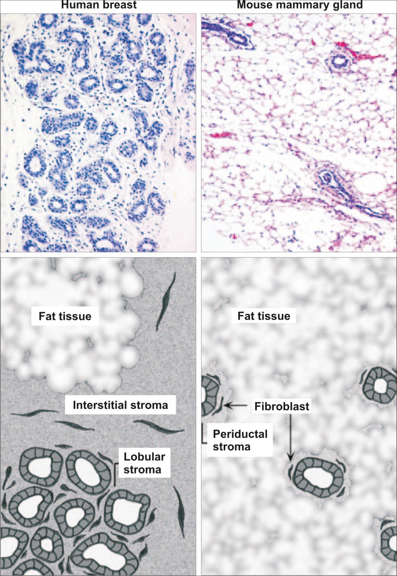
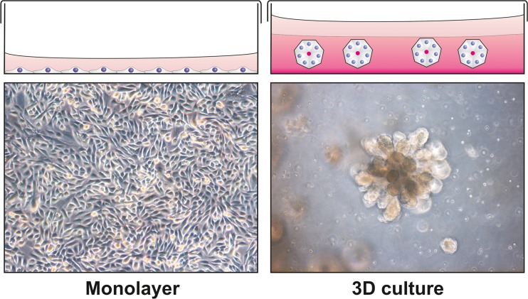
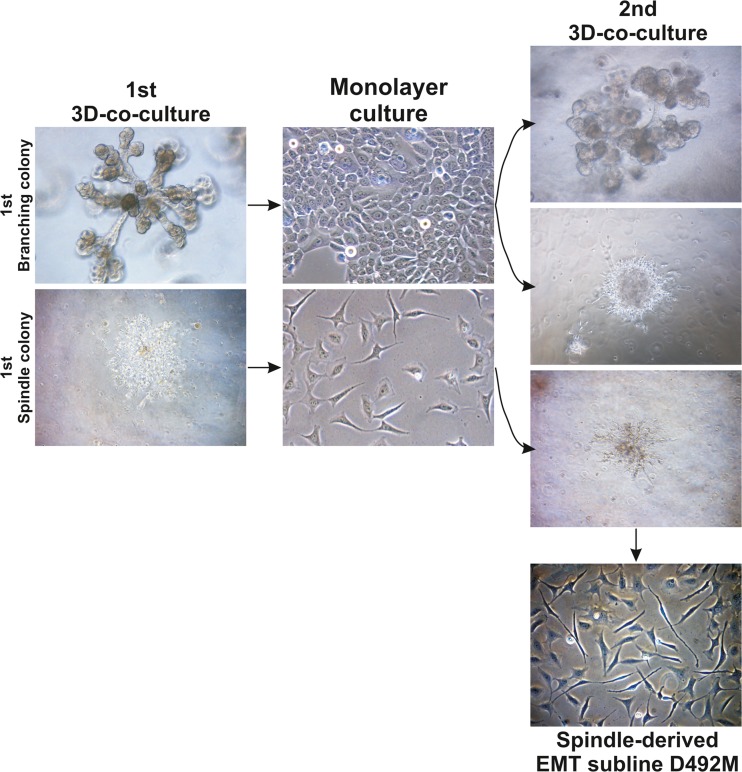
Understanding the complex events leading to formation of an epithelial-based organ such as the breast requires a detailed insight into the crosstalk between epithelial and stromal compartments. These interactions occur both through heterotypic cellular interactions and between cells and matrix components. While in vivo models may partially capture these complex interactions, there is a need for in- vitro models to study these events. In this review we discuss cell-cell interactions in breast development focusing on the stem cell niche and branching morphogenesis. Given the recent understanding that the basic developmental events underlying branching morphogenesis are closely related to pathways important to cancer progression, i.e. epithelial plasticity and epithelial to mesenchymal transition (EMT), we will also discuss aspects relevant to cancer progression. In cancer, the adoption of mesenchymal phenotype by the malignant cells allows stromal invasion and subsequent intravasation to blood- or lymphatic vessels, a route that is a prerequisite for metastasis. A number of publications have demonstrated that tumor initiating cells, sometimes referred to as cancer stem cells adapt an EMT phenotype that renders them more resistant to apoptosis and drug therapy. The mechanism behind this phenomenon is currently unknown but this may partially explain relapse in breast cancer patients. Increased understanding of branching morphogenesis in the breast gland and the regulation of EMT and its reverse process mesenchymal to epithelial transition (MET) may hold the keys for future development of methods/drugs that neutralize the invading properties of cancer cells.

Keywords: 3D cell culture; Breast cancer; EMT; Plasticity; Stem cells.


Figures

Fig. 1
Fig. 1
Histological differences between the human breast and mouse mammary gland. The functional unit of the human breast gland is the terminal duct lobular unit (TDLU). This structure is composed of multiple acini and small ductules and is embedded in a collagenous stroma. The stroma can be divided in two, the interstitial stroma, between TDLUs and ducts, and the looser cellular-rich lobular stroma, within TDLUs. The Human mammary tissue along with the stroma, is in turn embedded in fat tissue. In mice, the mammary gland is composed of a series of branching ducts, terminating in the terminal end bud (TEB). Compared to human breast tissue, the collagenous stroma surrounding the mouse mammary epithelium has a much smaller volume (periductal stroma), so the epithelial structure is enclosed by fat tissue. Adapted from Parmar et al. [13] with permission
Fig. 2
Fig. 2
Breast morphogenesis in 3D cell culture. During conventional cell culture, cells proliferate in a monolayer forming a sheet of cells at the bottom of the culture dish. This is an environment that is highly abstract to cells that in vivo find them self in a 3D environment. This may have great effects on gene expression resulting in altered phenotype. When seeded in a 3D matrix such as reconstituted basement membrane (rBM) matrix the same cells can undergo drastically different morphogenesis, forming polarized spheres with a central lumen, or as for certain cell lines, branching morphogenesis
Fig. 3
Fig. 3
Generation of the D492M cell line. The epithelial cell line D492 was established from normal breast tissue [23]. When cultured in 3D, it forms branching colonies reminiscent of the TDLU. In co-culture with breast endothelial cells, this cell line can form mesenchymal colonies. When branching colonies are isolated from 3D culture and re-plated, they form both branching and mesenchymal colonies during secondary co-culture, while the mesenchymal colonies only give rise to additional mesenchymal colonies. A mesenchymal cell line (D492M) was established from a mesenchymal colony using single cell cloning. Adapted from Sigurdsson et al. [46] with permission
Fig. 4
Fig. 4
Cancer progression and metastasis. Breast cancer arises as localized lesions, most often within ducts and termed ductal carcinoma in situ (DCIS). These tumors are encapsulated by the myoepithelial cells (green) and a basement membrane (black line). When tumors progress to invasive disease the myoepithelial cells are progressively lost and the tumor breaks into the surrounding stroma, coming in contact with stromal cellular components, such as fibroblasts and microvessels. Changes to the microenvironment can induce cancer cells to undergo an epithelial to mesenchymal transition (EMT) a process where cells acquire increased survival and mobility. Increased mobility can lead to cells entering lymphoid or blood vessels and forming metastases in lymph nodes or distant organs. After forming metastases, established tumors often re-acquire epithelial characteristics through mesenchymal to epithelial transition (MET). “Lungs Diagram Simple” and “Brain Human Lateral View” by Patrick J. Lynch, licensed under Creative Commons Attribution 2.5

References

    1. Blanpain C, Fuchs E. Stem cell plasticity. Plasticity of epithelial stem cells in tissue regeneration. Science. 2014;344(6189):1242281. doi: 10.1126/science.1242281. - DOI - PMC - PubMed
    1. Reya T, et al. Stem cells, cancer, and cancer stem cells. Nature. 2001;414(6859):105–11. doi: 10.1038/35102167. - DOI - PubMed
    1. Gudjonsson T, Magnusson MK. Stem cell biology and the cellular pathways of carcinogenesis. APMIS. 2005;113(11–12):922–9. doi: 10.1111/j.1600-0463.2005.apm_371.x. - DOI - PubMed
    1. Milanese TR, et al. Age-related lobular involution and risk of breast cancer. J Natl Cancer Inst. 2006;98(22):1600–7. doi: 10.1093/jnci/djj439. - DOI - PubMed
    1. Hutson SW, Cowen PN, Bird CC. Morphometric studies of age related changes in normal human breast and their significance for evolution of mammary cancer. J Clin Pathol. 1985;38(3):281–7. doi: 10.1136/jcp.38.3.281. - DOI - PMC - PubMed

Publication types