

Evaluation and Endoscopic Management of Esophageal Submucosal Tumor
- PMID: 27817183
- PMCID: PMC5475506
- DOI: 10.5946/ce.2016.109
Evaluation and Endoscopic Management of Esophageal Submucosal Tumor
Abstract
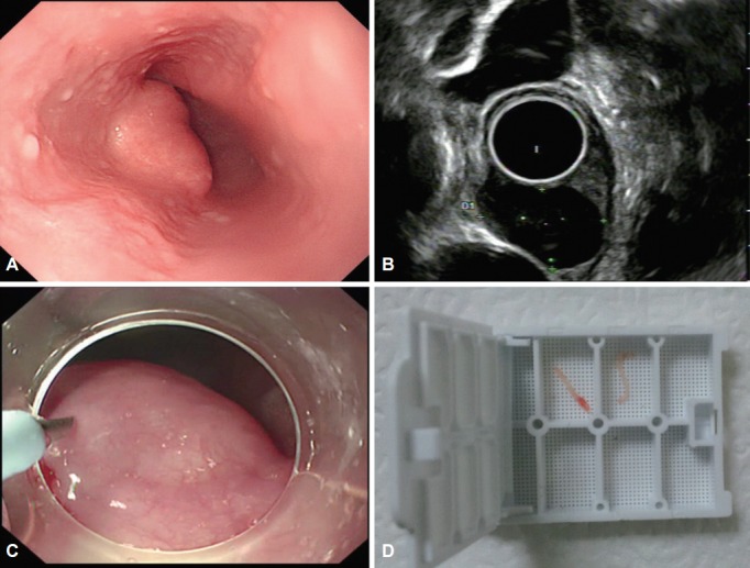
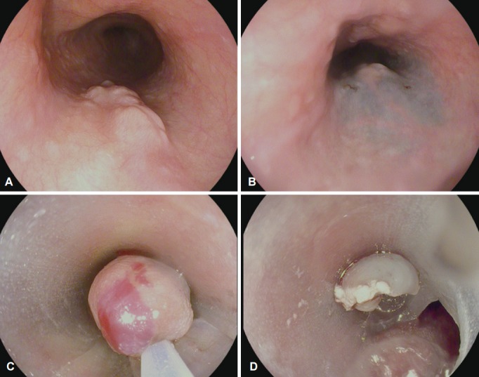
Submucosal tumors (SMTs) originate from tissues that constitute the submucosal layer and muscularis propria, and are covered by normal mucosa. Esophageal SMTs are rare, accounting for <1% of all esophageal tumors. However, the recent widespread use of endoscopy has led to a rapid increase in incidental detection of SMTs in Korea. Esophageal SMTs are benign in ≥90% of cases, but the possibility of malignancies such as gastrointestinal stromal tumor and malignant leiomyosarcoma still exists. Therefore, patients undergo resection in the presence of symptoms or the possibility of a malignant tumor. For resection of esophageal SMTs, surgical resection was the only option available in case of possible malignancy, but minimally invasive surgery by endoscopic resection is becoming more preferable to surgical resection with the development of endoscopic ultrasonography, endoscopic techniques, and other devices.
Keywords: Endoscopic resection; Endosonography; Submucosal tumor.
Conflict of interest statement
Figures
References
-
- Emory TS, Sobin LH, Lukes L, Lee DH, O’Leary TJ. Prognosis of gastrointestinal smooth-muscle (stromal) tumors: dependence on anatomic site. Am J Surg Pathol. 1999;23:82–87. - PubMed
-
- Mutrie CJ, Donahue DM, Wain JC, et al. Esophageal leiomyoma: a 40-year experience. Ann Thorac Surg. 2005;79:1122–1125. - PubMed
-
- Kim MC. Endoscopic incidence of upper gastrointestinal submucosal tumors and endosonographic findings [dissertation] Suwon: Ajou University School of Medicine; 2008.
-
- Ha TI, Kim GH, Eum JS, et al. Catheter probe endoscopic ultrasonography using the jelly-filled method for esophageal subepithelial lesions. Korean J Gastrointest Endosc. 2008;36:125–131.
-
- Gress F, Schmitt C, Savides T, et al. Interobserver agreement for EUS in the evaluation and diagnosis of submucosal masses. Gastrointest Endosc. 2001;53:71–76. - PubMed
Publication types
LinkOut - more resources
Full Text Sources
Other Literature Sources
