Comparative Study
. 2016 Nov 7;18(1):77.
doi: 10.1186/s12968-016-0297-3.

Dark blood late enhancement imaging

Affiliations
Comparative Study

Dark blood late enhancement imaging

Peter Kellman et al. J Cardiovasc Magn Reson. .

Abstract

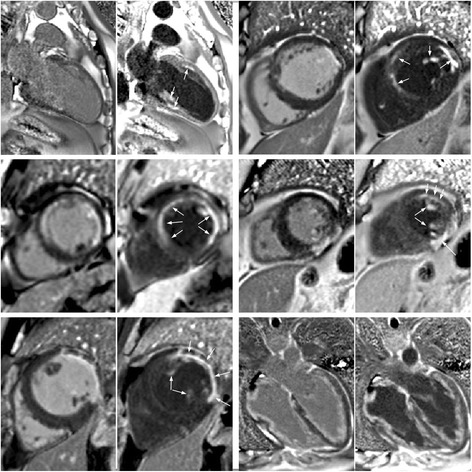
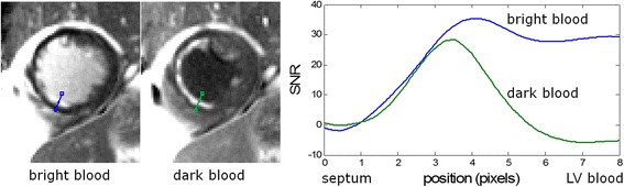
Background: Bright blood late gadolinium enhancement (LGE) imaging typically achieves excellent contrast between infarcted and normal myocardium. However, the contrast between the myocardial infarction (MI) and the blood pool is frequently suboptimal. A large fraction of infarctions caused by coronary artery disease are sub-endocardial and thus adjacent to the blood pool. It is not infrequent that sub-endocardial MIs are difficult to detect or clearly delineate.

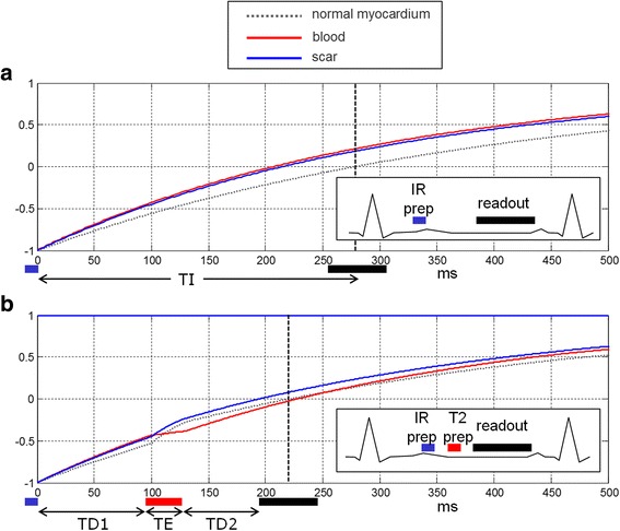
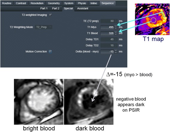
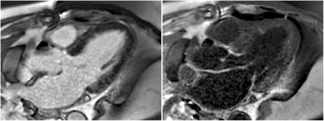
Methods: In this present work, an inversion recovery (IR) T2 preparation was combined with single shot steady state free precession imaging and respiratory motion corrected averaging to achieve dark blood LGE images with good signal to noise ratio while maintaining the desired spatial and temporal resolution. In this manner, imaging was conducted free-breathing, which has benefits for image quality, patient comfort, and clinical workflow in both adults and children. Furthermore, by using a phase sensitive inversion recovery reconstruction the blood signal may be made darker than the myocardium (i.e., negative signal values) thereby providing contrast between the blood and both the MI and remote myocardium. In the proposed approach, a single T1-map scout was used to measure the myocardial and blood T1 using a MOdified Look-Locker Inversion recovery (MOLLI) protocol and all protocol parameters were automatically calculated from these values within the sequence thereby simplifying the user interface.

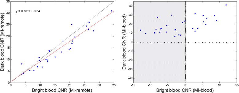
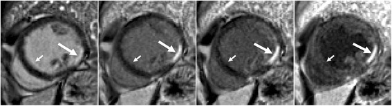
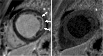
Results: The contrast to noise ratio (CNR) between MI and remote myocardium was measured in n = 30 subjects with subendocardial MI using both bright blood and dark blood protocols. The CNR for the dark blood protocol had a 13 % loss compared to the bright blood protocol. The CNR between the MI and blood pool was positive for all dark blood cases, and was negative in 63 % of the bright blood cases. The conspicuity of subendocardial fibrosis and MI was greatly improved by dark blood (DB) PSIR as well as the delineation of the subendocardial border.

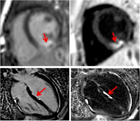
Conclusions: Free-breathing, dark blood PSIR LGE imaging was demonstrated to improve the visualization of subendocardial MI and fibrosis in cases with low contrast with adjacent blood pool. The proposed method also improves visualization of thin walled fibrous structures such as atrial walls and valves, as well as papillary muscles.

Keywords: Ablation; Dark blood; Gadolinium; LGE; Late enhancement; MOCO; Myocardial infarction; PSIR; Scar.


Figures

Fig. 1
Fig. 1
a inversion recovery for bright blood PSIR LGE in case with scar signal (blue) less than blood (red) resulting in poor contrast. b inversion recovery for Dark Blood (DB) PSIR using combined IR and T2 preparation to shift the null time of blood relative to the normal myocardium. In this case the delays are chosen such that the blood signal (red) is less than the myocardium (dashed gray) resulting in dark blood using PSIR reconstruction, which preserves the signal polarity. Inversion times to null the normal myocardium are depicted by vertical dashed lines. The loss in SNR due to the T2 preparation is mitigated by increased respiratory motion corrected averaging (MOCO)
Fig. 2
Fig. 2
Calculation of delays TD1 and TD2 for various TE for specified myocardial and blood T1 of 500 ms and 350 ms respectively. TD1 and TD2 are calculated to null the normal myocardium (point on blue line) to achieve a specified blood suppression. In this case, dark blood may be achieved for TE ≥ 20 ms. The spacing between the blue (myocardial null) and red (blood null) indicated the degree of blood suppression with dark blood (blood < myocardium) when red line is above the blue line. Calculations are performed interactively on the scanner in response to protocol changes (TR, FA, matrix size, etc.)
Fig. 3
Fig. 3
User Interface for Dark Blood (DB) Protocol: (1) User acquires T1 map and measures ROI values for remote blood and myocardium. (2) a enter ROI values in sequence user interface, b enter degree of blood suppression as “delta” value in user interface. (All other parameters (TE, TD1, TD2) are calculated automatically). (3) Acquire images free-breathing
Fig. 4
Fig. 4
CNR measurements between MI and remote (left) and MI and blood pool (right) for N = 30 patients. Note that using the dark blood PSIR LGE the contrast between the MI and blood (CNRMI-blood) is positive for all cases, whereas a large number of conventional bright blood PSIR have poor contrast between the MI and blood (CNRMI-blood < 0) as region shaded in gray
Fig. 5
Fig. 5
Bright blood PSIR LGE (left) and dark blood PSIR LGE with varying blood suppression. The subendocardial MI (long arrow) has increasing contrast with the blood pool as the blood suppression is increased. Gibb’s ringing (short arrow) of LV blood pool at septal border in bright blood LGE (left) is not present in DB LGE
Fig. 6
Fig. 6
Example of bright and dark blood PSIR LGE for 6 adult patient studies with subendocardial LGE
Fig. 7
Fig. 7
Example of dark blood PSIR LGE (right) showing that a possible lateral infarct on the bright blood (left) is in fact just blood pool signal in the trabeculae
Fig. 8
Fig. 8
Free-breathing PSIR MOCO LGE acquired immediately following ablation in subject 1 under sedation (2 year old male) using bright blood (left) and dark blood (right) protocols shows acute ablation lesion (top row) and subject 2 (14 year old female) in 2-week follow-up study with ablation lesion in LV septum (bottom row)
Fig. 9
Fig. 9
PSIR LGE images in 3 chamber view illustrating the improved visualization of thin walled atria and valve structures using DB (right). Note that the thin walled structures have greater enhancement than the myocardium due to fibrous composition leading to higher gadolinium concentration with shorter T1 than myocardium
Fig. 10
Fig. 10
Profiles of SNR vs position thru subendocardial MI for bright blood (blue) and dark blood (green) PSIR LGE protocols

References

    1. Fieno DS, Kim RJ, Chen E, Lomasney JW, Klocke FJ, Judd RM. Contrast-Enhanced Magnetic Resonance Imaging of Myocardium at Risk-Distinction Between Reversible and Irreversible Injury Throughout Infarct Healing. J Am Coll Cardiol. 2000;36:1985–1991. doi: 10.1016/S0735-1097(00)00958-X. - DOI - PubMed
    1. Kim RJ, Fieno DS, Parrish TB, Harris K, Chen E-L, Simonetti O, et al. Relationship of MRI Delayed Contrast Enhancement to Irreversible Injury, Infarct Age, and Contractile Function. Circulation. 1999;100:1992–2002. doi: 10.1161/01.CIR.100.19.1992. - DOI - PubMed
    1. Hunold P, Schlosser T, Vogt FM, Eggebrecht H, Schmermund A, Bruder O, et al. Myocardial Late Enhancement in Contrast-Enhanced Cardiac MRI: Distinction Between Infarction and Non-Infarction-Related Disease. AJR Am J Roentgenol. 2005;184:1420–1426. doi: 10.2214/ajr.184.5.01841420. - DOI - PubMed
    1. Bohl S, Wassmuth R, Abdel-Aty H, Rudolph A, Messroghli D, Dietz R, et al. Delayed enhancement cardiac magnetic resonance imaging reveals typical patterns of myocardial injury in patients with various forms of non-ischemic heart disease. Int J Cardiovasc Imaging. 2008;24:597–607. doi: 10.1007/s10554-008-9300-x. - DOI - PubMed
    1. Vergara GR, Marrouche NF. Tailored management of atrial fibrillation using a LGE-MRI based model: from the clinic to the electrophysiology laboratory. J Cardiovasc Electrophysiol. 2011;22:481–487. doi: 10.1111/j.1540-8167.2010.01941.x. - DOI - PubMed

MeSH terms