. 2016 Dec 5;26(23):3202-3208.
doi: 10.1016/j.cub.2016.09.044. Epub 2016 Nov 3.

Auxin Acts through MONOPTEROS to Regulate Plant Cell Polarity and Pattern Phyllotaxis

Affiliations

Auxin Acts through MONOPTEROS to Regulate Plant Cell Polarity and Pattern Phyllotaxis

Neha Bhatia et al. Curr Biol. .

Abstract

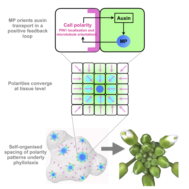
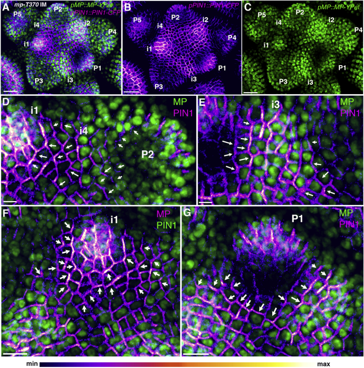
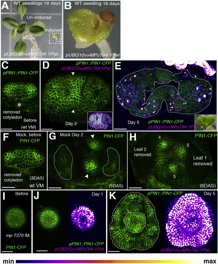
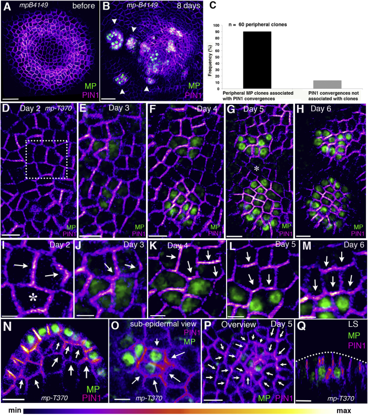
The periodic formation of plant organs such as leaves and flowers gives rise to intricate patterns that have fascinated biologists and mathematicians alike for hundreds of years [1]. The plant hormone auxin plays a central role in establishing these patterns by promoting organ formation at sites where it accumulates due to its polar, cell-to-cell transport [2-6]. Although experimental evidence as well as modeling suggest that feedback from auxin to its transport direction may help specify phyllotactic patterns [7-12], the nature of this feedback remains unclear [13]. Here we reveal that polarization of the auxin efflux carrier PIN-FORMED 1 (PIN1) is regulated by the auxin response transcription factor MONOPTEROS (MP) [14]. We find that in the shoot, cell polarity patterns follow MP expression, which in turn follows auxin distribution patterns. By perturbing MP activity both globally and locally, we show that localized MP activity is necessary for the generation of polarity convergence patterns and that localized MP expression is sufficient to instruct PIN1 polarity directions non-cell autonomously, toward MP-expressing cells. By expressing MP in the epidermis of mp mutants, we further show that although MP activity in a single-cell layer is sufficient to promote polarity convergence patterns, MP in sub-epidermal tissues helps anchor these polarity patterns to the underlying cells. Overall, our findings reveal a patterning module in plants that determines organ position by orienting transport of the hormone auxin toward cells with high levels of MP-mediated auxin signaling. We propose that this feedback process acts broadly to generate periodic plant architectures.

Keywords: ARF5; Arabidopsis; MONOPTEROS; PIN1; auxin; cell polarity; organ positioning; organogenesis; pattern formation; phyllotaxis.


Figures

None
Graphical abstract
Figure 1
Figure 1
MP Expression Patterns Predict PIN1 Polarity Changes (A) pMP::MP-YPet (green) and pPIN1::PIN1-CFP (magenta) expression and localization in the mp-T370 inflorescence meristem (IM). (B) Meristem in (A) showing pPIN1::PIN1-CFP alone. (C) Meristem in (A) showing pMP::MP-YPet expression alone. (D) Magnified view of i4 before PIN1 polarity convergence. (E) Magnified view of i3 after PIN1 convergence. (F) Magnified view of i1. Note the low MP expression surrounding i1 prior to PIN1 polarity reversal. (G) Magnified view of P1 showing reduced MP expression surrounding the primordium and PIN1 polarity oriented away from low-MP-expressing cells. The arrows indicate the estimated PIN1 polarity direction within the cells. Primordium (P) and incipient primordium (i) stages are numbered i4–P5. Scale bars, 30 μm (A–C), 5 μm (D and E), and 10 μm (F and G). See also Figure S1 and Movie S1.
Figure 2
Figure 2
Localized Organogenesis Requires Localized MP Activity (A) Wild-type seedling 18 days after induction of pUBQ10≫MPc794-YPet (dotted rectangle) in comparison to an un-induced plant. (B) Magnified view of an induced plant from (A). Note the fusion of the first two leaves. (C–H) pPIN1::PIN1-CFP expression and polarity (green) after induction of pUBQ10≫MPc794-YPet (C–E) compared to mock-treated seedlings (F–H) before treatment (C and F), 2 days later (D and G), and 5 days later (E and H). Note the absence of PIN1 convergences (arrowheads) in (D) compared to (G). Five days after MPc794-YPet induction a ring of organ tissue has developed (outlined by dotted line in E) that is absent in mock-treated control (H). The inset in (D) shows MPc794-YPet expression (magenta). The inset in (E) shows the phenotype after 11 days of MPc794-YPet induction (66%, n = 21). (I–K) pPIN1::PIN1-CFP expression and polarity (green) before (I) and after induction of pUBQ10≫MPc794-YPet (magenta) (J and K) in mp IM. Note the ring-shaped organ present 5 days after MPc794-YPet induction (K). The asterisk in (C) marks the removed cotyledon. Scale bars, 30 μm (C, D, and F–K) and 50 μm (E).
Figure 3
Figure 3
MP Polarizes Cells Non-Cell Autonomously (A and B) Confocal projection showing the mp-B4149 apex expressing pPIN1::PIN1-GFP (magenta) before (A) and 8 days after induction of MP-YPet clones (B) (green); arrowheads indicate initiating organs coinciding with MP expression. (C) Frequency of peripheral MP clones associated with PIN1 convergence patterns (n = 60 peripheral clones of MP). (D–M) Time series showing epidermal MP-VENUS clones in mp-T370 and associated changes in PIN1-CFP localization. (D–H) Overview showing two independent clones. Note the gradual increase in expression of both MP and PIN1 within the clones indicating an increase in local auxin levels compared to neighboring regions (asterisk in G). (I–M) Magnified view of the dotted rectangle in (D). The asterisk in (I) marks the cell before MP-VENUS induction, corresponding to the future clone. (N) Longitudinal optical section showing a sub-epidermal polarity response to the epidermal clone. (O) Cross-section showing lateral PIN1 polarity toward the MP clone in the sub-epidermal layer. (P) PIN1-CFP convergence in the epidermal cell layer in response to a sub-epidermal MP clone. (Q) Longitudinal reconstructed section of (P) showing a sub-epidermal MP-VENUS clone. The arrows indicate the estimated PIN polarity direction within the cells. Scale bars, 30 μm (A and B), 10 μm (D–H and N–Q), 4 μm (I), and 5 μm (J–M). See also Figures S3 and S4 and Movies S2 and S3.
Figure 4
Figure 4
Restriction of MP Activity to the Epidermis Results in Mobile Auxin Maxima (A and B) pML1::MP-YPet expression (magenta) in wild-type before (A) and 6 hr after auxin treatment (B) (n > 20). The inset in (A) shows a longitudinal optical section; white arrowheads in (A) point to regions of low MP-YPet expression, as also seen when MP is driven by its own promoter. (C and D) pML1::2X-CFP-N7 expression (magenta) before (C) and 6 hr after auxin treatment (D) (n = 6). The inset in (C) shows a longitudinal optical section showing epidermal localization. (E) Photograph of the mp-T370 mutant expressing pML1::MP-YPet. (F) Confocal projection of an mp-T370 mutant seedling with fused leaves (white arrowhead), with pML1::MP-YPet (magenta) and pPIN1::PIN1-CFP (green). (G and H) IM of mp-T370 expressing pML1::MP-YPet; photograph (G) and confocal projection, with pML1::MP-YPet (magenta) and pPIN1::PIN1-CFP (green) (H). (I) Magnified view of the dotted rectangle in (H) showing PIN1-CFP forming a convergence pattern. The arrows indicate the estimated PIN1 polarity directions within the cells. (J and K) Time series of mp-T370 IMs expressing pML1::MP-YPet and pPIN1::PIN1-CFP before (J) and 12 hr later (K). Note the change in the position of maximum MP expression (white arrowheads) and PIN1 convergences (yellow arrowheads). The asterisks mark the same cells at 0 hr and at 12 hr time point. (L and M) Longitudinal reconstructed optical sections of mp-T370 IM showing pPIN1::PIN1-CFP and pMP::MP-YPet expression (L) and pML1::MP-YPet (magenta) and pPIN1::PIN1-CFP (M). Arrowheads indicate PIN1-CFP signal in sub-epidermal layers/pro-vasculature (L) or the absence thereof (M). Scale bars, 30 μm (A and B), 40 μm (C and D), 50 μm (F and H), and 20 μm (I–M). See also Movie S4.

Comment in

References

    1. Jean R.V., Barabé D. World Scientific; 1998. Symmetry in Plants.
    1. Okada K., Ueda J., Komaki M.K., Bell C.J., Shimura Y. Requirement of the auxin polar transport system in early stages of Arabidopsis floral bud formation. Plant Cell. 1991;3:677–684. - PMC - PubMed
    1. Reinhardt D., Mandel T., Kuhlemeier C. Auxin regulates the initiation and radial position of plant lateral organs. Plant Cell. 2000;12:507–518. - PMC - PubMed
    1. Reinhardt D., Pesce E.R., Stieger P., Mandel T., Baltensperger K., Bennett M., Traas J., Friml J., Kuhlemeier C. Regulation of phyllotaxis by polar auxin transport. Nature. 2003;426:255–260. - PubMed
    1. Benková E., Michniewicz M., Sauer M., Teichmann T., Seifertová D., Jürgens G., Friml J. Local, efflux-dependent auxin gradients as a common module for plant organ formation. Cell. 2003;115:591–602. - PubMed

Publication types

MeSH terms