

Review
doi: 10.1016/j.rboe.2016.08.007.
eCollection 2016 Sep-Oct.
Diagnosis and treatment of osteochondral lesions of the ankle: current concepts
Affiliations
- PMID: 27818968
- PMCID: PMC5091026
- DOI: 10.1016/j.rboe.2016.08.007
Item in Clipboard
Review
Diagnosis and treatment of osteochondral lesions of the ankle: current concepts
Rev Bras Ortop.
.
Abstract
We conducted a wide-ranging review of the literature regarding osteochondral lesions of the ankle, with the aim of presenting the current concepts, treatment options, trends and future perspectives relating to this topic.
Os autores fazem uma revisão ampla da literatura a respeito das lesões osteocondrais do tornozelo, com o intuito de expor os conceitos atuais sobre o tema, as opções de tratamento, as tendências e as perspectivas.
Keywords: Ankle injuries/diagnosis; Ankle injuries/therapy; Osteochondritis/diagnosis; Osteochondritis/therapy; Talus.
Figures
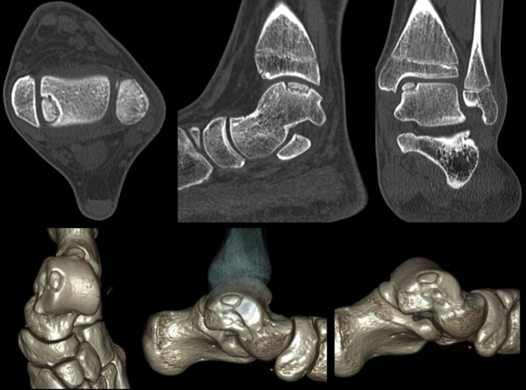
Axial computed tomography allows for the identification, measurement, and accurate classification of osteochondral lesions of the talus. The lower images correspond to three-dimensional reconstruction.
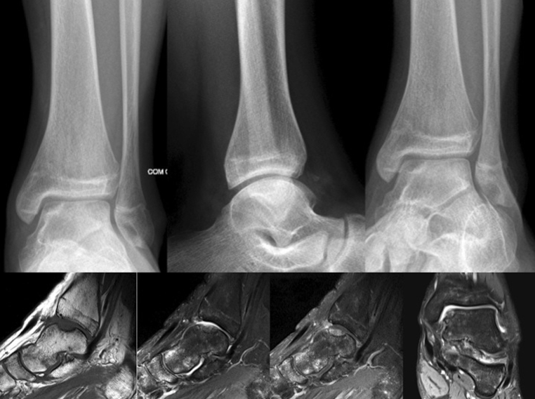
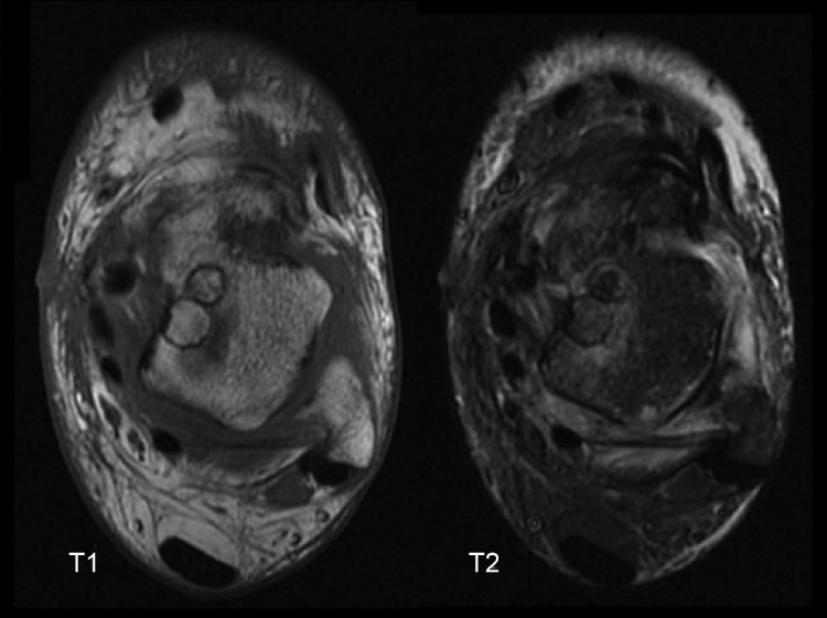
X-rays of the ankle and magnetic resonance imaging of a patient who underwent arthroscopic treatment with debridement and microfractures.
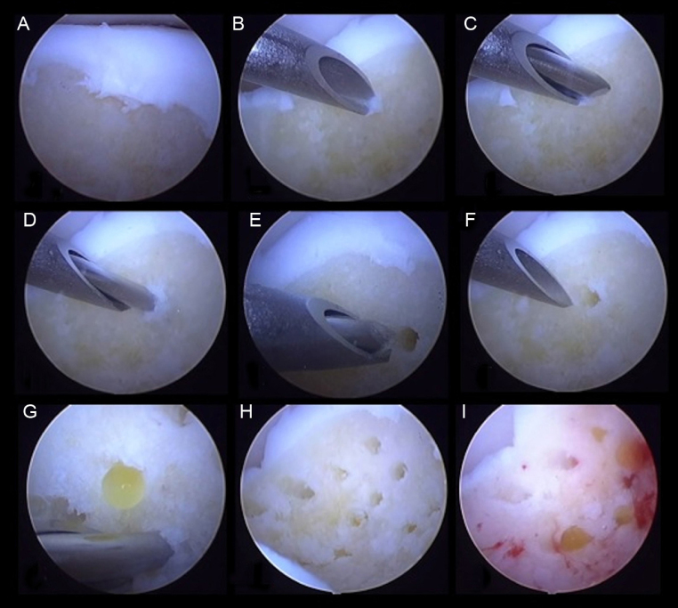
Drilling in the base of osteochondral lesions of the talus.
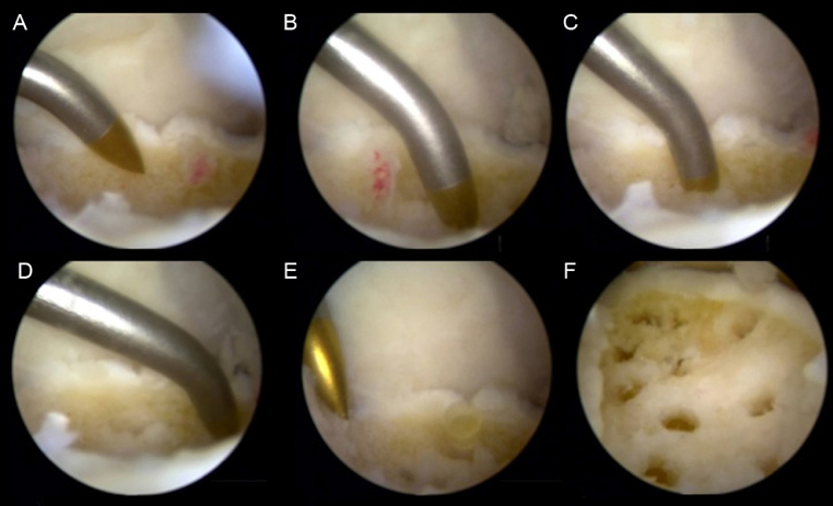
Microfractures in the base of osteochondral lesions of the talus.
MRI scans obtained six months after autologous osteochondral graft.
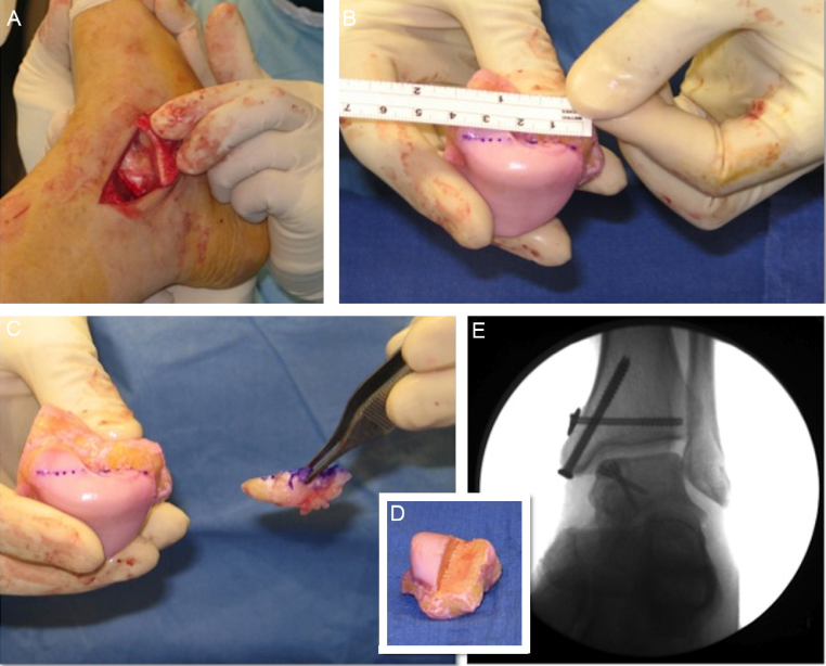
Osteochondral homograft used for the treatment of extensive medial talar shoulder injury (The authors thank Dr. Mark S. Myerson for the authorization to use these figures).
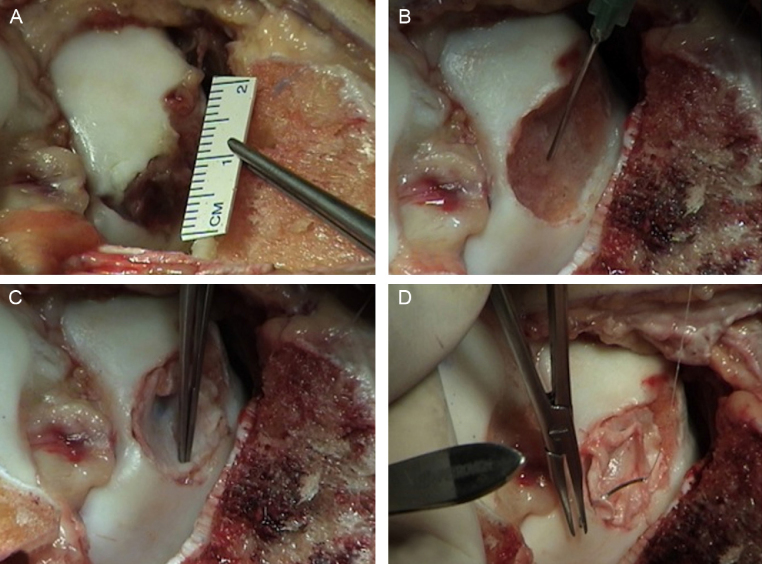
Autologous chondrocyte implantation (sandwich technique).
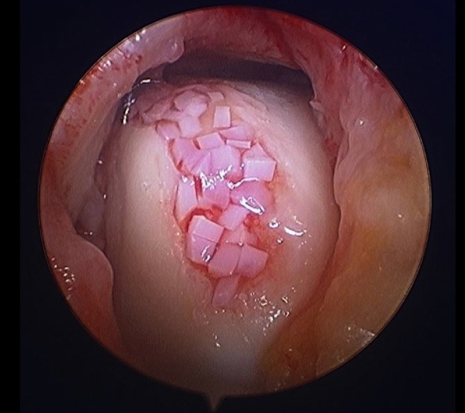
Particulated juvenile cartilage. (The authors thank Dr. Rebecca Cerrato – Mercy Hospital, Baltimore, USA – for the authorization to use these figures).

Diagnostic and treatment flowchart for osteochondral lesions of the talus, based on the literature.
References
-
- Baltzer A.W., Arnold J.P. Bone-cartilage transplantation from the ipsilateral knee for chondral lesions of the talus. Arthroscopy. 2005;21(2):159–166. - PubMed
-
- Dheer S., Khan M., Zoga A.C., Morrison W.B. Limitations of radiographs in evaluating non-displaced osteochondral lesions of the talus. Skeletal Radiol. 2012;41(4):415–421. - PubMed
-
- Ferkel R.D., Flannigan B.D., Elkins B.S. Magnetic resonance imaging of the foot and ankle: correlation of normal anatomy with pathologic conditions. Foot Ankle. 1991;11(5):289–305. - PubMed
-
- Mintz D.N., Tashjian G.S., Connell D.A., Deland J.T., O’Malley M., Potter H.G. Osteochondral lesions of the talus: a new magnetic resonance grading system with arthroscopic correlation. Arthroscopy. 2003;19(4):353–359. - PubMed
-
- Lüsse S., Claassen H., Gehrke T., Hassenpflug J., Schünke M., Heller M. Evaluation of water content by spatially resolved transverse relaxation times of human articular cartilage. Magn Reson Imaging. 2000;18(4):423–430. - PubMed
Publication types
LinkOut - more resources
Full Text Sources
Other Literature Sources