. 2017 Apr 20;36(16):2265-2274.
doi: 10.1038/onc.2016.380. Epub 2016 Nov 7.

LMP1 signaling pathway activates IRF4 in latent EBV infection and a positive circuit between PI3K and Src is required

Affiliations

LMP1 signaling pathway activates IRF4 in latent EBV infection and a positive circuit between PI3K and Src is required

L Wang et al. Oncogene. .

Abstract

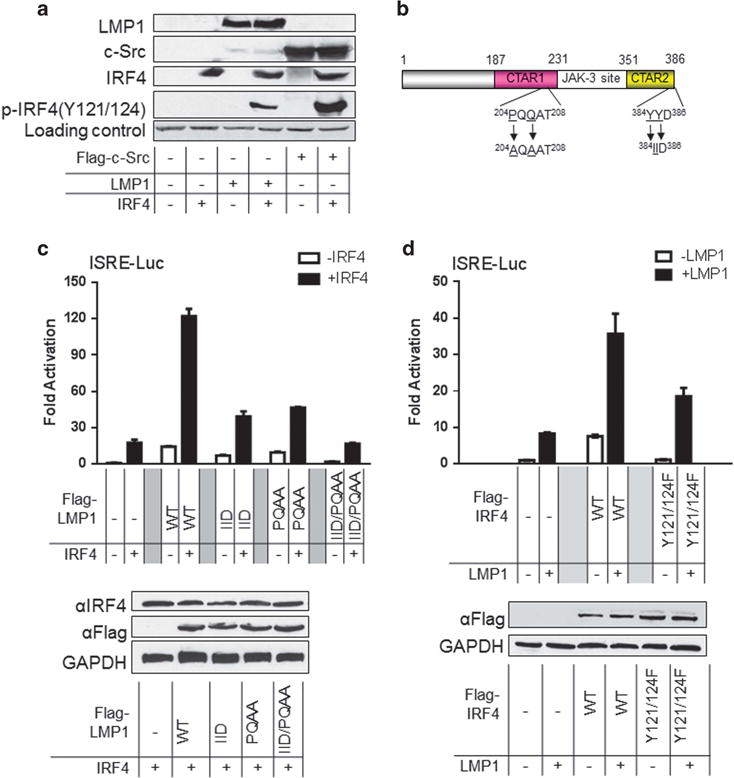
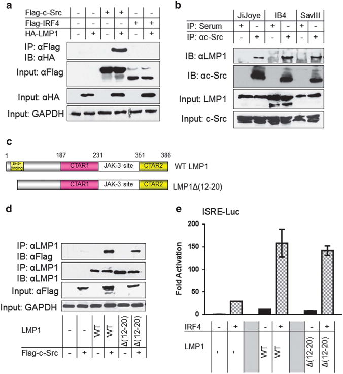
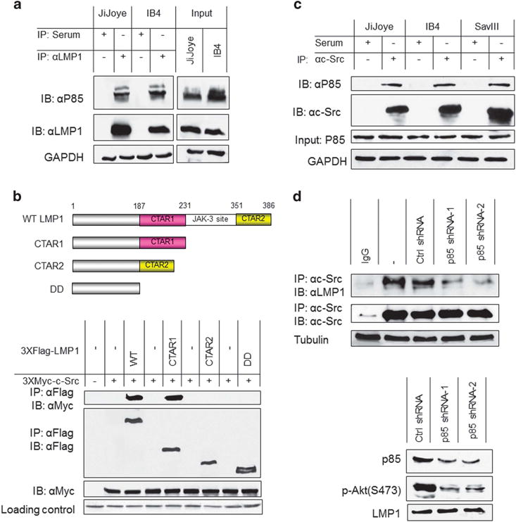
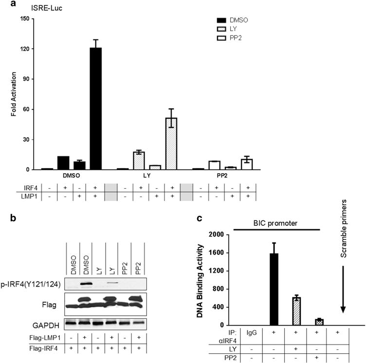
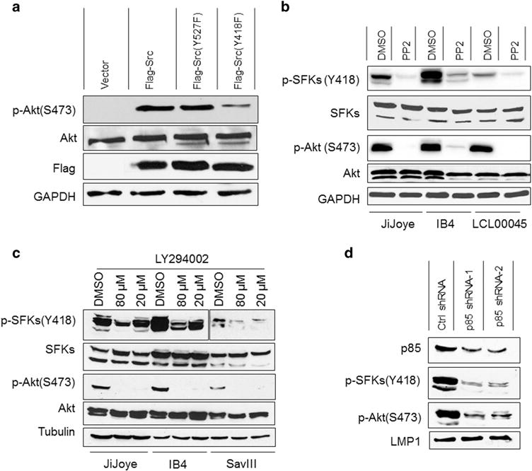
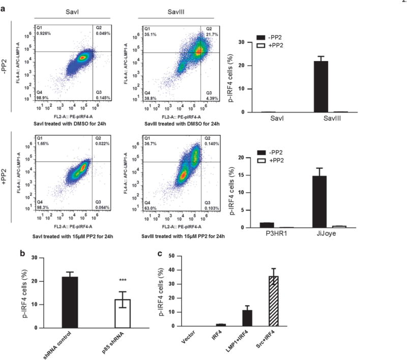
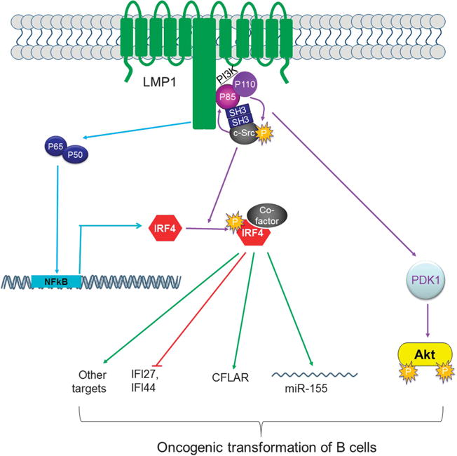
Interferon (IFN) regulatory factors (IRFs) have crucial roles in immune regulation and oncogenesis. We have recently shown that IRF4 is activated through c-Src-mediated tyrosine phosphorylation in virus-transformed cells. However, the intracellular signaling pathway triggering Src activation of IRF4 remains unknown. In this study, we provide evidence that Epstein-Barr virus (EBV) latent membrane protein 1 (LMP1) promotes IRF4 phosphorylation and markedly stimulates IRF4 transcriptional activity, and that Src mediates LMP1 activation of IRF4. As to more precise mechanism, we show that LMP1 physically interacts with c-Src, and the phosphatidylinositol 3 kinase (PI3K) subunit P85 mediates their interaction. Depletion of P85 by P85-specific short hairpin RNAs disrupts their interaction and diminishes IRF4 phosphorylation in EBV-transformed cells. Furthermore, we show that Src is upstream of PI3K for activation of both IRF4 and Akt. In turn, inhibition of PI3K kinase activity by the PI3K-speicfic inhibitor LY294002 impairs Src activity. Our results show that LMP1 signaling is responsible for IRF4 activation, and further characterize the IRF4 regulatory network that is a promising therapeutic target for specific hematological malignancies.


Conflict of interest statement

CONFLICT OF INTEREST

The authors declare no conflict of interest.

Figures

Figure 1
Figure 1
LMP1 promotes IRF4 transcriptional activity and phosphorylation. (a) LMP1 stimulates IRF4 phosphorylation. 293 cells in 60-mm dishes were transfected with 1 μg LMP1 (or Flag-c-Src) and 1 μg IRF4 expression plasmids. Cells were collected 48 h post-transfection and analyzed for IRF4 phosphorylation with p-IRF4(Y121/124). (b) A diagram showing the LMP1 point mutants. (c) and (d) LMP1 promotes IRF4 transcriptional activity. 293 cells in 24-well plates were transfected with 50 ng IRF4 or the mutant IRF4(Y121/124 F) and 50 ng LMP1 or its mutants expression plasmids, 40 ng pGL3/Interferon-Stimulated Responsive Element (ISRE)-Luc and 10 ng Renilla. Dual luciferase assay was performed. Results are the averages ± s.e. of duplicates. Representative results from at least three independent experiments are shown. The ability of the vector control to activate the promoter construct was set to 1.
Figure 2
Figure 2
LMP1 interacts with Src. (a) LMP1 interacts with Src in vitro. 293T cells in 60-mm dishes were transfected with 1 μg HA-LMP1 (or Flag-c-Src) and 1 μg Flag-IRF4 expression plasmids. Cells were collected 48 h post-transfection and then subjected to immunoprecipitation with the Flag antibody clone M2. Beads were washed extensively and then subjected to immunoblotting with the HA antibody clone HA-7. Inputs (5%) were subjected to immunoblotting with the indicated antibodies. (b) Endogenous LMP1 and Src interact in EBV latency. Cell lysates (approximately 1 mg of total proteins in each sample) prepared from JiJoye, IB4 and SavIII cells were subjected to immunoprecipitation with the c-Src antibody clone N16 (or rabbit serum as control). Beads were washed extensively and probed with LMP1 and Src antibodies. Inputs (5%) were also probed with these antibodies. (c) A diagram showing the LMP1 deletion mutant LMP1Δ(12–20). (d) The LMP1 SH3-binding region is not required for Src interaction. 293T cells in 60-mm dishes were transfected with 1 μg LMP1 or LMP1Δ(12–20) and 1 μg Flag-c-Src expression plasmids. Cells were collected 48 h post-transfection and then subjected to immunoprecipitation with the LMP1 antibody clone CS1-4. Beads were washed extensively and then subjected to immunoblotting with the Flag antibody clone M2 and the LMP1 antibody. Inputs (5% of total lysates) were subjected to immunoblotting with indicated antibodies. (e) The LMP1 SH3-binding region is not required for IRF4 activation. The 293 cells in 24-well plates were transfected with 50 ng IRF4 and 50 ng LMP1 (or LMP1Δ(12–20)) expression plasmids, 40 ng pGL3/IFNβ-Luc and 10 ng Renilla. Dual luciferase assay was performed. Results are the averages ± s.e. of duplicates. Representative results from at least three independent experiments are shown. The ability of the vector control to activate the promoter construct was set to 1.
Figure 3
Figure 3
P85 mediates LMP1 and Src interaction. (a) Cell lysates (approximately 1 mg of total proteins in each sample) prepared from JiJoye and IB4 cells were subjected to immunoprecipitation with the LMP1 antibody clone CS1-4 (or mouse serum as control). Beads were washed extensively and probed with P85 and LMP1 antibodies. Inputs (5%) were also probed with these antibodies. (b) LMP1 CTAR1 is responsible for its interaction with Src. 293T cells in 60-mm dishes were transfected with 1 μg 3XMyc-Src and 1 μg Flag-LMP1 or its deletion mutants as indicated. Cells were collected 48 h post-transfection and then subjected to immunoprecipitation with 1.5 μg Myc antibody 9E10 (Roche). Beads were washed extensively and then subjected to immunoblotting with the Flag antibody clone M2 and Myc antibody 9E10. Loading control is Ponceau S staining. Upper panel shows a diagram showing the LMP1 protein structure. (c) Endogenous P85 and Src interact in EBV latency. Cell lysates (approximately 1 mg of total proteins in each sample) prepared from JiJoye, IB4 and SavIII cells were subjected to immunoprecipitation with the c-Src antibody clone N16 (or rabbit serum as control). Beads were washed extensively and probed with P85 and Src antibodies. (d) P85 depletion disrupts the interaction between LMP1 and Src. IB4 cells were infected with retrovirus expressing P85-specific shRNAs (or control). Stable transfectants were selected with 1 μg/ml puromycin for 2 weeks. shRNA expression was induced by 1 μg/ml doxycycline for 3 days. Knockdown efficiency is shown in the lower panel. Cells were then harvested and subjected to IP with the Src antibody clone N16, and then probed with the LMP1 antibody clone CS1-4. Representative results from at least three independent experiments are shown.
Figure 4
Figure 4
PI3K and Src are required for LMP1 activation of IRF4. (a) The PI3K-specific inhibitor LY294002, and the Src-specific inhibitor PP2, inhibit LMP1 activation of IRF4. 293 cells in 24-well plates were transfected with 50 ng IRF4 and 50 ng LMP1 expression plasmids, 40 ng pGL3/ISRE-Luc and 10 ng Renilla. Five hours later, cells were treated with the following inhibitors for 20 h: 20 μM LY or 40 μM PP2 (or DMSO as control). Dual luciferase assay was performed. Results are the averages ± s.e. of duplicates. Representative results from at least five independent experiments are shown. The ability of the vector control to activate the promoter construct in each treatment group was set to 1. (b) Inhibition of LMP1-promoted IRF4 phosphorylation by LY and PP2. 293T cells in six-well plates were transfected with 0.2 μg Flag-LMP1 and 0.2 μg Flag-IRF4 expression plasmids. Cells were then treated with 20 μM LY or 40 μM PP2 (or DMSO as control) for 20 h before harvest for immunoblotting with the indicated antibodies. (c) IB4 cells (2 × 10⁶ for each treatment) were treated with 20 μM LY or 20 μM PP2 for 24 h. Cells were then subjected to ChIP assay. For each sample, DNA pellets were dissolved in 200 μl ddH2O, and 15 μl was used for quantitative PCR using the B cell Integration Cluster (BIC) and β-actin promoter primers. Representative results from at least three independent experiments are shown.
Figure 5
Figure 5
A positive circuit between Src and PI3K for LMP1 activation of Akt and IRF4. (a) Src triggers Akt phosphorylation and activation. DG75 cells (1 × 10⁶) were transfected with Flag-Src or its mutants Flag-Src(Y527F) or Flag-Src(Y418F), or vector control. Cells were subjected to serum-free treatment for 24 h. Cells were then harvest for immunoblotting with the indicated antibodies. (b) Inhibition of Src activity abolishes Akt activity in EBV-transformed cells. JiJoye, IB4 and LCL00045 cells were treated with 20 μM Src-specific inhibitor PP2 for 24 h in serum-free medium before subjected to immunoblotting analysis. (c) LY inhibits endogenous Src Y418 phosphorylation. JiJoye, IB4 and SavIII were treated with 10 μM 20 or 80 μM LY (or DMSO control) for 48 h. Cell lysates were then prepared and probed with the indicated antibodies. (d) P85 depletion diminishes Src phosphorylation and activity. IB4 cells were infected with retrovirus expressing P85-specific shRNAs (or control). Stable transfectants were selected with 1 μg/ml puromycin for 2 weeks. shRNA expression was induced by 1 μg/ml doxycycline for 3 days. Cells were then harvested and subjected to immunoblotting with the indicated antibodies. Representative results from at least three independent experiments are shown.
Figure 6
Figure 6
The level of IRF4 phosphorylation correlates with LMP1 and is dampened by the Src-specific inhibitor PP2 in EBV+ cells. (a) Cells were collected, washed and fixed after treatment with 15 μM PP2 or DMSO (control) for 24 h. Cells were then permeabilized, and incubated with mouse anti-LMP1 antibody (Dako) and rabbit anti-p-IRF4 antibody (21st Century Biochemicals). After wash, cells were incubated with anti-mouse IgG APC (eBioscience) and anti-rabbit PE (eBioscience) before subjected to flow cytometry analysis. Q4 represents the portion of LMP1+/p-IRF4+ cells. (b) SavIII cells were transfected with p85 shRNA (#1) (or control). shRNA expression was induced by 1 μg/ml doxycycline for 3 days. p-IRF4 was then evaluated by flow. P = 0.0007 (unpaired t-test). (c) BJAB cells were transfected with IRF4, IRF4 plus LMP1 (or c-Src as a positive control). Cells were subjected to flow analysis after 48 h. Statistical analysis was performed for three independent flow analyses. Results are the averages ± s.d. Representative results from at least three independent experiments are shown.
Figure 7
Figure 7
A diagram showing LMP1 signaling transduction to IRF4 activation. LMP1 is known to induce IRF4 expression via NFκB. In our current study, we show that LMP1 recruits Src through P85 that is a subunit of the PI3K kinase complex, and that these two kinases constitutes a positive regulatory circuits, leading to Akt and IRF4 activation. Activated IRF4, in cooperation with a co-factor, regulates expression of its targets, such as IFI27, IFI44, CFLAR and BIC/miR-155.

Similar articles

Cited by

References

    1. Boshoff C, Weiss R. Aids-related malignancies. Nat Rev Cancer. 2002;2:373–382. - PubMed
    1. Carbone A, Gloghini A, Dotti G. EBV-associated lymphoproliferative disorders: classification and treatment. Oncologist. 2008;13:577–585. - PubMed
    1. Pagano JS, Blaser M, Buendia MA, Damania B, Khalili K, Raab-Traub N, et al. Infectious agents and cancer: criteria for a causal relation. Semin Cancer Biol. 2004;14:453–471. - PubMed
    1. Saddawi-Konefka R, Crawford JR. Chronic viral infection and primary central nervous system malignancy. J Neuroimmun Pharmacol. 2010;5:387–403. - PMC - PubMed
    1. Ning S, Pagano J, Barber G. IRF7: activation, regulation, modification, and function. Genes Immun. 2011;12:399–414. - PMC - PubMed

Publication types

MeSH terms

Substances