. 2016 Dec 1;540(7631):124-128.
doi: 10.1038/nature20558. Epub 2016 Nov 7.

RIPK1 counteracts ZBP1-mediated necroptosis to inhibit inflammation

Affiliations

RIPK1 counteracts ZBP1-mediated necroptosis to inhibit inflammation

Juan Lin et al. Nature. .

Abstract

Receptor-interacting protein kinase 1 (RIPK1) regulates cell death and inflammation through kinase-dependent and -independent functions. RIPK1 kinase activity induces caspase-8-dependent apoptosis and RIPK3 and mixed lineage kinase like (MLKL)-dependent necroptosis. In addition, RIPK1 inhibits apoptosis and necroptosis through kinase-independent functions, which are important for late embryonic development and the prevention of inflammation in epithelial barriers. The mechanism by which RIPK1 counteracts RIPK3-MLKL-mediated necroptosis has remained unknown. Here we show that RIPK1 prevents skin inflammation by inhibiting activation of RIPK3-MLKL-dependent necroptosis mediated by Z-DNA binding protein 1 (ZBP1, also known as DAI or DLM1). ZBP1 deficiency inhibited keratinocyte necroptosis and skin inflammation in mice with epidermis-specific RIPK1 knockout. Moreover, mutation of the conserved RIP homotypic interaction motif (RHIM) of endogenous mouse RIPK1 (RIPK1mRHIM) caused perinatal lethality that was prevented by RIPK3, MLKL or ZBP1 deficiency. Furthermore, mice expressing only RIPK1mRHIM in keratinocytes developed skin inflammation that was abrogated by MLKL or ZBP1 deficiency. Mechanistically, ZBP1 interacted strongly with phosphorylated RIPK3 in cells expressing RIPK1mRHIM, suggesting that the RIPK1 RHIM prevents ZBP1 from binding and activating RIPK3. Collectively, these results show that RIPK1 prevents perinatal death as well as skin inflammation in adult mice by inhibiting ZBP1-induced necroptosis. Furthermore, these findings identify ZBP1 as a critical mediator of inflammation beyond its previously known role in antiviral defence and suggest that ZBP1 might be implicated in the pathogenesis of necroptosis-associated inflammatory diseases.


Conflict of interest statement

The authors declare no competing financial interest.

Figures
Extended Data Figure 1
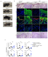
Extended Data Figure 1. ZBP1 deficiency strongly delays and ameliorates skin inflammation in RIPK1E-KO mice.
a, Photographs of mice with the indicated genotypes at the age of 4 weeks. Images shown are representative of n≥60 RIPK1E-KO and n≥40 RIPK1E-KO Zbp1-/- mice. b, Photographs of mice with the indicated genotypes and age. Images shown are representative of n≥4 RIPK1E-KO mice at the age of 5-7 weeks and n≥20 RIP1E-KO Zbp1-/- mice at the age of 17-35 weeks. c, Table summarizing the macroscopically observed skin lesions and time of sacrifice of 21 aged RIPK1E-KO Zbp1-/- mice. d, Representative images of skin sections from RIPK1E-KO Zbp1-/- mice and their respective controls stained with H&E (n≥18) or immunostained with the indicated antibodies (n≥4) at the age of 17-35 weeks. Nuclei stained with DAPI. Scale bars, 50 μm. e, Representative images of skin sections from 4-5 week old RIPK1E-KO (n≥6) and RIPK1E-KO Zbp1-/- (n≥3) and their respective control mice stained with TUNEL or immunostained with anti-CC3 antibodies. Nuclei stained with DAPI. Scale bars, 50 μm. f, Microscopic quantification of CC3 and TUNEL positive cells on skin sections from 4-5 week old mice with the indicated genotypes. Epi, Epidermis; Der, Dermis.
Extended Data Figure 2
Extended Data Figure 2. ZBP1 is not required for keratinocyte necroptosis and skin inflammation in mice with epidermis-specific FADD deficiency.
a, Representative photographs depicting the macroscopically observed phenotype of FADDE-KO (n≥10) and FADDE-KO Zbp1-/- (n=5) mice at the age of 6 days. b, Representative images of skin sections from 6 day old mice with the indicated genotypes stained with H&E (n=6) or immunostained with the indicated antibodies (n=3). Nuclei stained with DAPI. Scale bars, 50 μm. c, qRT-PCR analysis of the mRNA expression of the indicated cytokines and chemokines in total skin RNA from 6 day old mice with the indicated genotypes.
Extended Data Figure 3
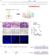
Extended Data Figure 3. CRISPR/Cas9 mediated generation of Ripk1mRHIM and Mlkl-/-mice.
a, Schematic depiction of the generation of Ripk1mRHIM/mRHIM mice indicating the sequence of the sgRNA and the single stranded oligo used for mutating the RHIM domain that were introduced by pronuclear injection into mouse zygotes and the sequencing result of one of the two obtained founders. b, Small intestinal sections from E18.5 pups were stained with H&E or TUNEL or immunostained with anti-CC3 antibodies. Representative images shown (WT n=6 for H&E, n=3 for TUNEL and anti-CC3; Ripk1mRHIM/mRHIM n=5 for H&E and n=3 for TUNEL and anti-CC3; Ripk1-/- n=3 for H&E, TUNEL and anti-CC3). Nuclei stained with DAPI. Scale bars, 50 µm. c, Microscopic quantification of CC3 and TUNEL positive cells on gut sections from E18.5 pups with the indicated genotypes. d, Diagram indicating the sgRNA target sequence (capital letters) used to generate a mutation in exon 2 downstream of the ATG of the Mlkl gene. The PAM sequence is indicated in red. Sequencing showing the 2 base pair deletion found in #97 at position chr8:111,333,648-111,333,649 (mm10), which results in a frameshift after amino acid 34 and a premature stop codon at amino acid position 55 of MLKL. This Mlkl knockout allele was used throughout this study.
Extended Data Figure 4
Extended Data Figure 4. Rescue of perinatal lethality of Ripk1mRHIM/mRHIM mice by deficiency of ZBP1, MLKL, or RIPK3.
a, Representative photographs and body weights of the indicated mice. b, Representative H&E stainings of skin, liver, spleen, colon and small intestine sections from 5-month-old Ripk3wt/-(n=4), Ripk1mRHIM/mRHIM Ripk3-/- (n=4) and Ripk1mRHIM/mRHIM Zbp1-/- mice (n=3).
Extended Data Figure 5
Extended Data Figure 5. MLKL or ZBP1-deficiency prevents skin inflammation in Ripk1mRHIM/E-KO mice.
a, Representative photographs of RIPK1mRHIM/E-KO (n=9), RIPK1mRHIM/E-KO Mlklwt/- (n=11), RIPK1mRHIM/E-KO Mlkl-/- (n=16) and RIPK1mRHIM/E-KO Zbp1-/- (n=7) at the age of 9-11 weeks. b, Representative images of skin sections from 9-11 week old RIPK1mRHIM/E-KO Mlklwt/- (n=11) and the respective control mice stained with H&E or immunostained with the indicated antibodies. Nuclei stained with DAPI. Scale bars, 50 μm. c, Representative images of skin sections from 9-11 week old RIPK1mRHIM/E-KO (n≥6), RIPK1mRHIM/E-KO Mlkl-/-(n=3), RIPK1mRHIM/E-KO Zbp-1- (n=3) and their respective control mice stained with TUNEL or immunostained with anti-CC3 antibodies. Nuclei stained with DAPI. Scale bars, 50 μm. d, Microscopic quantification of CC3 and TUNEL positive cells on skin sections from 4-5 week old mice with the indicated genotypes. Epi, Epidermis; Der, Dermis.
Extended Data Figure 6
Extended Data Figure 6. Expression of ZBP1 in primary keratinocytes.
a, Immunoblot analysis of lysates of primary keratinocytes derived from mice with the indicated genotypes. Each lane represents keratinocytes from individual mice. Cell lysates of wildtype FLM was used as a positive control. For gel source data, see Supplementary Figure 1. b, Immunoblot analysis of primary keratinocytes derived from mice with the indicated genotypes were left untreated (Medium) or stimulated with TNF or IFNβ for 18h. For gel source data, see Supplementary Figure 1.
Figure 1
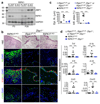
Figure 1. ZBP1 induces keratinocyte necroptosis and skin inflammation in RIPK1E-KO mice.
a, Immunoblot analysis of ZBP1, RIPK1 and GAPDH in epidermal protein extracts from wild type (Ripk1FL/WT) and RIPK1E-KO mice at postnatal day 3 (P3) and P28. Lanes represent samples from individual mice. For gel source data, see Supplementary Figure 1. b, Skin sections from 4 – 5 week old mice were stained with H&E or immunostained with the indicated antibodies. Representative images shown (RIPK1E-KO n=9 for H&E and n≥6 for immunostainings; RIPK1E-KO Zbp1-/- n=10 for H&E and n≥3 for immunostainings). Scale bars, 50 μm. c, Microscopic quantification of epidermal thickness (Epi. th.) and inflamed skin area (Infl. area) in 4 – 5 week old mice with the indicated genotypes. d, qRT-PCR analysis of the mRNA expression of the indicated cytokines and chemokines in RNA isolated from total skin from 4 – 5 week old mice with the indicated genotypes.
Figure 2
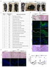
Figure 2. Mutation of the RIPK1 RHIM domain causes perinatal lethality and inflammatory skin hyperplasia in mice.
a, Table showing the numbers of weaned offspring of Ripk1mRHIM/wt parents from two independently generated knock-in lines (L1 & L2). b, Skin sections from E18.5 pups were stained with H&E or TUNEL or immunostained for CC3 or F4/80. Representative images shown (WT n=6 for H&E, n=3 for TUNEL, anti-CC3 and anti-F4/80; Ripk1mRHIM/mRHIM n=5 for H&E and anti-CC3, n=4 for TUNEL and n=3 for anti-F4/80; Ripk1-/- n=3 for H&E, TUNEL, anti-F4/80 and anti-CC3). Scale bars, 50 µm. c, Microscopic quantification of CC3 and TUNEL positive cells on skin sections from E18.5 pups with the indicated genotypes. Epi, Epidermis; Der, Dermis. d, Immunoblot analysis of total skin lysates from E18.5 pups of the indicated genotypes. Lanes represent samples from individual embryos. For gel source data, see Supplementary Figure 1.
Figure 3
Figure 3. Mutation of the RIPK1 RHIM domain prevents TNF-induced association of RIPK1 with RIPK3 and necroptosis.
a, Immunoblot analysis with the indicated antibodies of RIPK1 immunoprecipitates and total lysates from primary WT and Ripk1mRHIM/mRHIM MEFs treated with TNF (20ng/ml), CHX (1μg/ml) and z-VAD-fmk (20μM) for the indicated periods of time. h, hours. Representative data shown from two independent experiments. For gel source data, see Supplementary Figure 1. b, c, Primary MEFs (b) or FLMs (c) from mice with the indicated genotypes were treated with combinations of TNF (20ng/ml), CHX (1μg/ml), z-VAD-fmk (20μM) and Nec-1 (30μM) for 18 hours. Cell viability was determined by neutral red assay. Graphs show mean ± SEM from pooled data from 5 (b) and 3 (c) independent experiments. *P ≤ 0.05; **P ≤ 0.01; ***P ≤ 0.005. d, Immunoblot analysis of primary MEFs or FLMs from WT or Ripk1mRHIM/mRHIM mice with the indicated antibodies. Lanes represent primary cells from individual mice. For gel source data, see Supplementary Figure 1. e, f, Primary MEFs (e) or FLMs (f) from WT or Ripk1mRHIM/mRHIM mice were stimulated with TNF (20ng/ml) for the indicated periods of time and NF-κB activation was assessed by immunoblotting with the indicated antibodies. Representative data shown from three independent experiments. For gel source data, see Supplementary Figure 1.
Figure 4
Figure 4. RHIM-dependent RIPK1 function prevents MLKL/ZBP1-mediated necroptosis and skin inflammation.
a, Skin sections from 9-11 week old mice were stained with H&E or immunostained with the indicated antibodies. Representative images shown (RIPK1mRHIM/E-KO n=9 for H&E and n=3 for immunostainings; RIPK1mRHIM/E-KO Mlkl-/- n=5 for H&E and n=3 for immunostainings; RIPK1mRHIM/E-KO Zbp1-/- n=4 for H&E and n=3 for immunostainings). Scale bars, 50 μm. b, c, Microscopic quantification of epidermal thickness (Epi. th.) and inflamed skin area (Infl. area) (b) and qRT-PCR analysis of the mRNA expression of the indicated cytokines and chemokines (c) in 9-11 week old mice with the indicated genotypes. d, e, Immunoblot analysis of ZBP1 and MLKL expression in epidermal lysates (d) and qRT-PCR analysis of Zbp1 and Ifnb1 mRNA levels in total skin (e) from 4 week-old mice of the indicated genotypes. Lanes represent samples from individual mice. For gel source data, see Supplementary Figure 1. f, g, Immunoblot analysis with the indicated antibodies of anti-FLAG (f) or anti-RIPK1 (g)immunoprecipitates and total lysates from primary WT and Ripk1mRHIM/mRHIM (mR) MEFs transduced with lentiviruses expressing FLAG or FLAG-tagged ZBP1. Representative data shown from two independent experiments. For gel source data, see Supplementary Figure 1.

References

    1. Christofferson DE, Li Y, Yuan J. Control of life-or-death decisions by RIP1 kinase. Annu Rev Physiol. 2014;76:129–150. doi: 10.1146/annurev-physiol-021113-170259. - DOI - PubMed
    1. Newton K. RIPK1 and RIPK3: critical regulators of inflammation and cell death. Trends Cell Biol. 2015;25:347–353. doi: 10.1016/j.tcb.2015.01.001. - DOI - PubMed
    1. Pasparakis M, Vandenabeele P. Necroptosis and its role in inflammation. Nature. 2015;517:311–320. doi: 10.1038/nature14191. - DOI - PubMed
    1. Silke J, Rickard JA, Gerlic M. The diverse role of RIP kinases in necroptosis and inflammation. Nat Immunol. 2015;16:689–697. doi: 10.1038/ni.3206. - DOI - PubMed
    1. Weinlich R, Green DR. The two faces of receptor interacting protein kinase-1. Mol Cell. 2014;56:469–480. doi: 10.1016/j.molcel.2014.11.001. - DOI - PMC - PubMed

Publication types

MeSH terms