. 2016 Dec;22(12):1465-1469.
doi: 10.1038/nm.4223. Epub 2016 Nov 7.

Influenza immunization elicits antibodies specific for an egg-adapted vaccine strain

Affiliations

Influenza immunization elicits antibodies specific for an egg-adapted vaccine strain

Donald D Raymond et al. Nat Med. 2016 Dec.

Abstract

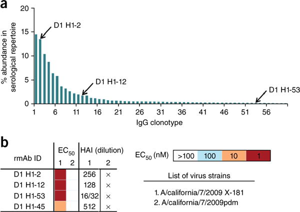
For broad protection against infection by viruses such as influenza or HIV, vaccines should elicit antibodies that bind conserved viral epitopes, such as the receptor-binding site (RBS). RBS-directed antibodies have been described for both HIV and influenza virus, and the design of immunogens to elicit them is a goal of vaccine research in both fields. Residues in the RBS of influenza virus hemagglutinin (HA) determine a preference for the avian or human receptor, α-2,3-linked sialic acid and α-2,6-linked sialic acid, respectively. Transmission of an avian-origin virus between humans generally requires one or more mutations in the sequences encoding the influenza virus RBS to change the preferred receptor from avian to human, but passage of a human-derived vaccine candidate in chicken eggs can select for reversion to avian receptor preference. For example, the X-181 strain of the 2009 new pandemic H1N1 influenza virus, derived from the A/California/07/2009 isolate and used in essentially all vaccines since 2009, has arginine at position 226, a residue known to confer preference for an α-2,3 linkage in H1 subtype viruses; the wild-type A/California/07/2009 isolate, like most circulating human H1N1 viruses, has glutamine at position 226. We describe, from three different individuals, RBS-directed antibodies that recognize the avian-adapted H1 strain in current influenza vaccines but not the circulating new pandemic 2009 virus; Arg226 in the vaccine-strain RBS accounts for the restriction. The polyclonal sera of the three donors also reflect this preference. Therefore, when vaccines produced from strains that are never passaged in avian cells become widely available, they may prove more capable of eliciting RBS-directed, broadly neutralizing antibodies than those produced from egg-adapted viruses, extending the established benefits of current seasonal influenza immunizations.


Conflict of interest statement

COMPETING FINANCIAL INTERESTS

The authors declare competing financial interests: details are available in the online version of the paper.

Figures

Figure 1
Figure 1
B cell clonal lineage CL6515 from Siena patient 7. The tree was derived, using Clonalyst18,19, from single-cell, paired heavy- and light-chain sequences. The genes encoding the variable domains are: IGHV5-51, IGHJ6*02, IGKV3-20, IGKJ1*01. The antibodies are designated by heavy-chain sequence number, preceded by ‘D8’ or ‘D22’ for cells obtained either 8 or 22 d after vaccination, respectively. Inset: the subtree that includes Ab6639. Antibodies defining that subtree are marked with asterisks in the main figure.
Figure 2
Figure 2
Structure of Fab 6639 bound with uncleaved HA (HA0) of the X-181 vaccine strain. (a) Ribbon representation of the structure. The HA trimer dissociates after removal of the C-terminal foldon tag, causing part of the stem of the HA0 monomer to become disordered; the complex that crystallizes contains one Fab bound with one HA0 monomer. CDR-H3 of the Fab projects into the RBS on the ‘head’ of the HA molecule. Fab heavy chain is in blue; Fab light chain is in yellow; the HA1 (receptor-binding) part of the HA0 chain is in green; and the HA2 (fusogenic) part is in magenta. The initial N-acetylglucosamine of five glycans, shown in stick representation, were visible in the density map. (b) Contacts between residues at the tip of CDR-H3 and residues in the RBS, showing the role of Arg226. The view is roughly 90° (clockwise about a vertical axis) from the view in a. Hydrogen bonds are yellow dashed lines. The hydrogen bonds that buttress the Arg226 side chain position it to donate two hydrogen bonds to carbonyls on the CDR-H3 loop of the Fab. (c) View 180° from the view in b. (d) Contacts with residues in the 190-helix. View direction is from ‘below’ in a.
Figure 3
Figure 3
Serum antibody repertoire and analysis of three antibodies from donor 1 of the 2011–12 cohort. (a) Abundance of individual antibody clonotypes in the anti-H1 repertoire, as determined from mass spectrometric analysis of serum 28 d after vaccination. Each bar denotes a unique antibody clonotype, and the three antibodies analyzed represent high-, moderate- and low-abundance clonotypes (second, twelfth, and fifty-third ranked, respectively). (b) Characterization of the three antibodies with HA from X-181 and pdm2009. HAI, hemagglutination inhibition assayed with the virus strains listed; cross (×) indicates no activity even at the lowest dilution. IC50 values were determined by ELISA with the HAs from the listed strains.

Comment in

References

    1. Wu X, et al. Rational design of envelope identifies broadly neutralizing human monoclonal antibodies to HIV-1. Science. 2010;329:856–861. - PMC - PubMed
    1. Wu X, et al. Maturation and diversity of the VRC01 antibody lineage over 15 years of chronic HIV-1 infection. Cell. 2015;161:470–485. - PMC - PubMed
    1. Zhou T, et al. Structural basis for broad and potent neutralization of HIV-1 by antibody VRC01. Science. 2010;329:811–817. - PMC - PubMed
    1. Ekiert DC, et al. Cross-neutralization of influenza A viruses mediated by a single antibody loop. Nature. 2012;489:526–532. - PMC - PubMed
    1. Hong M, et al. Antibody recognition of the pandemic H1N1 influenza virus hemagglutinin receptor-binding site. J Virol. 2013;87:12471–12480. - PMC - PubMed

Publication types

MeSH terms