. 2016 Nov 7;11(11):e0166106.
doi: 10.1371/journal.pone.0166106. eCollection 2016.

Fast-to-Slow Transition of Skeletal Muscle Contractile Function and Corresponding Changes in Myosin Heavy and Light Chain Formation in the R6/2 Mouse Model of Huntington's Disease

Affiliations

Fast-to-Slow Transition of Skeletal Muscle Contractile Function and Corresponding Changes in Myosin Heavy and Light Chain Formation in the R6/2 Mouse Model of Huntington's Disease

Tanja Hering et al. PLoS One. .

Abstract

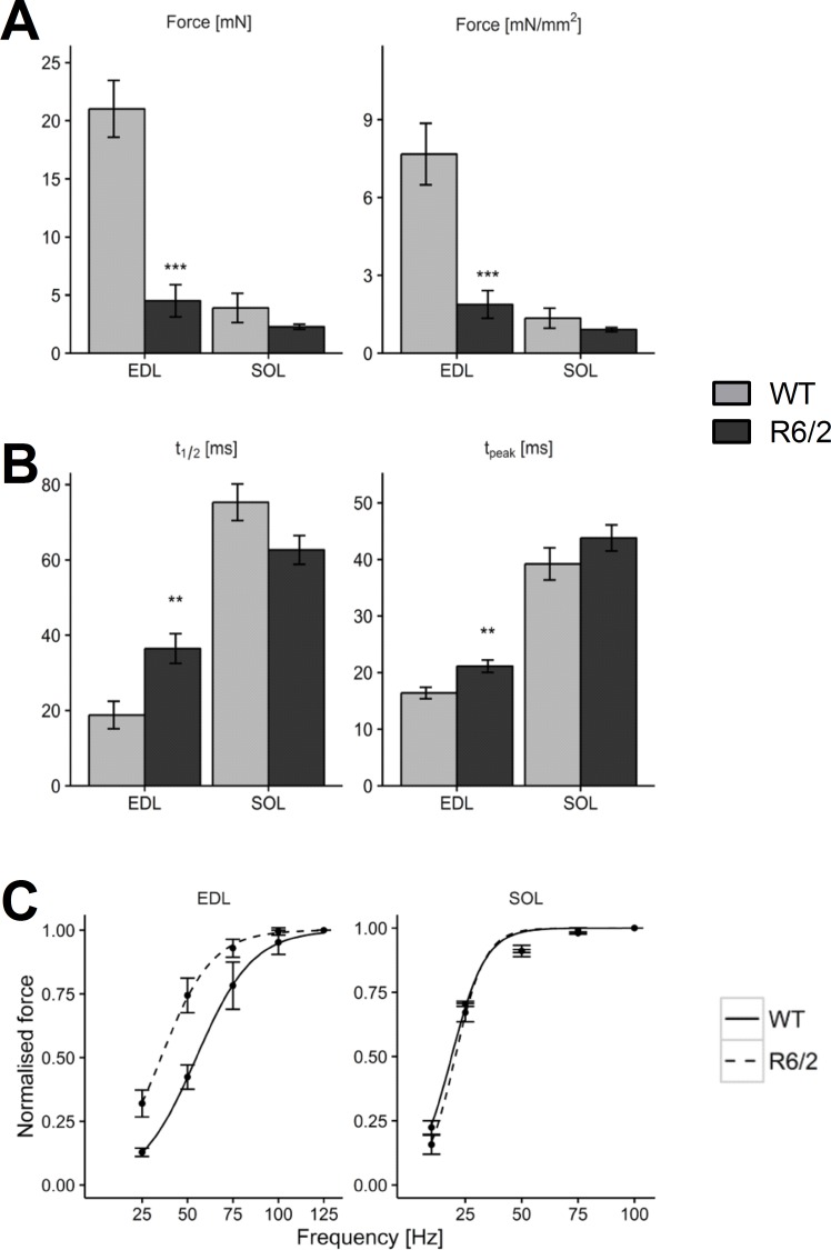
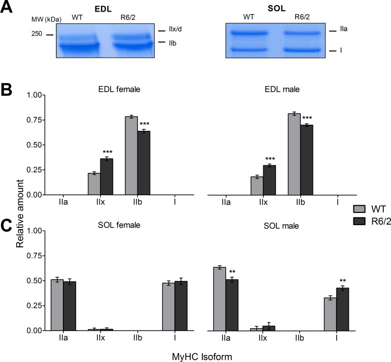
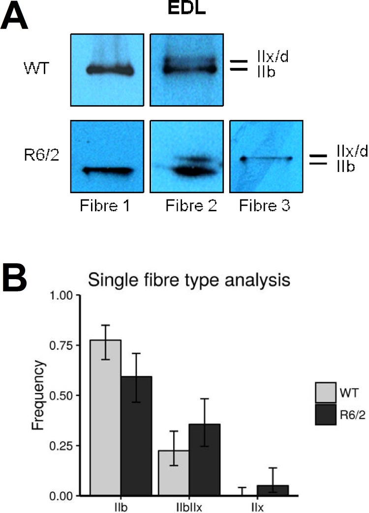
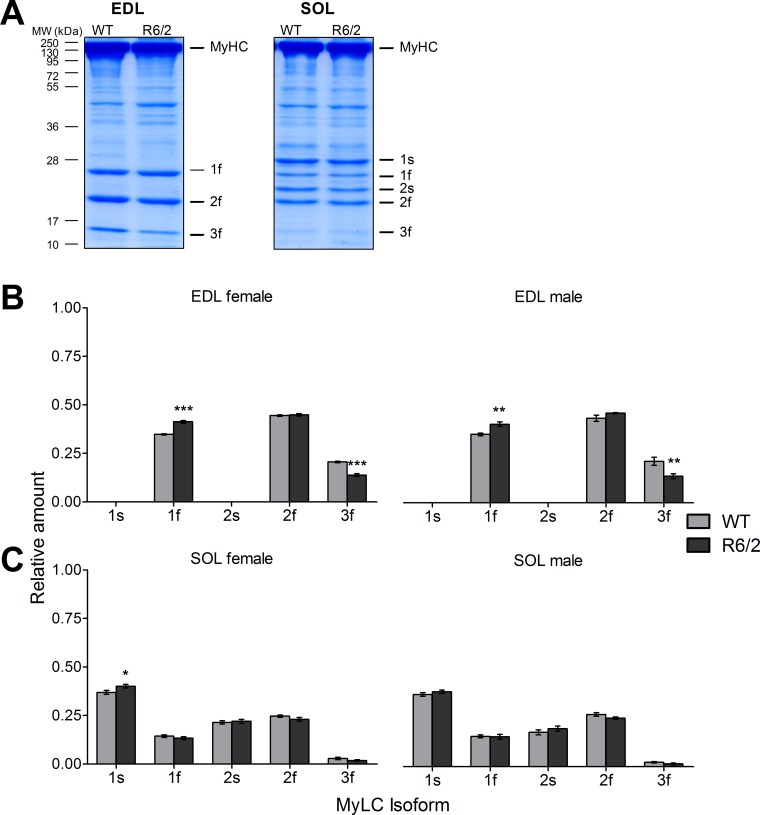
Huntington´s disease (HD) is a hereditary neurodegenerative disease resulting from an expanded polyglutamine sequence (poly-Q) in the protein huntingtin (HTT). Various studies report atrophy and metabolic pathology of skeletal muscle in HD and suggest as part of the process a fast-to-slow fiber type transition that may be caused by the pathological changes in central motor control or/and by mutant HTT in the muscle tissue itself. To investigate muscle pathology in HD, we used R6/2 mice, a common animal model for a rapidly progressing variant of the disease expressing exon 1 of the mutant human gene. We investigated alterations in the extensor digitorum longus (EDL), a typical fast-twitch muscle, and the soleus (SOL), a slow-twitch muscle. We focussed on mechanographic measurements of excised muscles using single and repetitive electrical stimulation and on the expression of the various myosin isoforms (heavy and light chains) using dodecyl sulfate polyacrylamide gel electrophoresis (SDS-PAGE) of whole muscle and single fiber preparations. In EDL of R6/2, the functional tests showed a left shift of the force-frequency relation and decrease in specific force. Moreover, the estimated relative contribution of the fastest myosin isoform MyHC IIb decreased, whereas the contribution of the slower MyHC IIx isoform increased. An additional change occurred in the alkali MyLC forms showing a decrease in 3f and an increase in 1f level. In SOL, a shift from fast MyHC IIa to the slow isoform I was detectable in male R6/2 mice only, and there was no evidence of isoform interconversion in the MyLC pattern. These alterations point to a partial remodeling of the contractile apparatus of R6/2 mice towards a slower contractile phenotype, predominantly in fast glycolytic fibers.


Conflict of interest statement

The authors have declared that no competing interests exist.

Figures

Fig 1
Fig 1. Lower force and slowed kinetics of contraction in R6/2.
(A) Mean twitch force (left panel) and specific force (i.e. normalized by cross sectional area; right panel) compared in EDL and SOL muscles of WT (n = 15 and n = 15, respectively) and R6/2 (n = 10 and n = 10, respectively). (B) Comparison of half time of relaxation (t1/2, left panel) and time to peak (tpeak, right panel). (C) Comparison of force frequency relations. Error bars indicate SEM.
Fig 2
Fig 2. Relative content of myosin heavy chain isoforms in WT and R6/2 muscle.
(A) Examples showing sections of Roti®-Blue-stained gels exhibiting MyHC bands that were evaluated in the analysis. See also S1 Fig. (B) Relative amounts of the indicated MyHC isoforms in EDL of WT and R6/2, respectively, presented separately as results from female (left panel) and male specimen (right panel). (C) Relative amounts of the indicated MyHC isoforms in SOL of WT and R6/2, respectively, presented separately as results from female (left panel) and male (right panel) specimen. Error bars indicate SEM. n = 11 to 13 mice.
Fig 3
Fig 3. MyHC determination in single muscle fibers.
Muscle fiber composition for EDL was assessed on a single fiber basis. For this MyHC were extracted from 6 to 15 randomly selected, intact fibers per muscle sample. (A) Examples showing Roti®-Blue-stained MyHC bands from selected single fiber SDS PAGE gels. Fibers were classified as Type IIB and IIX when a single band could be detected and as mixed IIB/X when both bands were detectable, irrespective of staining intensity. (B) Relative contribution (fractional number) of pooled fibers (59 fibers of 5 R6/2 mice and 89 fibers of 7 WT mice) exhibiting expression of MyHC IIb, IIx or both. Distributions of fibers amongst the types IIB, IIX and mixed type IIB/X differed significantly between WT and R6/2 mice (p = 0.02) indicating a different muscle fiber composition of R6/2 EDL muscle with more mixed-type fibers. Bars show relative frequencies; error bars show confidence intervals for binomial proportions (i.e. fibers of respective type vs. all other fibers) (95% CI).
Fig 4
Fig 4. Relative content of myosin light chain isoforms in WT and R6/2 muscle.
(A) Examples showing Roti®-Blue-stained gels exhibiting MyLC bands that were evaluated in the analysis. See also S2 Fig. (B) Relative amounts of the indicated MyLC isoforms in EDL of WT and R6/2, respectively, separated in results from female (left panel) and male specimen (right panel). (C) Relative amounts of the indicated MyLC isoforms in SOL of WT and R6/2, respectively, separated in results from female (left panel) and male (right panel) specimen. Error bars indicate SEM. n = 10 to 12 mice.

References

    1. Walker FO. Huntington's disease. The Lancet 2007; 369(9557):218–28. - PubMed
    1. Ross CA, Pantelyat A, Kogan J, Brandt J. Determinants of functional disability in Huntington's disease: role of cognitive and motor dysfunction. Movement disorders 2014; 29(11):1351–8. 10.1002/mds.26012 - DOI - PMC - PubMed
    1. Imarisio S, Carmichael J, Korolchuk V, Chen C-W, Saiki S, Rose C, et al. Huntington's disease: from pathology and genetics to potential therapies. Biochem. J. 2008; 412(2):191–209. 10.1042/BJ20071619 - DOI - PubMed
    1. Cepeda C, Wu N, Andre V, Cummings D, Levine M. The corticostriatal pathway in Huntington's disease. Progress in Neurobiology 2007; 81(5–6):253–71. 10.1016/j.pneurobio.2006.11.001 - DOI - PMC - PubMed
    1. Ross CA, Tabrizi SJ. Huntington's disease: from molecular pathogenesis to clinical treatment. The Lancet. Neurology 2011; 10(1):83–98. 10.1016/S1474-4422(10)70245-3 - DOI - PubMed

MeSH terms