

Comparing culture and molecular methods for the identification of microorganisms involved in necrotizing soft tissue infections
- PMID: 27821087
- PMCID: PMC5100109
- DOI: 10.1186/s12879-016-1976-2
Comparing culture and molecular methods for the identification of microorganisms involved in necrotizing soft tissue infections
Abstract
Background: Necrotizing soft tissue infections (NSTIs) are a group of infections affecting all soft tissues. NSTI involves necrosis of the afflicted tissue and is potentially life threatening due to major and rapid destruction of tissue, which often leads to septic shock and organ failure. The gold standard for identification of pathogens is culture; however molecular methods for identification of microorganisms may provide a more rapid result and may be able to identify additional microorganisms that are not detected by culture.
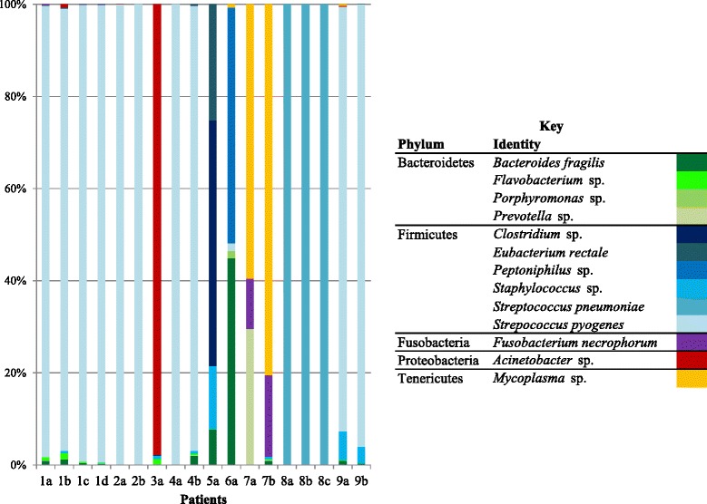
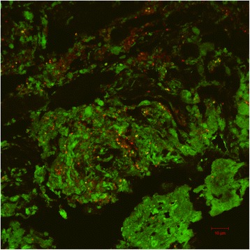
Methods: In this study, tissue samples (n = 20) obtained after debridement of 10 patients with NSTI were analyzed by standard culture, fluorescence in situ hybridization (FISH) and multiple molecular methods. The molecular methods included analysis of microbial diversity by 1) direct 16S and D2LSU rRNA gene Microseq 2) construction of near full-length 16S rRNA gene clone libraries with subsequent Sanger sequencing for most samples, 3) the Ibis T5000 biosensor and 4) 454-based pyrosequencing. Furthermore, quantitative PCR (qPCR) was used to verify and determine the relative abundance of Streptococcus pyogenes in samples.
Results: For 70 % of the surgical samples it was possible to identify microorganisms by culture. Some samples did not result in growth (presumably due to administration of antimicrobial therapy prior to sampling). The molecular methods identified microorganisms in 90 % of the samples, and frequently detected additional microorganisms when compared to culture. Although the molecular methods generally gave concordant results, our results indicate that Microseq may misidentify or overlook microorganisms that can be detected by other molecular methods. Half of the patients were found to be infected with S. pyogenes, but several atypical findings were also made including infection by a) Acinetobacter baumannii, b) Streptococcus pneumoniae, and c) fungi, mycoplasma and Fusobacterium necrophorum.
Conclusion: The study emphasizes that many pathogens can be involved in NSTIs, and that no specific "NSTI causing" combination of species exists. This means that clinicians should be prepared to diagnose and treat any combination of microbial pathogens. Some of the tested molecular methods offer a faster turnaround time combined with a high specificity, which makes supplemental use of such methods attractive for identification of microorganisms, especially for fulminant life-threatening infections such as NSTI.
Keywords: 16S rRNA; 454 pyrosequencing; Cloning; Direct Sanger sequencing; FISH; Ibis T5000 biosensor; Microorganisms; Necrotizing soft tissue infections; qPCR.
Figures

References
Publication types
MeSH terms
Substances
Grants and funding
LinkOut - more resources
Full Text Sources
Other Literature Sources
Molecular Biology Databases
