Review
. 2016 Dec 19;371(1710):20150401.
doi: 10.1098/rstb.2015.0401.

Nodal signalling and asymmetry of the nervous system

Affiliations
Review

Nodal signalling and asymmetry of the nervous system

Iskra A Signore et al. Philos Trans R Soc Lond B Biol Sci. .

Abstract

The role of Nodal signalling in nervous system asymmetry is still poorly understood. Here, we review and discuss how asymmetric Nodal signalling controls the ontogeny of nervous system asymmetry using a comparative developmental perspective. A detailed analysis of asymmetry in ascidians and fishes reveals a critical context-dependency of Nodal function and emphasizes that bilaterally paired and midline-unpaired structures/organs behave as different entities. We propose a conceptual framework to dissect the developmental function of Nodal as asymmetry inducer and laterality modulator in the nervous system, which can be used to study other types of body and visceral organ asymmetries. Using insights from developmental biology, we also present novel evolutionary hypotheses on how Nodal led the evolution of directional asymmetry in the brain, with a particular focus on the epithalamus. We intend this paper to provide a synthesis on how Nodal signalling controls left-right asymmetry of the nervous system.This article is part of the themed issue 'Provocative questions in left-right asymmetry'.

Keywords: Nodal; asymmetry; context-dependency; epithalamus; evo-devo; nervous system.


Figures

Figure 1.
Figure 1.
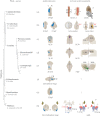
Phylogenetic distribution of asymmetric Nodal expression associated with the development of nervous system asymmetries across metazoans. For each phylum and species, the asymmetric pattern of Nodal expression is shown on the left while the observed morphological asymmetry of the nervous system is on the right. (a) Nodal expression in the left side of the future head of the cephalochordate Branchiostoma floridae precedes and controls the development of asymmetries in the bilaterally paired somatic neurons and peripheral nerves (arrows) associated with the somites. The role of Nodal in the asymmetry of the adult brain involving the right infundibulum (arrowhead) and related brain lobe (B) is yet to be determined (#). Notochord (N), Hatcheck's pit (H), hours post-fertilization (hpf). (b) Nodal expression in the left epidermis and sensory vesicle (SV) of the ascidian embryo precedes and controls the right-sided asymmetric positioning of the SV lumen and photoreceptor cell (PR, yellow). C. intestinalis, Ciona intestinalis. (ce) Nodal expression in the left epithalamus of different craniate species precedes and controls the development of asymmetries in the bilaterally paired habenular nuclei (Hb) and the midline-unpaired parapineal organ (PpO). The right Hb (RHb) is larger than the left Hb (LHb) in the lamprey Petromyzon marinus (c) while the opposite is seen in the elasmobranch Scyliorhinus canicula (d) and in the teleost zebrafish (Danio rerio) (e). In zebrafish, the PpO also locates on the left side of the epithalamus. Somites (som). (f) In the sea urchin Paracentrotus lividus, Nodal is expressed in the right ciliary band (CB), in a cluster of prospective neurons located on the right side of the apical tuft (AT). However, this expression has not been yet related to nervous system asymmetry. (g) In the hemichordate Saccoglossus kowalevskii, Nodal is asymmetrically expressed in the right ectoderm along the entire dorsoventral axis, a site where diffuse neurogenesis occurs. However, asymmetries in this region have not been described. (h) In snails, sided expression of Nodal in either the left or right ectoderm controls the chirality of shell rotation. Chirality of shell rotation, in turn, associates with the side where the parietal and visceral ganglia will fuse in the nervous system of Lymnaea stagnalis. Direct control of Nodal in this type of nervous system asymmetry has yet to be examined (#). Dextral (D), left parietal ganglia (L-pg), right parietal ganglia (R-pg), left visceral ganglia (L-vg), right visceral ganglia (R-vg), left parietal-visceral ganglia (L-pvg), right parietal-visceral ganglia (R-pvg), sinistral (S). Figures correspond to dorsal views with anterior to the top and left to the left, with the exception of the Hb of P. marinus and S. canicula, where the Hb is shown in cross-sections of the brain, with dorsal to the top and left to the left (right panels of (c) and (d), respectively). The stage of analysis is shown at the bottom of each panel. ‘?’ represents an unknown feature (no information available). The animal phylogeny of this figure is based on [9]. See the text for additional details.
Figure 2.
Figure 2.
Nodal and nervous system asymmetries in ascidians. Two different types of asymmetry in the ascidian nervous system where Nodal acts as asymmetry inducer. For each panel, the pattern of Nodal expression in the left sensory vesicle (SV) and epidermis is shown on the left, while the resulting asymmetric phenotype is on the right. The results of three experimental conditions are compared: wild-type left-sided Nodal expression (top), pharmacological inhibition of Nodal function that is equivalent to absent Nodal (middle) and gain of Nodal function that induces bilateral Nodal expression (bottom). (a) Asymmetry of the photoreceptor cell (PR, yellow) surrounding the ocellus (black), in the SV (red circle). The PR domain can be regarded as a bilaterally paired structure concerning the bilateral competence to form PR cells. Asymmetry involves L–R differences in PR differentiation. Nodal inhibits photoreceptor cell fate; therefore the PR in the wild-type is located on the right side. Loss and gain of Nodal function result in right and left isomerism, respectively. Hours post-fertilization (hpf). (b) Asymmetry of the lumen of the SV. The lumen of the SV (red circle) containing the ocellus (black dot) can be regarded as a midline-unpaired structure of initial midline position. Asymmetry of the SV involves clockwise rotation of the neural tube in which Nodal is presumably involved. Asymmetric Nodal expression probably promotes morphogenetic transformations that result in neural tube rotation. Therefore, loss of Nodal function generates a midline-positioned SV (symmetry). By contrast, differences in Nodal expression between left and right sides, which are normally observed in bilateral Nodal expression, are presumably responsible for generating a randomized phenotype that combines left-, midline- and right-positioned SV. See §§3 and 4 for additional details.
Figure 3.
Figure 3.
Nodal and nervous system asymmetry in the epithalamus of fishes. Asymmetries in the epithalamic bilaterally paired habenular nuclei (Hb) in different species of fishes in which Nodal acts as asymmetry inducer and/or laterality modulator. For each panel, the pattern of Nodal expression in the prospective epithalamus is shown on the left, while the resulting habenular phenotype is on the right. The results of three experimental conditions are shown: wild-type left-sided Nodal expression (top), genetically induced or pharmacologically mediated inhibition of Nodal function (absent Nodal) (middle), and bilateral Nodal expression (bottom). Asymmetries of the Hb can be classified into two types according to their dependency on the midline-unpaired parapineal organ (PpO). In zebrafish, the PpO can direct the development of a sub-type of habenular asymmetries (PpO-dependent asymmetries) (d). Other subtypes of habenular asymmetries in zebrafish (c), and the asymmetries described in the Hb of lampreys and catshark (a,b) are independent of the PpO (PpO-independent asymmetries). (a) Asymmetry in the Hb of the lamprey Petromyzon marinus. The right Hb is usually larger than the left Hb and expresses phospho-ERK (red). Nodal functions as asymmetry inducer. Therefore, loss of Nodal signalling (Nodal absent) induces right isomerism with both sides of the Hb showing a large size and expressing phospho-ERK. Gain of function experiments (Nodal bilateral) have not been performed in this species. (b) Asymmetry in the Hb of the catshark S. canicula. The left Hb is usually larger and shows a more-extended pattern of Kct12b expression (purple) compared to the right Hb. Nodal functions as asymmetry inducer. Therefore, absence of Nodal results in right isomerism, with both sides of the Hb showing a small size and a right-type pattern of Kct12b expression. Gain of function experiments (Nodal bilateral) have not been performed in this species. (c) Parapineal-independent asymmetry of the Hb in zebrafish (Danio rerio). At early stages of development, the left Hb contains more cells expressing elav3/HuC (orange) than the right Hb. Nodal functions as asymmetry inducer. Therefore, loss and gain of Nodal function induce right and left isomerism, with both sides of the Hb showing a symmetric pattern of elav3 expression of right and left characteristics, respectively. Hours post-fertilization (hpf), somites (som). (d) Parapineal-dependent asymmetry of the Hb in zebrafish. The parapineal organ (PpO, green) typically locates on the left side and induces the elaboration of asymmetry in the Hb. This sub-type of habenular asymmetry is characterized (among other features) by a larger left Hb with an extended Kctd12.1 expression domain (purple) compared with the right Hb. Nodal functions as laterality modulator in this type of habenular asymmetry, although indirectly, through modulating the laterality of PpO asymmetric migration. Therefore, loss (Nodal absent) and gain (Nodal bilateral) of function approaches both unmask an antisymmetry of the PpO (and as a consequence induce antisymmetry of the Hb), with equal frequencies of left (50%) and right (50%) asymmetry phenotypes in the population. See §4 for additional details.
Figure 4.
Figure 4.
Ontogeny of epithalamic asymmetry in zebrafish. (a) Developmental paths leading to the development of asymmetries in the Hb. Two distinct paths classified according to their dependence on the parapineal organ (PpO) develop in parallel, under the control of asymmetric left-sided Nodal signalling (i). In the PpO-independent path (top), Nodal functions as asymmetry inducer and generates an enhanced level of neurogenesis in the left Hb, with increased number of elav3/HuC and cxcr4b positive cells compared with the right Hb (ii). At later stages, subtle asymmetries develop in the habenular neuropil and axonal terminal morphology in the interpeduncular nucleus (IPN), which become evident after ablation of the PpO (iii). In the PpO-dependent path (bottom), Nodal functions as laterality modulator directing PpO migration to the side of Nodal expression (iv). As a consequence of PpO asymmetric positioning, the Hb then develops striking structural and functional differences between the left and right sides. These asymmetries involve: gene expression (v); morphology (size and neuropil content); afferent connectivity from the olfactory bulb (ob) to the right Hb and from the PpO to the left Hb (vi); sub-nuclear organization, with enlarged dorsolateral (Hb-dl) and dorsomedial (Hb-dm) sub nuclei in the left and right Hb, respectively (vii); and efferent connectivity towards the midbrain IPN, with left and right habenular neurons projecting primarily to dorsal and ventral domains of the IPN, respectively (viii). Also, activation of habenular neurons to visual and olfactory stimuli are asymmetric and mostly involve the left and right sides of the Hb, respectively (not shown). Schemes are based on references [–49] and correspond to dorsal views of the epithalamus, with anterior to the top. Time is in hours post fertilization. (b) Model of PpO antisymmetric migration. In the absence of Nodal signalling, a bi-stable cell migratory event dependent on fibroblast growth factor (FGF) signalling establishes PpO antisymmetry, with equal left- (50%) and right- (50%) sided migration [50]. PpO cells (dark green) express the FGF receptor fgfr4, while cells along the path of PpO migration express the ligand fgf8 (light green). The migratory state of the PpO is unstable at the midline. Small differences in FGF signalling between left and right sides, probably owing to stochastic lateral differences in the level of FGF8 protein, break this unstable state and induce the PpO to migrate to either the left or right side with a random frequency (green arrows). Autocatalytic events then amplify the initial differences in PpO asymmetric migration. (c) In the presence of asymmetric Nodal signalling (blue), the FGF-dependent PpO antisymmetry becomes biased towards the side of Nodal expression [50,51]. Once the PpO adopts an initial left-sided position, autocatalytic events then reinforce the lateral migration as in (b).
Figure 5.
Figure 5.
A conceptual framework to study the role of Nodal signalling in the development of asymmetry. For details, see the text in §7.
Figure 6.
Figure 6.
Evolutionary routes to directional asymmetry in the epithalamus. (a) Model of the evolution of directional asymmetry from a symmetric ancestral condition proposed by Palmer [71]. Two routes can be distinguished. In the direct route (red arrow, 1) both asymmetry and laterality of asymmetry evolve together in a single step likely by conventional evolution (genotype precedes phenotype). In the indirect or two-step route (blue arrows, 2), a non-inheritable antisymmetric phenotype evolves first by mutation or developmental mechanisms under the control of environmental and/or behavioural factors (left). Then, one of the asymmetric phenotypes is fixed by the appearance of a laterality mechanism (right), resulting in directional asymmetry. (b) Proposed evolutionary route leading to directional asymmetry of the habenula (Hb). As a bilaterally paired structure, directional asymmetry of the Hb probably evolved by a direct route through Nodal acting as asymmetry inducer (red arrow, 1). (c) Proposed models of evolutionary paths leading to directional asymmetry in the position of the parapineal organ (PpO). As the PpO is an initially midline-unpaired structure, both direct and indirect routes are equally possible. In the indirect two-step route (blue arrows, 2), an antisymmetric positioning of the PpO first evolved independently from Nodal (e.g. mediated by FGF signalling) (left). In a second step, asymmetric Nodal might have been co-opted to direct the antisymmetric mechanism of PpO migration to the side of Nodal expression, thus transforming antisymmetry into directional asymmetry (right). Alternatively, directional asymmetry of the PpO evolved along the direct route (red arrow, 1) by Nodal acting as asymmetry inducer. In this scenario, Nodal directly induced the asymmetric positioning of the PpO towards the left side. This initial asymmetry might have then co-opted antisymmetric mechanisms of PpO migration, or coexisted with them, with Nodal gaining a new function as laterality modulator. (d) Model for the evolution of epithalamic asymmetry based on Hb–PpO interactions. The hypothetical ancestor had a symmetric Hb and a medially positioned PpO with no left-sided projection to the Hb (or bilateral projection) (top). Left-sided Nodal then acted as asymmetry inducer to generate directional asymmetry of the Hb (i). Then, the establishment of interactions between the Hb and PpO led to the recruitment of afferent axonal connectivity from the PpO (ii). Finally, intimate Hb–PpO interactions promoted asymmetric positioning of the PpO, which became influenced by asymmetric Nodal signalling during ontogeny. See §8 for additional details.

References

    1. Quail DF, Siegers GM, Jewer M, Postovit LM. 2013. Nodal signalling in embryogenesis and tumourigenesis. Int. J. Biochem. Cell Biol. 45, 885–898. (10.1016/j.biocel.2012.12.021) - DOI - PubMed
    1. Shiratori H, Hamada H. 2014. TGFβ signaling in establishing left–right asymmetry. Semin. Cell Dev. Biol. 32, 80–84. (10.1016/j.semcdb.2014.03.029) - DOI - PubMed
    1. Pauklin S, Vallier L. 2015. Activin/nodal signalling in stem cells. Development 142, 607–619. (10.1242/dev.091769) - DOI - PubMed
    1. Sampath K, Robertson EJ. 2016. Keeping a lid on nodal: transcriptional and translational repression of nodal signalling. Open Biol. 6, 150200 (10.1098/rsob.150200) - DOI - PMC - PubMed
    1. Geschwind N, Levitsky W. 1968. Human brain: left–right asymmetries in temporal speech region. Science N.Y. 161, 186–187. (10.1126/science.161.3837.186) - DOI - PubMed

Publication types

LinkOut - more resources