Review
. 2016 Dec 19;371(1710):20150403.
doi: 10.1098/rstb.2015.0403.

Cell chirality: its origin and roles in left-right asymmetric development

Affiliations
Review

Cell chirality: its origin and roles in left-right asymmetric development

Mikiko Inaki et al. Philos Trans R Soc Lond B Biol Sci. .

Abstract

An item is chiral if it cannot be superimposed on its mirror image. Most biological molecules are chiral. The homochirality of amino acids ensures that proteins are chiral, which is essential for their functions. Chirality also occurs at the whole-cell level, which was first studied mostly in ciliates, single-celled protozoans. Ciliates show chirality in their cortical structures, which is not determined by genetics, but by 'cortical inheritance'. These studies suggested that molecular chirality directs whole-cell chirality. Intriguingly, chirality in cellular structures and functions is also found in metazoans. In Drosophila, intrinsic cell chirality is observed in various left-right (LR) asymmetric tissues, and appears to be responsible for their LR asymmetric morphogenesis. In other invertebrates, such as snails and Caenorhabditis elegans, blastomere chirality is responsible for subsequent LR asymmetric development. Various cultured cells of vertebrates also show intrinsic chirality in their cellular behaviours and intracellular structural dynamics. Thus, cell chirality may be a general property of eukaryotic cells. In Drosophila, cell chirality drives the LR asymmetric development of individual organs, without establishing the LR axis of the whole embryo. Considering that organ-intrinsic LR asymmetry is also reported in vertebrates, this mechanism may contribute to LR asymmetric development across phyla.This article is part of the themed issue 'Provocative questions in left-right asymmetry'.

Keywords: cell chirality; cortical inheritance; f actin; left–right asymmetry; myosin I.


Figures

Figure 1.
Figure 1.
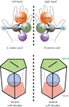
Chirality in hands, molecules and cells. Chirality is a property of an item that cannot be superimposed on its mirror image, as seen in the left and right hands. Most biological molecules, such as amino acids, are chiral. Cells can also be chiral if they have LR asymmetry and apico-basal polarity.
Figure 2.
Figure 2.
Chirality in ciliates. Right: ciliates show chirality in their global cortical structures, including the ciliary rows, oral apparatus and contractile vacuole. Left: the cortical unit of ciliates, which includes the ciliary rootlet and basal body, is chiral (adapted from [7]). In wild-type ciliates, the ciliary rootlet (cr) extends anteriorly and is positioned to the right relative to the basal body and the cell itself. The transversal microtubule ribbon (tmr) is on the left side of the basal body, and the post-ciliary microtubule ribbon (p-cmr) points posteriorly. Basal bodies are seen from the outside of the cell, and the viewer's right corresponds to the cell's left. Schema is adopted from Beisson [7]. A, anterior; P, posterior; R, right; L, left. (Online version in colour.)
Figure 3.
Figure 3.
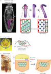
Cell chirality and LR asymmetric morphogenesis in Drosophila. (a) The Drosophila embryonic hindgut shows sinistral looping as the consequence of an LR asymmetric rotation. Before the onset of the rotation, hindgut epithelial cells show chirality with more frequent leftward-tilted cell boundaries than rightward-tilted ones. The chirality disappears when the rotation is completed. Distribution of DE-cadherin (green) also shows chirality. (b) The Drosophila male genitalia undergo a 360° clockwise rotation during the pupal stages. Epithelial cells in the A8a segment of male genitalia show chirality just before and during the LR asymmetric rotation. These cells have more frequent rightward-tilted cell boundaries and a higher expression of Myosin II along the rightward-tilted cell boundaries. Schema is adopted from Sato K et al. [23]. (c) Drosophila adult gut shows LR directional looping. The adult gut develops from larval primordia called the imaginal ring, consisting of H1 and H2 segments. The cell chirality determinant Myo31DF is required only in the H1 segment during larval stages. Cell chirality is observed in the H2 segment only after the H1 segment is eliminated. The handedness determined by Myo31DF in the H1 segment might be conveyed to the H2 segment through atypical cadherins, Dachsous and Fat. Schema is adopted from González-Morales et al. [24].
Figure 4.
Figure 4.
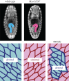
Cell chirality is an intrinsic property of individual cells, and Myo31DF switches the direction of cell chirality. Left: wild-type embryos show rightward looping of the hindgut and dextral cell chirality. Middle: in Myo31DF mutant embryos, both the hindgut looping and cell chirality are inverted. Right: when cells expressing wild-type Myo31DF are randomly introduced into the Myo31DF hindgut, only the cells expressing wild-type Myo31DF show the normal dextral chirality, indicating that cell chirality is formed intrinsically in each cell.
Figure 5.
Figure 5.
Cell chirality in snails and C. elegans. (a) Upper: in Lymnaea, the blastomere spindles slant clockwise as viewed from the animal pole at the four-cell stage, and blastomeres are rearranged clockwise at the eight-cell stage. Bottom: Pulmonata is a snail species with counter clockwise-coiling shells and internal organs that mirror those of Lymnaea. In Pulmonata, blastomere spindles slant anticlockwise as viewed from the animal pole, resulting in a counter clockwise blastomere rearrangement at the eight-cell stage. In both cases, blastomere chirality determines the shell coiling direction and LR asymmetry of the body. Schema is adopted from Shibazaki et al. [39]. (b) Top: in C. elegans, mitotic spindles are skewed during the transition from four cells to six cells. Bottom: at the six-cell stage, changing the LR-asymmetric arrangement of blastomeres to their mirror-image positions results in situs inversus. Thus, in both snails and C. elegans, blastomere chirality is completely responsible for the subsequent LR-asymmetric development. Schema is adopted from Wood & Kershaw [40].
Figure 6.
Figure 6.
Chiral shape and swirling of cultured mammalian cells. Left: cultured murine myoblasts (top) and vascular mesenchymal cells (bottom) demonstrate intrinsic chirality when plated on a substrate with a ring or stripe micropattern. Schemas are adopted from Wan et al. [49] and Chen et al. [50]. Right: fibroblasts from human foreskin seeded on a micropattern show anticlockwise chiral swirling. Radial actin fibres initially situated in a radial pattern eventually start to tilt rightward (top). The clockwise rotation of actin filaments in radial fibres generated by formin may cause the rightward tilting (bottom). Schemas are adopted from Tee et al. [42].
Figure 7.
Figure 7.
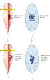
The ‘F cell’ concept and LR asymmetric development in the absence of an LR axis. Left: in vertebrates, LR morphogenesis occurs according to an established body LR axis. Right: in Drosophila, chiral cells may behave like an F cell, which is analogous to the F molecule—a hypothetical LR determinant—at the cellular level and drive LR asymmetric development in individual organs, without establishing an LR axis of the whole embryo.

References

    1. Mothet JP, et al. 2000. D-serine is an endogenous ligand for the glycine site of the N-methyl-d-aspartate receptor. Proc. Natl Acad. Sci. USA 97, 4926–4931. (10.1073/pnas.97.9.4926) - DOI - PMC - PubMed
    1. Pizzarello S, Cronin JR. 2000. Non-racemic amino acids in the Murray and Murchison meteorites. Geochim. Cosmochim. Acta 64, 329–338. (10.1016/S0016-7037(99)00280-X) - DOI - PubMed
    1. Blackmond DG. 2010. The origin of biological homochirality. Cold Spring Harb. Perspect. Biol. 2, a002147 (10.1101/cshperspect.a002147) - DOI - PMC - PubMed
    1. Leiman PG, Arisaka F, van Raaij MJ, Kostyuchenko VA, Aksyuk AA, Kanamaru S, Rossmann MG. 2010. Morphogenesis of the T4 tail and tail fibers. Virol. J. 7, 355 (10.1186/1743-422X-7-355) - DOI - PMC - PubMed
    1. Macnab RM. 2003. How bacteria assemble flagella. Annu. Rev. Microbiol. 57, 77–100. (10.1146/annurev.micro.57.030502.090832) - DOI - PubMed

Publication types

LinkOut - more resources