. 2016 Dec 23;291(52):26886-26898.
doi: 10.1074/jbc.M116.756098. Epub 2016 Nov 7.

Role for the MED21-MED7 Hinge in Assembly of the Mediator-RNA Polymerase II Holoenzyme

Affiliations

Role for the MED21-MED7 Hinge in Assembly of the Mediator-RNA Polymerase II Holoenzyme

Shigeo Sato et al. J Biol Chem. .

Abstract

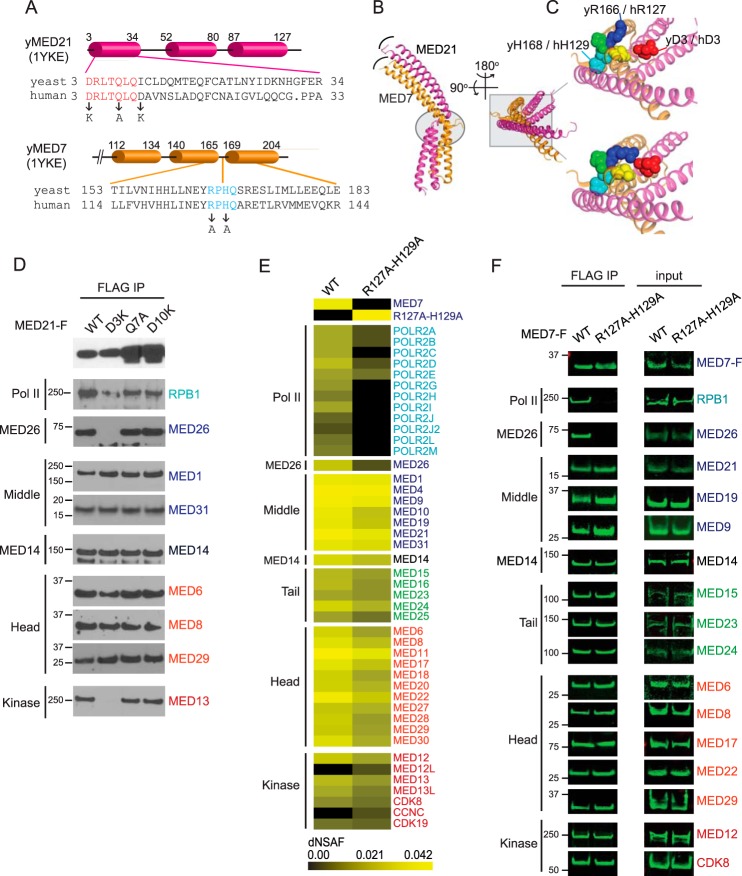
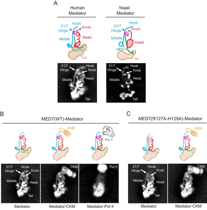
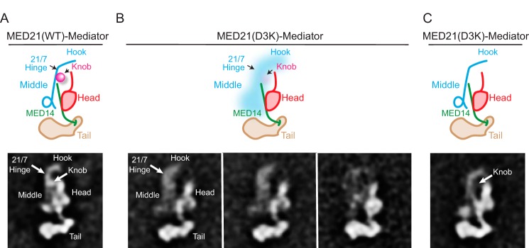
Mediator plays an integral role in activation of RNA polymerase II (Pol II) transcription. A key step in activation is binding of Mediator to Pol II to form the Mediator-Pol II holoenzyme. Here, we exploit a combination of biochemistry and macromolecular EM to investigate holoenzyme assembly. We identify a subset of human Mediator head module subunits that bind Pol II independent of other subunits and thus probably contribute to a major Pol II binding site. In addition, we show that binding of human Mediator to Pol II depends on the integrity of a conserved "hinge" in the middle module MED21-MED7 heterodimer. Point mutations in the hinge region leave core Mediator intact but lead to increased disorder of the middle module and markedly reduced affinity for Pol II. These findings highlight the importance of Mediator conformation for holoenzyme assembly.

Keywords: Mediator; RNA polymerase II; general transcription factor (GTF); transcription; transcription coregulator; transcription regulation.


Figures

FIGURE 1.
FIGURE 1.
A, comparison of yeast and human Mediator architecture. Left, ∼18 Å cryo-EM map of S. cerevisiae core Mediator; right, ∼30 Å map of human core Mediator, calculated from images of single particles preserved in stain (6). Positions of the middle, head, and tail modules are shown in blue, red, and yellow, respectively. B, yeast Mediator core complex; an X-ray structure of the yeast head module bound to CTD peptide (22) and a model of the middle module, based on partial X-ray structures and protein cross-linking (34), docked into the yeast cryo-EM map. C, structural model of the yeast Mediator-Pol II holoenzyme based on cryo-EM maps of a Mediator-PIC complex (17) and of a core yeast Mediator-ITC (18). Pol II subunits Rpb3/Rpb11 contact the movable jaw of Mediator, and the CTD (CTD peptide in cyan) is expected to bind between the head and the knob.
FIGURE 2.
FIGURE 2.
MED21 depletion disrupts the interaction of Mediator complex and RNA polymerase II. A, anti-MED21 immunoblot of lysates from untreated or siRNA-treated HeLa S3 cells from one of three biological replicates. 200 μg of lysate protein from untreated cells (none) or cells treated with non-targeting (NT) control or MED21 siRNA were applied to each lane. None, no siRNA; NT, non-targeting control siRNA; MED21, MED21 siRNA. In this and subsequent figures, positions of molecular mass markers (kDa) are indicated at the side of each immunoblot. B, effect of MED21 depletion on gene activation. Firefly luciferase activity was measured in lysates from HeLa cells with a stably integrated, NF-κB-responsive luciferase reporter, transfected with non-targeting control siRNA or MED21 siRNA. ● and ■, luciferase activity detected in lysates of cells treated with non-targeting or MED21 siRNA, respectively, from three independent experiments, each performed in duplicate. Horizontal lines, median. C, effect of MED21 depletion on Mediator-Pol II interactions. HeLa S3 cells stably expressing FLAG-RPB9 were transfected with non-targeting or MED21 siRNA, and Pol II-associated proteins were detected by immunoblotting of anti-FLAG immunoprecipitates (IP) using the indicated antibodies. Middle, Tail, and Head, Mediator modules to which individual subunits have been assigned.
FIGURE 3.
FIGURE 3.
Identification of Mediator and Pol II subunits associated with full-length or mutant forms of MED21. Heat maps summarize estimates of relative recovery of Mediator and Pol II subunits after FLAG immunopurification through the indicated FLAG epitope-tagged MED21 protein from HeLa S3 (A) or FlpIN-TREx-293 (B) cell nuclear extracts, measured by MudPIT mass spectrometry and expressed as a normalized spectral abundance factor (dNSAF). In a MudPIT data set, the number of spectra derived from peptides of most proteins is a function of the protein's length and abundance. Thus, the relative amount of a particular protein in different samples can be estimated from dNSAF. Supporting MudPIT information is provided in supplemental Table S1. F-WT, N-terminally FLAG-tagged MED21; WT-F, C-terminally FLAG-tagged MED21. MED21-F, C-terminally FLAG-tagged WT MED21 or the indicated C-terminally tagged MED21 mutants. Mediator subunits were assigned to head, middle, tail, or kinase modules based on previously defined interactions and sequence similarity to yeast Mediator complex subunits (6, 25).
FIGURE 4.
FIGURE 4.
The MED21 N terminus contributes to Mediator assembly and is required for binding of Pol II and kinase module to Mediator core. Nuclear extracts from HeLa S3 (A) or Flp-In T-REx HEK293 (B) cells expressing either no exogenously expressed MED21 (None) or C-terminally FLAG-tagged WT or the indicated MED21 mutants were subjected to anti-FLAG M2 immunoaffinity purification and analyzed by immunoblotting using antibodies directed against Mediator or Pol II subunits. The first three lanes of A show 3-fold serial dilutions of immunopurified proteins from cells expressing wild type MED21-FLAG. C, immunoblots of cell lysates (input) used for FLAG immunoaffinity purification in B. D, MED21 lacking the first 5 amino acids is defective in TNFα-induced gene activation. Firefly luciferase activity was measured in lysates from Flp-InTM T-RExTM-293 cells stably expressing wild type or mutant MED21.
FIGURE 5.
FIGURE 5.
MED21 and MED7 mutations in or near the MED21-MED7 hinge interfere with holoenzyme assembly. A, MED21 and MED7C secondary structure defined by X-ray crystallography of yeast MED21-MED7C heterodimer (Protein Data Bank entry 1YKE (30)) and sequence alignments of conserved MED21 N-terminal and MED7 hinge regions and positions of human MED21 and MED7 mutations. Cylinders, α-helices. B, superposition of two crystal forms of the yeast MED21 (magenta)-MED7C (orange) heterodimer, one containing chains B (MED21) and A (MED7C) and the other chains D (MED21) and C (MED7C), all from Protein Data Bank entry 1YKE. The four-helix bundles (which include α-helices 1 and 2 of MED21 and MED7C) from each crystal form are superimposed. Curved lines show difference in positions of the C-terminal α-helices of MED21 and MED7C in the two crystal forms. The MED21-MED7 hinge region is indicated by the oval. C, enlarged views of the region of the MED21-MED7C heterodimer outlined by the box in B, with the two crystal forms shown separately (top, chains D and C; bottom, chains B and A). Residues near the flexible hinge are shown as spheres. MED21 D3 (red) and MED7 residues Arg-166, Pro-167, His-168, and Gln-169 are blue, green, cyan, and yellow, respectively; in the more closed form of the MED21-MED7 heterodimer, MED21 Asp-3 is closer to Arg-166 of yeast MED7. y, yeast; h, human. D, nuclear extracts from Flp-In T-REx HEK293 cells expressing C-terminally FLAG-tagged WT or the indicated MED21 mutants were subjected to anti-FLAG M2 immunoaffinity purification and analyzed by immunoblotting using antibodies directed against Mediator or Pol II subunits. E, heat map showing number of spectra detected for Mediator and Pol II subunits after FLAG immunopurification through FLAG epitope-tagged wild type or mutant MED7 protein from HeLa S3 cell nuclear extracts, measured by MudPIT. Supporting MudPIT data are shown in supplemental Table S2. F, immunoblots showing Mediator and Pol II subunits that co-purified with FLAG-tagged wild type or mutant MED7. Immunoblots were visualized using an Odyssey infrared imaging system (LI-COR). IP, immunoprecipitation.
FIGURE 6.
FIGURE 6.
EM analysis of wild-type and MED7(R127A/H129A) human Mediators. A, a 2D class average obtained from stained particle images of wild-type human Mediator and a corresponding diagram summarizing the organization of the head, middle, and tail modules, the hook, and MED14 (left), based on comparison with wild-type yeast mediator (right). B, 2D class averages obtained after alignment and clustering of stained particle images from a wild-type human Mediator preparation. Free Mediator (left) and Mediator associated with kinase module (Mediator-CKM, center) or Pol II (Mediator-Pol II, right) were observed. The head, middle, and tail modules and the MED19-containing “hook” region at top of the middle module can be readily identified. The arrowhead marks the approximate position of the MED21-MED7 hinge region and the MED7(R127A-H129A) mutation, also inferred by comparison with the yeast Mediator structure (6). The Mediator-Pol II average is blurred by variability in the relative position of Mediator and Pol II, exacerbated by disruption of the complex after preservation in stain. C, class averages of free (left) and CKM-associated MED7(R127A/H129A) human Mediator. As illustrated in the diagrams, the MED7(R127A/H129A) mutation increases mobility of the hook and knob portions of the Middle module.
FIGURE 7.
FIGURE 7.
EM analysis of wild-type and MED21(D3K) yeast Mediators. A, a 2D class average from stained images and corresponding diagram showing that the structure and arrangement of head, middle, and tail modules is unaffected by introduction of a FLAG tag at the MED21 C terminus. The approximate position of the D3K mutation is indicated. B, 2D image class averages and corresponding diagrams showing variability in the position of the middle module in yeast Mediator containing MED21(D3K). C, a MED21(D3K) yeast Mediator class average in which the Middle module is well resolved shows that density corresponding to the MED7N-MED31 “knob” is absent. This agrees with what seems to be comparatively weak MED7N-MED31 density in the averages shown in B.

References

    1. Kim Y. J., Björklund S., Li Y., Sayre M. H., and Kornberg R. D. (1994) A multiprotein mediator of transcriptional activation and its interaction with the C-terminal repeat domain of RNA polymerase II. Cell 77, 599–608 - PubMed
    1. Myers L. C., and Kornberg R. D. (2000) Mediator of Transcriptional Regulation. Annu. Rev. Biochem. 69, 729–749 - PubMed
    1. Malik S., and Roeder R. G. (2010) The metazoan Mediator co-activator complex as an integrative hub for transcriptional regulation. Nat. Rev. Genet. 11, 761–772 - PMC - PubMed
    1. Conaway R. C., and Conaway J. W. (2013) The Mediator complex and transcription elongation. Biochim. Biophys. Acta 1829, 69–75 - PMC - PubMed
    1. Allen B. L., and Taatjes D. J. (2015) The Mediator complex: a central integrator of transcription. Nat. Rev. Mol. Cell Biol. 16, 155–166 - PMC - PubMed

MeSH terms

Associated data

LinkOut - more resources