. 2017 Apr;28(4):1093-1105.
doi: 10.1681/ASN.2016050567. Epub 2016 Nov 7.

APOL1 Renal-Risk Variants Induce Mitochondrial Dysfunction

Affiliations

APOL1 Renal-Risk Variants Induce Mitochondrial Dysfunction

Lijun Ma et al. J Am Soc Nephrol. 2017 Apr.

Abstract

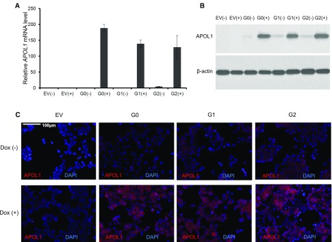
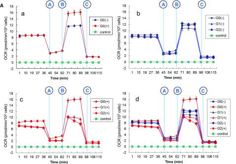
APOL1 G1 and G2 variants facilitate kidney disease in blacks. To elucidate the pathways whereby these variants contribute to disease pathogenesis, we established HEK293 cell lines stably expressing doxycycline-inducible (Tet-on) reference APOL1 G0 or the G1 and G2 renal-risk variants, and used Illumina human HT-12 v4 arrays and Affymetrix HTA 2.0 arrays to generate global gene expression data with doxycycline induction. Significantly altered pathways identified through bioinformatics analyses involved mitochondrial function; results from immunoblotting, immunofluorescence, and functional assays validated these findings. Overexpression of APOL1 by doxycycline induction in HEK293 Tet-on G1 and G2 cells led to impaired mitochondrial function, with markedly reduced maximum respiration rate, reserve respiration capacity, and mitochondrial membrane potential. Impaired mitochondrial function occurred before intracellular potassium depletion or reduced cell viability occurred. Analysis of global gene expression profiles in nondiseased primary proximal tubule cells from black patients revealed that the nicotinate phosphoribosyltransferase gene, responsible for NAD biosynthesis, was among the top downregulated transcripts in cells with two APOL1 renal-risk variants compared with those without renal-risk variants; nicotinate phosphoribosyltransferase also displayed gene expression patterns linked to mitochondrial dysfunction in HEK293 Tet-on APOL1 cell pathway analyses. These results suggest a pivotal role for mitochondrial dysfunction in APOL1-associated kidney disease.

Keywords: APOL1; chronic kidney disease; mitochondria.


Figures

Figure 1.
Figure 1.
APOL1 expression levels with Dox induction in HEK293 Tet-on G0, G1, and G2 cells are comparable. (A) Relative APOL1 mRNA levels in HEK293 Tet-on cells stably expressing G0, G1, or G2 APOL1 variants. HEK293 Tet-on cells with (+) or without (−) Dox induction were grown in complete DMEM growth media for 16 hours. Without Dox induction, APOL1 mRNA levels in HEK293 Tet-on cells were negligible compared with Dox induction. The ΔΔCT method was used to quantify relative APOL1 mRNA expression. Fold changes were normalized to mRNA levels of G0 cells without Dox induction. Data are expressed as mean±SD. APOL1 mRNA levels in G0 cells without Dox induction set at 1 (reference). (B) Relative APOL1 levels from total protein lysates in HEK293 Tet-on cells stably expressing G0, G1, or G2 APOL1 variants. HEK293 Tet-on cells with (+) or without (−) Dox induction were grown in complete DMEM growth media for 16 hours. Four micrograms of total cell lysate protein was loaded onto a 4%–20% SDS-PAGE gel and probed with APOL1 antibody. HEK293 Tet-on empty pTRE2hyg vector cells did not express APOL1 with or without Dox induction. HEK293 Tet-on APOL1 G0, G1, and G2 cells expressed similar amounts of APOL1 with Dox induction, whereas trace amounts of APOL1 were present in HEK293 Tet-on cells without Dox induction. Data are representative of three trials of immunoblotting with similar results. (C) APOL1 immunofluorescence in HEK293 Tet-on cells stably expressing G0, G1, or G2 APOL1 variants with and without Dox induction. HEK293 Tet-on cells grown with (+) or without (−) Dox for 16 hours. After washing with PBS and fixing with 4% paraformaldehyde, cells were stained for APOL1 (red) with Epitomics anti-APOL1 antibody and counterstained with DAPI (blue). Elevated APOL1 signal intensities were comparable in HEK293 Tet-on G0, G1, and G2 cells with Dox induction, whereas APOL1 was absent in HEK293 Tet-on empty pTRE2hyg vector cells. EV, HEK293 Tet-on cell line with empty pTRE2hyg plasmid; DAPI, 4′,6-diamidino-2-phenylindole.
Figure 2.
Figure 2.
Illumina human HT-12 v4 arrays identify significantly clustered gene expression patterns for HEK293 Tet-on APOL1 G0, G1, and G2 cells. From top to bottom are 1699 transcripts, in order from pattern 1 to pattern 14. (A) Gene expression patterns most altered in HEK293 Tet-on G0 (circle), G1 (triangle), and G2 (diamond) cells with Dox induction. Asterisk indicates that mitochondrial pathway enrichment is present (patterns 6, 7, and 13). (B) Heat map of clustered gene expression patterns: transcript expression in G0 shown in black; red and green correspond to up and downregulation, respectively, by G1 or G2. Lighter colors denote less differential expression.
Figure 3.
Figure 3.
APOL1 G1 and G2 renal-risk variants contribute to mitochondrial dysfunction. Affymetrix patterns 1 and 2 were analyzed using IPA for HEK293 Tet-on APOL1 G1 and G2 versus G0 cells. Results indicate marked downregulation of enzymes in mitochondrial complexes I–V.
Figure 4.
Figure 4.
APOL1 colocalizes with mitochondria in HEK293 Tet-on APOL1 cells with Dox induction. HEK293 Tet-on APOL1 cells with 8-hour Dox induction (stably expressing G0, G1, or G2 APOL1 variants) were washed with PBS and fixed with 4% paraformaldehyde. Cells were stained for APOL1 (red) with Epitomics anti-APOL1 antibody, mitochondria (green) with ATP synthase 5 A1 (ATP5A1) antibody, and counterstained with nuclear dye 4′,6-diamidino-2-phenylindole (DAPI; blue).
Figure 5.
Figure 5.
APOL1 G1 and G2 renal-risk variants decrease mitochondrial respiration. (A) a-d: Mitochondrial stress tests in HEK293 Tet-on APOL1 cells. Cells were seeded on a V7 cell culture plate (Seahorse Bioscience) with final density 100,000 cells/well before assay. Effects of G0, G1, and G2 with (+) and without (−) Dox induction were compared using a Seahorse XF-24 extracellular flux analyzer to measure oxygen consumption rate (OCR), a measure of oxidative phosphorylation, in the presence of a series of metabolic inhibitors and uncoupling agents. The first injection (shown as vertical blue line A) was oligomycin, an inhibitor of ATP synthesis via blockade of the proton channel of ATP synthase (complex V). Oligomycin distinguishes the OCR from ATP synthesis by blocking the oxygen consumption required to overcome proton leakage across the inner mitochondrial membrane, and basal respiration rate, by blocking nonmitochondrial respiration. The second injection (shown as vertical blue line B) was FCCP, an uncoupling agent that disrupts ATP synthesis by transporting hydrogen ions across the mitochondrial membrane instead of the ATP synthase proton channel (complex V). Collapse of the mitochondrial membrane potential leads to rapid energy and oxygen consumption without generation of ATP. FCCP is used to calculate the spare respiration capacity of cells, defined as the quantitative difference between maximal and basal respiration rates. The third injection (shown as vertical blue line C) was a combination of rotenone, a complex I inhibitor, and antimycin A, a complex III inhibitor; this combination blocks mitochondrial respiration and enables calculation of mitochondrial and nonmitochondrial cellular respiration. At least three wells were assigned for each type of HEK293 Tet-on cells with (+) or without (−) Dox induction on the same 24-well V7 cell culture plate. Data were expressed as mean±SEM of each time point, grouped by HEK293 Tet-on G0, G1, and G2 cells with or without Dox induction. (B) Bioenergetic profiles for HEK293 Tet-on APOL1 G0, G1, and G2 cells. APOL1 stably transfected HEK293 cells were incubated with (+) or without (−) Dox for 8 hours before assay on a Seahorse XF-24 extracellular flux analyzer to measure the OCR. After a 15 minute equilibration period, three measures of OCR were made for each stage, i.e., preoligomycin, between oligomycin and FCCP, between FCCP and rotenone/antimycin, and postrotenone (injections denoted as vertical blue lines, see A). The average of three measures was used to represent the OCR of the corresponding stage for each well. Data were grouped by APOL1 genotype and Dox treatment (+/−). Basal respiration rate, maximum respiration rate, and spare respiration capacity were expressed as mean±SD; n refers to replicated well number for each type of HEK293 Tet-on cells on the 24-well V7 cell culture plate. (a) For G0 cells, induction of APOL1 did not alter the basal respiration rate (t test P=0.68), but improved maximum respiration rate and spare respiration capacity (t test P values 0.005 and 0.005, respectively). (b) For G1 cells, induction of APOL1 did not alter the basal respiration rate (t test P=0.08), maximum respiration rate, and spare respiration capacity (t test P values 0.48 and 0.68, respectively). (c) For G2 cells, induction of APOL1 diminished the basal respiration rate, maximum respiration rate, and spare respiration capacity (t test P values 0.002, 0.003, and 0.01, respectively). (d) Without Dox induction, G0, G1, and G2 cells appeared to have similar basal respiration rate, maximum respiration rate, and spare respiration capacity (ANOVA P=0.78, 0.22, and 0.13, respectively). (e) With Dox induction, the basal respiration rate, maximum respiration rate, and spare respiration capacity for G1 and G2 were significantly reduced (ANOVA P=0.004, <0.0001, and 0.0007, respectively) compared with G0. (f) Overview of bioenergetic profiles for HEK293 Tet-on APOL1 G0, G1, and G2 cells.
Figure 5.
Figure 5.
APOL1 G1 and G2 renal-risk variants decrease mitochondrial respiration. (A) a-d: Mitochondrial stress tests in HEK293 Tet-on APOL1 cells. Cells were seeded on a V7 cell culture plate (Seahorse Bioscience) with final density 100,000 cells/well before assay. Effects of G0, G1, and G2 with (+) and without (−) Dox induction were compared using a Seahorse XF-24 extracellular flux analyzer to measure oxygen consumption rate (OCR), a measure of oxidative phosphorylation, in the presence of a series of metabolic inhibitors and uncoupling agents. The first injection (shown as vertical blue line A) was oligomycin, an inhibitor of ATP synthesis via blockade of the proton channel of ATP synthase (complex V). Oligomycin distinguishes the OCR from ATP synthesis by blocking the oxygen consumption required to overcome proton leakage across the inner mitochondrial membrane, and basal respiration rate, by blocking nonmitochondrial respiration. The second injection (shown as vertical blue line B) was FCCP, an uncoupling agent that disrupts ATP synthesis by transporting hydrogen ions across the mitochondrial membrane instead of the ATP synthase proton channel (complex V). Collapse of the mitochondrial membrane potential leads to rapid energy and oxygen consumption without generation of ATP. FCCP is used to calculate the spare respiration capacity of cells, defined as the quantitative difference between maximal and basal respiration rates. The third injection (shown as vertical blue line C) was a combination of rotenone, a complex I inhibitor, and antimycin A, a complex III inhibitor; this combination blocks mitochondrial respiration and enables calculation of mitochondrial and nonmitochondrial cellular respiration. At least three wells were assigned for each type of HEK293 Tet-on cells with (+) or without (−) Dox induction on the same 24-well V7 cell culture plate. Data were expressed as mean±SEM of each time point, grouped by HEK293 Tet-on G0, G1, and G2 cells with or without Dox induction. (B) Bioenergetic profiles for HEK293 Tet-on APOL1 G0, G1, and G2 cells. APOL1 stably transfected HEK293 cells were incubated with (+) or without (−) Dox for 8 hours before assay on a Seahorse XF-24 extracellular flux analyzer to measure the OCR. After a 15 minute equilibration period, three measures of OCR were made for each stage, i.e., preoligomycin, between oligomycin and FCCP, between FCCP and rotenone/antimycin, and postrotenone (injections denoted as vertical blue lines, see A). The average of three measures was used to represent the OCR of the corresponding stage for each well. Data were grouped by APOL1 genotype and Dox treatment (+/−). Basal respiration rate, maximum respiration rate, and spare respiration capacity were expressed as mean±SD; n refers to replicated well number for each type of HEK293 Tet-on cells on the 24-well V7 cell culture plate. (a) For G0 cells, induction of APOL1 did not alter the basal respiration rate (t test P=0.68), but improved maximum respiration rate and spare respiration capacity (t test P values 0.005 and 0.005, respectively). (b) For G1 cells, induction of APOL1 did not alter the basal respiration rate (t test P=0.08), maximum respiration rate, and spare respiration capacity (t test P values 0.48 and 0.68, respectively). (c) For G2 cells, induction of APOL1 diminished the basal respiration rate, maximum respiration rate, and spare respiration capacity (t test P values 0.002, 0.003, and 0.01, respectively). (d) Without Dox induction, G0, G1, and G2 cells appeared to have similar basal respiration rate, maximum respiration rate, and spare respiration capacity (ANOVA P=0.78, 0.22, and 0.13, respectively). (e) With Dox induction, the basal respiration rate, maximum respiration rate, and spare respiration capacity for G1 and G2 were significantly reduced (ANOVA P=0.004, <0.0001, and 0.0007, respectively) compared with G0. (f) Overview of bioenergetic profiles for HEK293 Tet-on APOL1 G0, G1, and G2 cells.

Comment in

  • Identifying the Intracellular Function of APOL1.
    Bruggeman LA, O'Toole JF, Sedor JR. Bruggeman LA, et al. J Am Soc Nephrol. 2017 Apr;28(4):1008-1011. doi: 10.1681/ASN.2016111262. Epub 2017 Feb 14. J Am Soc Nephrol. 2017. PMID: 28196842 Free PMC article. No abstract available.

References

    1. Genovese G, Friedman DJ, Ross MD, Lecordier L, Uzureau P, Freedman BI, Bowden DW, Langefeld CD, Oleksyk TK, Uscinski Knob AL, Bernhardy AJ, Hicks PJ, Nelson GW, Vanhollebeke B, Winkler CA, Kopp JB, Pays E, Pollak MR: Association of trypanolytic ApoL1 variants with kidney disease in African Americans. Science 329: 841–845, 2010 - PMC - PubMed
    1. Tzur S, Rosset S, Shemer R, Yudkovsky G, Selig S, Tarekegn A, Bekele E, Bradman N, Wasser WG, Behar DM, Skorecki K: Missense mutations in the APOL1 gene are highly associated with end stage kidney disease risk previously attributed to the MYH9 gene. Hum Genet 128: 345–350, 2010 - PMC - PubMed
    1. Freedman BI, Kopp JB, Langefeld CD, Genovese G, Friedman DJ, Nelson GW, Winkler CA, Bowden DW, Pollak MR: The apolipoprotein L1 (APOL1) gene and nondiabetic nephropathy in African Americans. J Am Soc Nephrol 21: 1422–1426, 2010 - PMC - PubMed
    1. Larsen CP, Beggs ML, Saeed M, Walker PD: Apolipoprotein L1 risk variants associate with systemic lupus erythematosus-associated collapsing glomerulopathy. J Am Soc Nephrol 24: 722–725, 2013 - PMC - PubMed
    1. Freedman BI, Langefeld CD, Andringa KK, Croker JA, Williams AH, Garner NE, Birmingham DJ, Hebert LA, Hicks PJ, Segal MS, Edberg JC, Brown EE, Alarcón GS, Costenbader KH, Comeau ME, Criswell LA, Harley JB, James JA, Kamen DL, Lim SS, Merrill JT, Sivils KL, Niewold TB, Patel NM, Petri M, Ramsey-Goldman R, Reveille JD, Salmon JE, Tsao BP, Gibson KL, Byers JR, Vinnikova AK, Lea JP, Julian BA, Kimberly RP; Lupus Nephritis–End‐Stage Renal Disease Consortium : End-stage renal disease in African Americans with lupus nephritis is associated with APOL1. Arthritis Rheumatol 66: 390–396, 2014 - PMC - PubMed