. 2016 Nov 22;113(47):13528-13533.
doi: 10.1073/pnas.1610973113. Epub 2016 Nov 7.

Engineering dynamical control of cell fate switching using synthetic phospho-regulons

Affiliations

Engineering dynamical control of cell fate switching using synthetic phospho-regulons

Russell M Gordley et al. Proc Natl Acad Sci U S A. .

Abstract

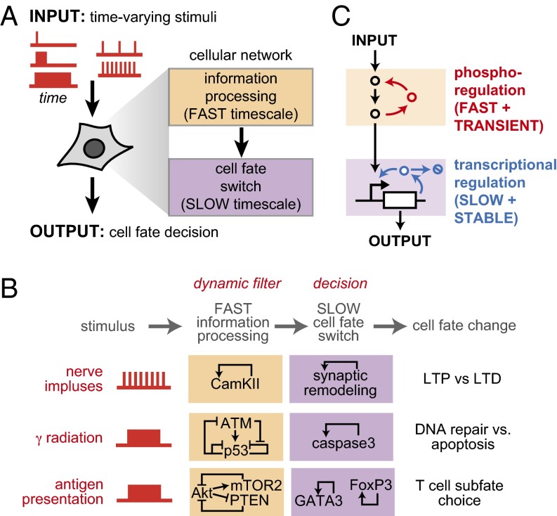
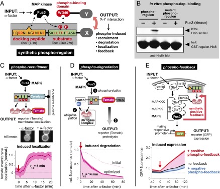
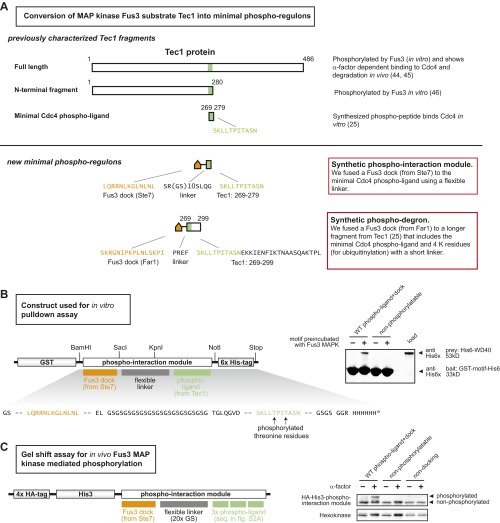
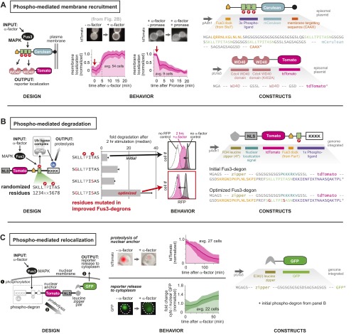
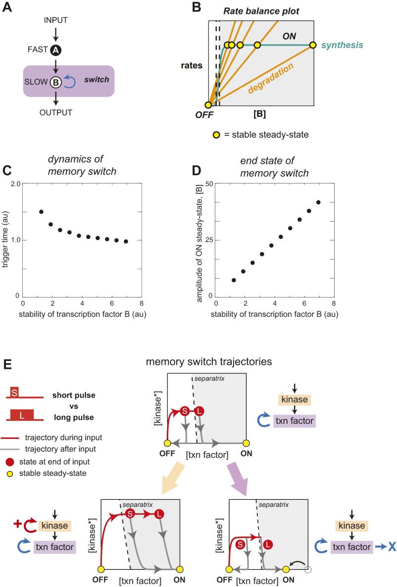
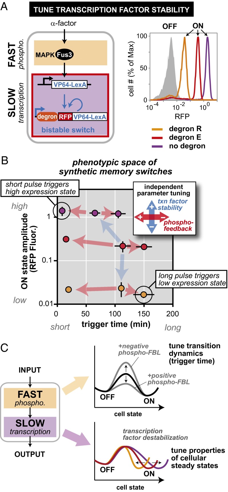
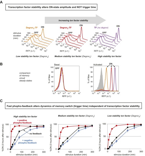
Many cells can sense and respond to time-varying stimuli, selectively triggering changes in cell fate only in response to inputs of a particular duration or frequency. A common motif in dynamically controlled cells is a dual-timescale regulatory network: although long-term fate decisions are ultimately controlled by a slow-timescale switch (e.g., gene expression), input signals are first processed by a fast-timescale signaling layer, which is hypothesized to filter what dynamic information is efficiently relayed downstream. Directly testing the design principles of how dual-timescale circuits control dynamic sensing, however, has been challenging, because most synthetic biology methods have focused solely on rewiring transcriptional circuits, which operate at a single slow timescale. Here, we report the development of a modular approach for flexibly engineering phosphorylation circuits using designed phospho-regulon motifs. By then linking rapid phospho-feedback with slower downstream transcription-based bistable switches, we can construct synthetic dual-timescale circuits in yeast in which the triggering dynamics and the end-state properties of the ON state can be selectively tuned. These phospho-regulon tools thus open up the possibility to engineer cells with customized dynamical control.

Keywords: dynamical control; phosphorylation; synthetic biology.


Conflict of interest statement

W.A.L. is a founder of Cell Design Labs and a member of its scientific advisory board.

Figures

Fig. 1.
Fig. 1.
Dual-timescale architecture is a common feature of regulatory networks that dynamically control cell fate decisions. (A) Dual-timescale regulation (fast and slow layers) has been implicated as an important regulatory mechanism in cellular dynamic gates. (B) This two-layer network architecture is shared by cell fate decision circuits used in neural plasticity (3, 4), apoptosis (7, 8), and adaptive immunity (9, 10). (C) Design principles of a dual-timescale dynamic gate—rapid timescale layer processes and filters inputs, determining what temporal patterns will be propagated to the slow-timescale memory layer.
Fig. 2.
Fig. 2.
Design of phospho-regulation modules that allow engineering of synthetic kinase–substrate relationships. (A) A synthetic phospho-regulated interaction module in which phosphorylation by a yeast MAP kinase (Fus3) triggers recruitment of a phospho-binding domain (WD40 domain of Cdc4). The synthetic phospho-regulon is composed of composite linear motifs optimized for three simultaneous recognition functions: MAPK docking, MAPK phosphorylation, and phospho-binding domain recognition. Key recognition residues are denoted in red. (B) GST pull-downs confirmed phospho-dependent binding of the synthetic phospho-regulon components in vitro. (C) Phospho-regulated plasma membrane recruitment, in yeast, triggered by mating pathway activation (α-factor). “+α-factor” image taken 20 min after induction. Time course of tdTomato reporter membrane recruitment, quantified by image analysis; mean ± SD (n = 54 cells; shaded region) are shown. (D) The phospho-regulon can be converted to a phospho-dependent degradation motif (induced with α-factor) by adding lysine (K) residues to serve as ubiquitinylation sites. Phosphorylation by Fus3-triggered binding by endogenous E3 ligase Cdc4 [a substrate targeting component of the SKP1–CUL1–F-box protein (SCF) ubiquitin ligase complex (25)], ubiquitination, and proteolysis. Fus3-induced degradation was enhanced by screening mutant phospho-regulons and combining enhancing mutations (Fig. S2). Time course of reporter decay in yeast after α-factor stimulation is shown (RFP fluorescence measured by flow cytometry, normalized to cell volume; see Materials and Methods). Mean ± SD (n = 3; shaded region) are shown. (E) Phospho-regulons can be used to build synthetic fast positive and negative feedback into the yeast mating pathway. Positive feedback was generated by using Fus3 phospho-regulon to induce interaction of Ste18 and Ste50N; this complex acts as a positive regulator of pathway activity, increasing the extent of Fus3 phosphorylation and downstream transcription (pFus1-GFP reporter). An analogous negative phospho-feedback loop was engineered by using the phospho-regulon to induce formation of a known inhibitory complex [Fus3 and Ste20, Fig. S5 (27)]. GFP fluorescence (measured by flow cytometry) is normalized to cell volume (Materials and Methods). Mean ± SD (n = 3; shaded region) are shown. Phospho-regulons mutated to prevent phosphorylation (pT→V) do not mediate in vivo recruitment, degradation, or feedback (Figs. S3–S5).
Fig. S1.
Fig. S1.
Strategy for designing Fus3 MAPK phospho-regulated protein interactions. (A) Approach to minimization of a known Fus3 phospho-regulated motif. Yeast Tec1 has been shown to be phosphorylated by Fus3 in vitro and degrades in response to α-factor in vivo (40, 41). A 280-residue N-terminal fragment of Tec1 has also been shown to be phosphorylated by Fus3 in vitro (42), but no shorter fragment has been shown to be phosphorylated by Fus3. An 11-residue fragment of Tec1 that, when synthesized as a phospho-peptide, acts as a Cdc4 phospho-ligand by itself (25) but cannot be phosphorylated by Fus3. We fused this sequence to a Fus3 dock (from Ste7) to create a minimal phospho-ligand that can be both phosphorylated by Fus3 and bound by a Cdc4 WD40 domain in vitro. We fused a longer Tec1 fragment that included 20 additional residues to a Fus3 dock (from Far1) to create a phospho-degron. (B) In vitro expression cassette for phospho-regulated interaction module. (C) A gel shift assay showed that in vivo phosphorylation of the minimal synthetic phospho-ligand required both known phospho-threonine residues (in the Tec1 fragment) and the docking motif for Fus3 MAP kinase (from Ste7). “+” samples treated with α-factor for 20 min; mutations that preclude phosphorylation (“nonphosphorylatable”) or docking (“nondocking”) described in SI Materials and Methods.
Fig. S2.
Fig. S2.
Phospho-recruitment can be used to engineer diverse response behaviors in yeast. Linear motifs and domains from yeast signaling proteins were recombined to create modular phospho-regulation tags (phospho-regulons). (A) Phospho-mediated interaction mediated by an inducible interaction between an engineered peptide and the Cdc4 WD40 domain (Fig. 2C). This synthetic interaction was reversible, and removal of α-factor (upon addition of Pronase) resulted in the rapid delocalization of tdTomato from the membrane. Functional optimization using the K402A affinity variant described in SI Materials and Methods. (B) Phospho-dependent degradation was achieved by fusing a longer sequence of Tec1 that contained four lysine residues to a Fus3 dock. This phospho-degron was then fused to tdTomato (Fig. 2D). Stimulation with the pathway inducer (α-factor) led to recognition by endogenous Cdc4 [a substrate targeting component of the SKP1–CUL1–F-box protein (SCF) ubiquitin ligase complex], ubiquitination, and proteolysis. In the context of this initial Fus3-degron, mutations of the Tec1 phospho-ligand were identified that improved fold degradation, and degron performance was further enhanced by creating a synthetic phospho-regulon that combined two enhancing mutations. Median fold degradation after α-factor stimulation (relative to an unstimulated control) ± SD (n = 3) are shown for selected degrons. Normalized histograms contain >10,000 cells. (C) Phospho-dependent change in subcellular localization was achieved by fusing the phospho-degron to tdTomato, a nuclear localization sequence (NLS), and a leucine zipper constitutive protein binding domain. GFP fused to the cognate leucine zipper domain was held in the nucleus by binding to the NLS containing protein. (1) Stimulation with the pathway inducer (α-factor) led to (2) degradation of the NLS containing anchor protein, which then (3) releases GFP into the cytoplasm. Mean ± SD (shaded region) are shown. For nuclear anchor proteolysis, “+α-factor” image taken 3 h after induction; pT→V negative control, Fig. S3 B and C. For reporter release, +α-factor image taken 1 h after induction; pT→V negative control, Fig. S3D.
Fig. S3.
Fig. S3.
Sample images for engineered phospho-regulons and corresponding negative controls (nonphosphorylatable mutants) (Fig. 2 and Fig. S2). Microscopy images showing larger fields of cells undergoing (A) phospho-mediated interaction and (B) degradation. In these panels, phospho-regulons mutated to prevent phosphorylation (pT→V) were used as negative controls for each mode of phospho-regulation. To confirm the ubiquitination of the phospho-degron, a mutant of the original (unoptimized) phospho-degron was made (“Ubi control”) in which all four lysine residues in the extended Tec1 fragment were mutated (K→R). Constructs that block either type of posttranslational modification show impaired α-factor–induced degradation [(C) liquid culture assay; mean ± SD (n = 3) are shown]. (D) The phospho-degron was used to engineer a phospho-regulated change in GFP localization (microscopy images showing larger fields of cells).
Fig. S4.
Fig. S4.
Addition of synthetic positive phospho-feedback loop enhances fast-timescale Fus3 MAP kinase response to α-factor stimulation (sustained or transient). (A) α-Factor (yeast mating pheromone) binding to receptor Ste2 triggers a canonical MAP kinase signaling cascade, leading to phosphorylation of the MAP kinase Fus3. In wild-type cells, Fus3pp activation peaks after ∼5 min, and then declines to an elevated steady-state level. (B) Addition of Pronase (a mixture of proteases that degrade α-factor) removes the stimulus and leads to rapid Fus3 dephosphorylation. When induced with a sustained α-factor stimulus, cells containing the synthetic positive phospho-feedback circuit, show higher levels of maximal Fus3pp that are sustained for at least 30 min. Once Pronase is added, the rate of Fus3 inactivation in cells with added feedback is consistent with fast-timescale phospho-regulation (<5-min t1/2; Fig. 2C). Phosphorylation sites in the negative-control phospho-regulon were mutated (pT→V) to block recruitment (shown as a black dotted line in each plot).
Fig. S5.
Fig. S5.
Engineered phospho-regulated feedback loops in the yeast mating pathway (Figs. 3 and 4). (A) Positive phospho-feedback was engineered by creating a Fus3-dependent interaction that promotes Ste5 recruitment to the membrane. (B) Negative phospho-feedback was engineered by linking Fus3 activity to the assembly of a complex known to inhibit the yeast mating pathway. This circuit may function by sequestering activated Fus3 at the membrane and/or by enhancing a native posttranslational negative-feedback loop mediated by Fus3 and membrane associated Sst2 (43). (C) Potency of synthetic phospho-feedback can be tuned by varying the strength of promoters driving expression of the two feedback components. Negative-feedback components coupled to a relatively weak promoter (pTef1m3) modestly reduces the rate of GFP synthesis induced by α-factor, whereas use of a stronger promoter (pTef1m10) effectively eliminates the response. The dotted line indicates the response of a phospho-regulon mutant in which the phospho-ligand has been mutated (pT→V) to preclude phosphorylation. Mean ± SD (n = 3) are shown. (D) Mutation of these phosphorylated residues (pT→V) also disrupts the synthetic positive phospho-feedback loop (shown in Fig. 2E). Mean ± SD (n = 3) are shown.
Fig. 3.
Fig. 3.
Engineering fast-timescale phospho-feedback yields yeast cell memory switch with highly tunable switching dynamics. (A) Design of a dual-timescale memory circuit: a core transcriptional bistable switch (with autoregulatory transcriptional positive feedback mediated by an synthetic fusion protein combining the VP64 activation domain and LexA DNA-binding domain) is combined with an upstream rapid-timescale MAPK signaling network that incorporates synthetic phospho-feedback loops. In deterministic computer simulations of the dual-time switch, the system flips from OFF to ON when the input duration is sufficient to build up enough transcription factor to pass the threshold for memory formation (dotted line). Addition of fast-timescale positive feedback accelerates the speed with which the system crosses this commitment threshold. (B) Pulses of varied duration can be experimentally applied to the mating pathway by adding α-factor and subsequently removing it by rapid proteolytic digestion (Pronase). After a 3-h memory consolidation phase (after end of input pulse), two cell populations emerge: memory ON cells with high levels of tdTomato (RFP), and OFF cells with basal fluorescence. ON cells stay RFP+ for >10 generations (Fig. S6). (C) Measuring trigger times for memory switch upon addition of synthetic positive or negative phospho-feedback. Normalized histograms contain >10,000 cells. Trigger times calculated as described in SI Materials and Methods, indicated by red arrows. Memory response are shown after 3-h consolidation time after transient exposure to α-factor. Addition of phospho-feedback tunes pulse trigger times by over an order of magnitude. (D) Phase plane diagrams depicting trajectories of kinase activation and transcription factor expression in response to short- and long-stimulus pulses (including how the cells relax after the end of the stimulus plus). Addition of fast phospho-feedback dramatically accelerates buildup of active kinase and leads to more rapid crossing of the commitment threshold (separatrix).
Fig. S6.
Fig. S6.
Characterization of the bistable transcriptional memory module (SLOW circuit) used in this paper. (A) A bistable cellular memory switch. α-Factor induces synthesis of a trigger transcription factor that, in turn, activates an autoregulated transcription factor (fluorescently labeled with tdTomato). (B) The self-sustaining positive-feedback loop creates a memory state that persists for multiple generations. Depicted cells were isolated from a culture that had been stimulated with α-factor overnight, and then, before imaging, treated with Pronase to remove the stimulus (Movie S1). (C) Memory cells (high fluorescence, ON state) continue to divide at rates comparable to untreated cells (low fluorescence, OFF state). The threshold for triggering cellular memory is determined by the rates of protein synthesis and elimination (largely due to dilution by cell division). Our finding that the synthetic circuit has only a modest effect on yeast doubling time confirms that the reduction in stimulus duration required to trigger memory formation (approximately eightfold; Fig. 3C) is primarily due to enhancement of Fus3 activation and synthesis of the downstream transcription factor. Mean + SD (n = 3) are shown. (D) Time course plot of average cellular fluorescence before, during, and after a 40-min pulse of α-factor stimulation. Values for ON and OFF populations given when these two states are readily resolved (E). tdTomato fluorescence (measured by flow cytometry) is normalized to cell volume (Materials and Methods). Mean + SD (n = 3) are shown. (E) Representative time-resolved histograms corresponding to the same 40-min stimulus pulse shown in D. Normalized histograms contain >5,000 cells.
Fig. S7.
Fig. S7.
Computational analysis: transcription factor stability is predicted to primarily alter ON-state amplitude (and NOT trigger time) in dual-timescale memory circuit. (A) Diagram of a model dual-time bistable switch in which an upstream kinase activates rapidly and a downstream transcription factor is synthesized and degraded on a slower timescale. Computational model equations and parameters described in SI Materials and Methods. (B) A rate balance plot shows that changes in the rate of transcription factor degradation shift the position of the activated stable steady state (ON) but have little effect on the switch’s separatrix (threshold for turning ON; dotted lines show the range of threshold positions). (C) Deterministic simulations show that changes in transcription factor stability have little effect on the duration of stimulus required to trigger a state switch. (D) The same set of transcription factor stabilities (from C) yielded a wide range of ON state amplitudes after stimulus was removed. Notably, these results are not altered in circuits that contain an additional intermediate trigger transcription factor—more closely matching the topology of our experimental cell memory switch. (E) Phase plane diagrams depicting trajectories of kinase and transcription factor activation in response to short and long stimulus pulses. Depicts time evolution of dual-timescale circuits with or without additional fast feedback or enhanced transcription factor degradation rate.
Fig. 4.
Fig. 4.
Independent tuning of cell fate switch dynamics and end states. (A) The ON-state steady-state amplitude of the dual-timescale circuit can be tuned by solely changing the rate of the autoregulatory transcription factor degradation [here, by adding N-end rule degron motifs: DegronE (weak) and DegronR (strong) (30)]. Normalized histograms contain >10,000 cells. (B) A small matrix of dual-time circuits illustrates how the trigger time and the steady-state amplitude of the memory ON state can be independently tuned. Changing ON state by altering stability of the transcription factor does not significantly change trigger time. Tuning trigger time by adding phospho-feedback does not significantly change ON-state amplitude. Trigger times and ON-state amplitude calculated as described in SI Materials and Methods; mean ± SD (n = 3) are shown. (C) Tuning the fast layer of a dual-timescale switch functions analogously to a catalyst by accelerating the stimulus time required to trigger cell fate change, but without changing the end OFF and ON states. Conversely, destabilizing the autoregulated transcription factor in the slow layer shifts the ON steady state but has little effect on the triggering dynamics.
Fig. S8.
Fig. S8.
Experimental analysis: independent tunability of memory switch trigger time (dynamics) and ON-state amplitude in dual-timescale memory circuit. (A) Degrons that destabilize the autoregulated transcription factor tune amplitude of the bistable system’s ON steady state but have little impact on the stimulus duration required to trigger memory. (B) Although all three transcriptional memory circuits share the same OFF (basal) state, their corresponding ON (activated) states span almost two orders of magnitude in transcription factor concentration. Histograms depict cell populations that were either untreated (basal) or induced with α-factor for a period of 5 h (activated), and then allowed to approach steady state for an additional 3 h. The gray shadow indicates the position of the OFF state (high-stability TF) for comparison. (C) Duration response plots used to calculate the trigger times (duration required to induce memory in 50% of cells) reported in Fig. 4C. Mean ± SD (n = 3) are shown.

References

    1. Purvis JE, Lahav G. Encoding and decoding cellular information through signaling dynamics. Cell. 2013;152(5):945–956. - PMC - PubMed
    1. Yosef N, Regev A. Impulse control: Temporal dynamics in gene transcription. Cell. 2011;144(6):886–896. - PMC - PubMed
    1. Lucchesi W, Mizuno K, Giese KP. Novel insights into CaMKII function and regulation during memory formation. Brain Res Bull. 2011;85(1-2):2–8. - PubMed
    1. Kandel ER, Dudai Y, Mayford MR. The molecular and systems biology of memory. Cell. 2014;157(1):163–186. - PubMed
    1. Murphy LO, Smith S, Chen R-H, Fingar DC, Blenis J. Molecular interpretation of ERK signal duration by immediate early gene products. Nat Cell Biol. 2002;4(8):556–564. - PubMed

Publication types

Substances