. 2016 Nov 29;7(48):79854-79868.
doi: 10.18632/oncotarget.13036.

TRIM71 suppresses tumorigenesis via modulation of Lin28B-let-7-HMGA2 signaling

Affiliations

TRIM71 suppresses tumorigenesis via modulation of Lin28B-let-7-HMGA2 signaling

Jinlong Yin et al. Oncotarget. .

Abstract

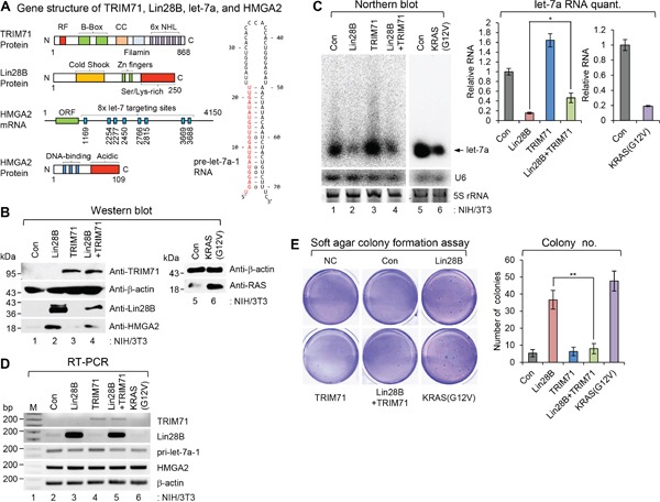
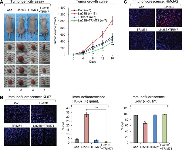
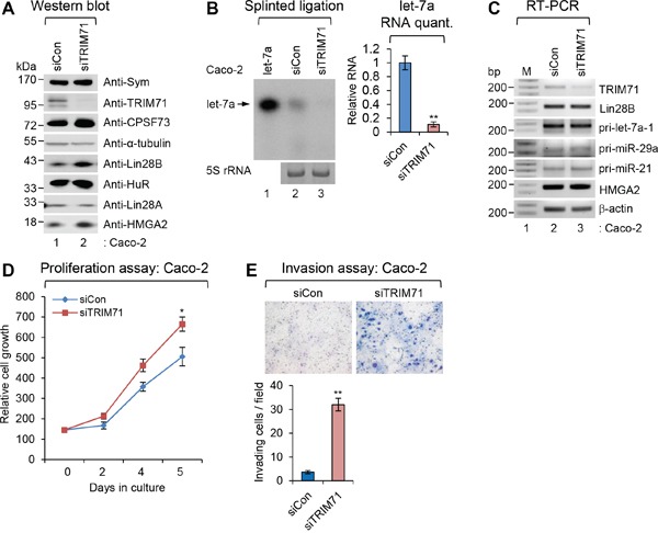
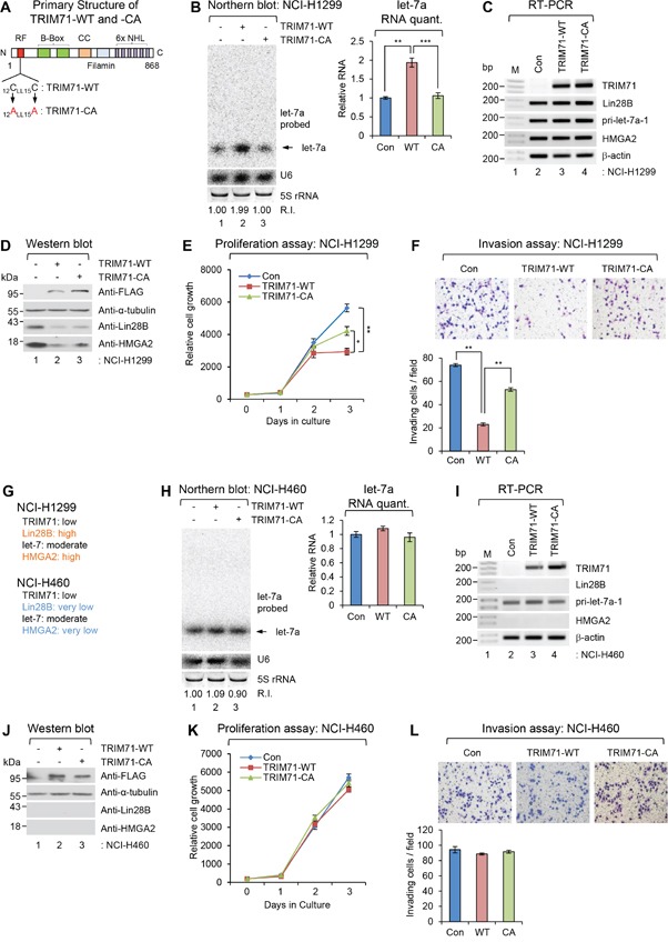
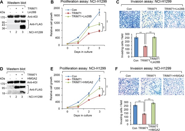
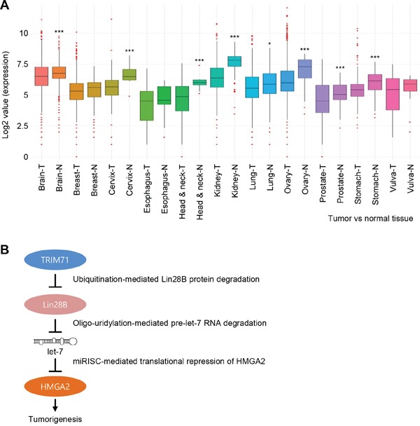
TRIM71 (tripartite motif-containing 71) belongs to the TRIM-NHL protein family, which plays a conserved role in regulating early development and differentiation. However, the molecular functions of TRIM71 have remained largely unknown. Here, we explored the role of TRIM71 together with modulation of Lin28B-let-7-HMGA2 (high-mobility group AT-hook 2) signaling in tumorigenesis. TRIM71 overexpression opposed Lin28B-induced transformation in primary cells and inhibited tumor formation in a mouse model. Specific knockdown of TRIM71 expression increased cancer cell proliferation and invasion. Conversely, overexpression of wild-type TRIM71 in non-small cell lung carcinoma (NSCLC) cells in which Lin28B-let-7-HMGA2 signaling was conserved decreased both cancer cell phenotypes. More importantly, overexpression of an ubiquitin transfer activity-deficient TRIM71 mutant in NSCLC cells had no effect on proliferation or invasion, regardless of the conservation status of Lin28B-let-7-HMGA2 signaling. The tumorigenic inhibitory action of TRIM71 was antagonized by overexpression of the TRIM71 downstream targets, Lin28B and HMGA2. Furthermore, a bioinformatics analysis revealed that TRIM71 expression was downregulated in various types of cancer tissue from patients. Taken together, these data indicate that TRIM71 acts through post-transcriptional repression of Lin28B and subsequent modulation of let-7-HMGA2 signaling during tumorigenesis to potentially function as a tumor suppressor.

Keywords: HMGA2; Lin28B; TRIM71; let-7; tumorigenesis.


Conflict of interest statement

CONFLICTS OF INTEREST

The authors declare no conflicts of interest.

Figures

Figure 1
Figure 1. TRIM71 suppresses the cellular-transforming activity of Lin28B
A. Gene structures of TRIM71, Lin28B, pre-let-7a-1, and HMGA2. Numbers within images represent amino acid or nucleotide position of each gene. Red colored nucleotides in the pre-let-7a-1 RNA represent mature let-7a sequence. RF, RING finger motif; CC, coiled-coil domain; ORF, open reading frame. B-D. Overexpression of Lin28B promotes cellular transformation in NIH/3T3 cells. However the transformation potential of Lin28B was abrogated by TRIM71 co-expression. NIH/3T3 cells infected with pMSCV-neo/pBABE-puro, pMSCV-neo/pBABE-puro-Lin28B, pMSCV-neo-TRIM71/pBABE-puro, pMSCV-neo-TRIM71/pBABE-puro-Lin28B, and pMSCV-neo/pBABE-puro-KRAS(G12V) and selected with puromycin and G418 together. Relative expression of each protein and RNA species determined by western blot (WB) (B), northern blot (NB) (C) [SD is indicated in the graph (*p<0.05). The p-value compares the Lin28B to Lin28B+TRIM71], and semi-quantitative RT-PCR (RT-PCR) (D). E. Soft agar colony formation assays were performed as described in MATERIALS AND METHODS. 5,000 cells/well of virus infected stable NIH/3T3 cells were plated on 6-well plates for soft agar colony formation assay. Colonies bigger than 50 μm were counted after 4 weeks. Colonies were counted from three wells and the average numbers are represented in graph. Data represent the mean values of at least three independent experiments performed in triplicate (**p<0.01). Error bars in the graph represent ± SD, and the p-value compares the Lin28B to Lin28B+TRIM71. NC; negative control without cell plating. Con; pMSCV-neo/pBABE-puro.
Figure 2
Figure 2. TRIM71 inhibits tumor growth initiated by Lin28B
A. Stable virus infected NIH/3T3 cell lines which expressing Lin28B, TRIM71, Lin28B+TRIM71 and KRAS(G12V) were injected subcutaneously into the hip area on both sides of nude mice. Stable virus infected NIH/3T3 cells were propagated in vivo for five weeks. Tumors were measured from a point of growing up to be counted as 0 day. KRAS(G12V) is positive control for tumor formation in mice. The tumor volume of KRAS(G12V) has grown to over 1,000 mm3 rapidly until for 14 days (data not shown). SDs are indicated in the graph (**p<0.01 and ***p<0.001). The p-value compares the Lin28B to Lin28B+TRIM71. B. Ki-67 staining in tumors derived from the subcutaneously injected with stable virus infected NIH/3T3 cells which expressing control, Lin28B, TRIM71, and Lin28B+TRIM71. Representative photos (left) and quantitative data (right) were shown. **p<0.01. C. HMGA2 staining in tumors derived from the subcutaneously injected with stable virus infected NIH/3T3 cells which expressing control, Lin28B, TRIM71, and Lin28B+TRIM71.
Figure 3
Figure 3. Depletion of TRIM71 promotes proliferation and invasion of colorectal carcinoma Caco-2 cells
A. The level of symplekin, TRIM71, CPSF73, α-tubulin, Lin28B, HuR, Lin28A, and HMGA2 proteins were confirmed by WB after specific knockdown of TRIM71 in colorectal carcinoma Caco-2 cells. B. Splinted ligation was performed with [32P] 5′-end-labeled oligonucleotide probe specific for mature let-7a as described in MATERIALS AND METHODS. Aliquot of total RNAs were resolved in 12% SequaGel together as the loading control (5S rRNA). The experiments were repeated at least three times with similar results. The images shown in panels are representative one. **p<0.01. C. The level of TRIM71, Lin28B, HMGA2, β-actin mRNAs, and various pri-miRNAs were analyzed with RT-PCR. D. Proliferation assay was performed in Caco-2 cells transfected with scrambled (siCon) or TRIM71 (siTRIM71) specific siRNA. *p<0.05 E. Invasion assay was performed in Caco-2 cells transfected with scrambled (siCon) or TRIM71 (siTRIM71) specific siRNA. Representative photos (top) and quantitative data of invaded cells (lower) are shown. **p<0.01.
Figure 4
Figure 4. Overexpression of wild-type TRIM71, but not its RING finger mutant, abrogates proliferation and invasion of NCI-H1299 NSCLC cells
A. Primary Structure of wild type TRIM71 and its RING finger mutant. Two essential cysteine residues located N-terminal RING finger motif were mutagenized as alanine residues (CA). B-D. Non-small cell lung cancer cell (NSCLC) NCI-H1299 was infected with control (Con), FLAG-TRIM71(WT), or its RING finger mutant [FLAG-TRIM71(CA)] expressing lentiviral construct. (B) The level of endogenous mature let-7a was monitored by NB with let-7a-specific [32P] 5′-end-labelled oligonucleotide probe. Arrow indicates position of mature let-7a. 5S rRNA was used as the loading control and U6 was used as hybridization control (lower left). The quantification of mature let-7a is shown in graph (right). The experiments were repeated at least three times with similar results. The images shown in panel are representative. **p<0.01 and ***p<0.001. (C) The level of TRIM71, Lin28B, HMGA2, β-actin mRNAs, and pri-let-7a-1 RNA were analyzed with RT-PCR in NCI-H1299 cells. M, 100bp ladder. (D) The protein level of FLAG-TRIM71s, α-tubulin, Lin28B, and HMGA2 were analyzed with WB in NCI-H1299 cells. E. Proliferation assay was performed in NCI-H1299 cells infected with control (Con), FLAG-TRIM71(WT), or its RING finger mutant [FLAG-TRIM71(CA)] expressing lentiviral construct. *p<0.05 and **p<0.01. F. Invasion assay was performed in NCI-H1299 cells infected with control (Con), FLAG-TRIM71(WT), or its RING finger mutant [FLAG-TRIM71(CA)] expressing lentiviral construct. Representative photos of invaded cells (upper) and quantitative data of invasion cells (lower) were shown. **p<0.01. G. Gene expression profiles of TRIM71, Lin28B, let-7, and HMGA2 in NSCLC NCI-H1299 and NCI-460. H. The level of endogenous mature let-7a was monitored by NB in NSCLC NCI-H460 cells infected with control (Con), FLAG-TRIM71(WT), or its RING finger mutant [FLAG-TRIM71(CA)] expressing lentiviral construct. I. The level of TRIM71, Lin28B, HMGA2, β-actin mRNAs, and pri-let-7a-1 RNA were analyzed with RT-PCR in NCI-H460 cells. J. The protein level of FLAG-TRIM71s, α-tubulin, Lin28B, and HMGA2 were analyzed with WB in NCI-H460 cells. K. Proliferation assay was performed in NCI-H1299 cells infected with control (Con), FLAG-TRIM71(WT), or its RING finger mutant [FLAG-TRIM71(CA)] expressing lentiviral construct. L. Proliferation assay was performed in NCI-H460 cells infected with control (Con), FLAG-TRIM71(WT), or its RING finger mutant [FLAG-TRIM71(CA)] expressing lentiviral construct.
Figure 5
Figure 5. Tumorigenesis-inhibitory function of TRIM71 is antagonized by overexpression of its downstream targets, Lin28B and HMGA2, in NCI-H1299 cells
A. WB was performed in NCI-H1299 cells infected with control (Con), FLAG-TRIM71(WT), or FLAG-TRIM71(WT) + FLAG-Lin28B(WT) expressing lentiviral construct. The protein level of eIF4GI, FLAG-TRIM71(WT), and FLAG-Lin28B were analyzed. B. Proliferation assay was performed in NCI-H1299 cells infected with control (Con), FLAG-TRIM71(WT), or FLAG-TRIM71(WT) + FLAG-Lin28B(WT) expressing lentiviral construct. **p<0.01. C. Invasion assay was performed in NCI-H1299 cells infected with control (Con), FLAG-TRIM71(WT), or FLAG-TRIM71(WT) + FLAG-Lin28B(WT) expressing lentiviral construct. Representative photos of invaded cells (upper) and quantitative data of invasion cells (lower) were shown. **p<0.01. D. WB was performed in NCI-H1299 cells infected with control (Con), FLAG-TRIM71(WT), or FLAG-TRIM71(WT) + HMGA2-FLAG expressing lentiviral construct. The protein level of eIF4GI, FLAG-TRIM71(WT), and HMGA2-FLAG were analyzed. E. Proliferation assay was performed in NCI-H1299 cells infected with control (Con), FLAG-TRIM71(WT), or FLAG-TRIM71(WT) + HMGA2-FLAG expressing lentiviral construct. **p<0.01. F. Invasion assay was performed in NCI-H1299 cells infected with control (Con), FLAG-TRIM71(WT), or FLAG-TRIM71(WT) + HMGA2-FLAG expressing lentiviral construct. Representative photos of invaded cells (upper) and quantitative data of invasion cells (lower) were shown. **p<0.01.
Figure 6
Figure 6. TRIM71 expression is downregulated in various cancer patient tissues
A. The expression level of TRIM71 obtained from Tumor (T) and Normal (N) in various cancers, including brain, breast, cervix, esophagus, head and neck, kidney, lung, ovary, prostate, stomach, and vulva cancers. TRIM71 gene expression in various cancer patient tissues was obtained from GENT (Gene Expression across Normal and Tumor tissue) database. The data was downloaded to normalized log2 value of TRIM71 gene in the database and the graph was re-drawn in R program. *p<0.05 and ***p<0.001. B. Proposed model of TRIM71-mediated modulation of Lin28B, let-7, and let-7 target gene HMGA2 during tumorigenesis. miRISC, miRNA-mediated silencing complex.

References

    1. Tocchini C, Ciosk R. TRIM-NHL proteins in development and disease. Semin Cell Dev Biol. 2015;47-48:52–59. - PubMed
    1. Ecsedi M, Grosshans H. LIN-41/TRIM71: emancipation of a miRNA target. Genes Dev. 2013;27:581–589. - PMC - PubMed
    1. Lipkowitz S, Weissman AM. RINGs of good and evil: RING finger ubiquitin ligases at the crossroads of tumour suppression and oncogenesis. Nat Rev Cancer. 2011;11:629–643. - PMC - PubMed
    1. Rybak A, Fuchs H, Hadian K, Smirnova L, Wulczyn EA, Michel G, Nitsch R, Krappmann D, Wulczyn FG. The let-7 target gene mouse lin-41 is a stem cell specific E3 ubiquitin ligase for the miRNA pathway protein Ago2. Nat Cell Biol. 2009;11:1411–1420. - PubMed
    1. Chen J, Lai F, Niswander L. The ubiquitin ligase mLin41 temporally promotes neural progenitor cell maintenance through FGF signaling. Genes Dev. 2012;26:803–815. - PMC - PubMed

MeSH terms

LinkOut - more resources