. 2016:2016:7176154.
doi: 10.1155/2016/7176154. Epub 2016 Oct 16.

Micro- and Macrostructured PLGA/Gelatin Scaffolds Promote Early Cardiogenic Commitment of Human Mesenchymal Stem Cells In Vitro

Affiliations

Micro- and Macrostructured PLGA/Gelatin Scaffolds Promote Early Cardiogenic Commitment of Human Mesenchymal Stem Cells In Vitro

Caterina Cristallini et al. Stem Cells Int. 2016.

Abstract

The biomaterial scaffold plays a key role in most tissue engineering strategies. Its surface properties, micropatterning, degradation, and mechanical features affect not only the generation of the tissue construct in vitro, but also its in vivo functionality. The area of myocardial tissue engineering still faces significant difficulties and challenges in the design of bioactive scaffolds, which allow composition variation to accommodate divergence in the evolving myocardial structure. Here we aimed at verifying if a microstructured bioartificial scaffold alone can provoke an effect on stem cell behavior. To this purpose, we fabricated microstructured bioartificial polymeric constructs made of PLGA/gelatin mimicking anisotropic structure and mechanical properties of the myocardium. We found that PLGA/gelatin scaffolds promoted adhesion, elongation, ordered disposition, and early myocardial commitment of human mesenchymal stem cells suggesting that these constructs are able to crosstalk with stem cells in a precise and controlled manner. At the same time, the biomaterial degradation kinetics renders the PLGA/gelatin constructs very attractive for myocardial regeneration approaches.


Figures
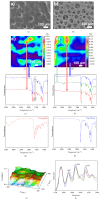
Figure 1
Figure 1
SEM images and Chemical Imaging analysis of nonmicrostructured PLGA/gelatin 70/30 material. (a) SEM image before and (b) after contact with water; (c) chemical map of the surface before and (d) after 15 days of degradation; (e) FT-IR spectra of the sample before and (f) after 15 days of degradation; (g) FT-IR spectrum of pure gelatin; (h) FT-IR spectrum of pure PLGA; (i) correlation map with respect to gelatin spectrum material; (j) deconvolution of amide I band of bioartificial material.
Figure 2
Figure 2
DMA analysis of nonmicrostructured PLGA/gelatin materials. (a) Curve stress-strain for PLGA pure and PLGA/gelatin 70/30 and 80/20 materials; (b) storage and loss modulus as function with deformation for pure PLGA sample; (c) storage and loss modulus as function with deformation for PLGA/gelatin 70/30 sample.
Figure 3
Figure 3
Degradation study for nonmicrostructured PLGA/gelatin materials at different composition in aqueous solution. (a) Trend of percentage weight loss versus time for PLGA/gelatin samples; (b) change of weight average molecular weight (M w) as function with time; (c) trend of number average molecular weight (M n) versus time; (d) polydispersity index (PDI) as function with time.
Figure 4
Figure 4
Schematic drawing (a) and SEM image (b) of a section of microstructured PLGA/gelatin 70/30 bilayer; mechanical behavior of microstructured PLGA/gelatin 70/30 monolayer (c) and bilayer (d) scaffold.
Figure 5
Figure 5
pH measurements in three different means of incubation (PBS, DMEM, and MilliQ water) for the sample microstructured PLGA/gel 70/30 at various incubation times up to 120 days (a); percentage residual weight for a microstructured PLGA/gel 70/30 sample and a microstructured PLGA pure sample, measured in PBS, DMEM, and MilliQ water after 7 days (b).
Figure 6
Figure 6
(a) SEM images of the surface of bilayer PLGA/gel 70/30, from left to right, before degradation, after 7,  15 and 30 days of incubation in DMEM; (b) SEM images of a bilayer PLGA/gel 70/30 from left to right in surface and section after 7 days, in surface after 15 and 30 days in PBS; (c) SEM images of a bilayer PLGA/gel 70/30 from left to right in surface and section after 7 days, in surface after 15 and 30 days in MilliQ water.
Figure 7
Figure 7
Stem cell colonization, growth, and elongation on monolayer and bilayer microstructured PLGA/gelatin scaffolds. (a) Stem cells stained with Calcein-AM and imaged using TPEF microscopy (green) visualized on a monolayer microstructured PLGA/gelatin scaffold imaged using CARS microscopy (blue) after 8 hours from seeding. Red arrows indicate a few hMSCs aligned in parallel inside the scaffold lanes. (b) Representative confocal microscopy images of living hMSCs cultured for either 1 or 15 days on bilayer scaffolds. Superposition of Calcein-AM (green) and bright field microscopy is shown. Magnification 20x, scale bar 50 μm. (c) Representative confocal microscopy image showing cytoskeletal organization of hMSCs cultured for 15 days on a bilayer scaffold. Actin filaments are stained with phalloidin in red, cytoplasm with Cell Mask Green, and nuclei with Hoechst in blue. Magnification 63x, scale bar 20 μm. (d) 3D reconstruction of a representative image showing hMSCs cultured for 24 h on a monolayer scaffold. Cells are stained with Calcein-AM. Magnification 20x, scale bar 100 μm. (e) Cell proliferation rates after 4, 8, 12, and 15 days of culture on bilayer scaffolds. Values were normalized on cell adhesion quantified at 24 hr. (f) Mean cell lengths of hMSCs cultured on monolayer and bilayer scaffolds. Elongation measurements were performed on living cells stained with Calcein-AM at 2, 4, and 8 days of culture. Lengths were not statistically different from the 2D control.
Figure 8
Figure 8
Analysis of stemness and cardiac differentiation markers. (a) qPCR results for hMSCs cultured for 3, 4, and 15 days on monolayer microstructured PLGA/gelatin scaffolds. The expression of the same genes in 2D control cultures was used for normalization. (b) Representative confocal immunofluorescence images of c-kit and GATA-4 proteins (red) and nuclei (blue) in hMSCs after either 2D control culture (top panel) or 15-day culture (bottom panel) on microstructured bilayer PLGA/gelatin scaffolds. Magnification 63x, scale bar 20 μm.

References

    1. Leor J., Landa N., Cohen S. Renovation of the injured heart with myocardial tissue engineering. Expert Review of Cardiovascular Therapy. 2006;4(2):239–252. doi: 10.1586/14779072.4.2.239. - DOI - PubMed
    1. Jana S., Tefft B. J., Spoon D. B., Simari R. D. Scaffolds for tissue engineering of cardiac valves. Acta Biomaterialia. 2014;10:2877–2893. - PubMed
    1. Kim D.-H., Lipke E. A., Kim P., et al. Nanoscale cues regulate the structure and function of macroscopic cardiac tissue constructs. Proceedings of the National Academy of Sciences of the United States of America. 2010;107(2):565–570. doi: 10.1073/pnas.0906504107. - DOI - PMC - PubMed
    1. Stevens M. M., George J. H. Exploring and engineering the cell surface interface. Science. 2005;310(5751):1135–1138. doi: 10.1126/science.1106587. - DOI - PubMed
    1. Park H., Cannizzaro C., Vunjak-Novakovic G., Langer R., Vacanti C. A., Farokhzad O. C. Nanofabrication and microfabrication of functional materials for tissue engineering. Tissue Engineering. 2007;13(8):1867–1877. doi: 10.1089/ten.2006.0198. - DOI - PubMed

LinkOut - more resources