. 2016 Oct 31;3(5):ENEURO.0220-16.2016.
doi: 10.1523/ENEURO.0220-16.2016. eCollection 2016 Sep-Oct.

Acute Axonal Degeneration Drives Development of Cognitive, Motor, and Visual Deficits after Blast-Mediated Traumatic Brain Injury in Mice

Affiliations

Acute Axonal Degeneration Drives Development of Cognitive, Motor, and Visual Deficits after Blast-Mediated Traumatic Brain Injury in Mice

Terry C Yin et al. eNeuro. .

Abstract

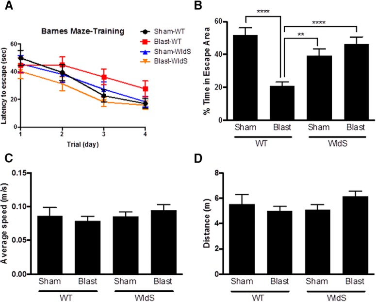
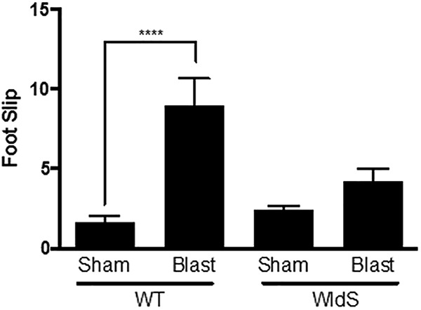
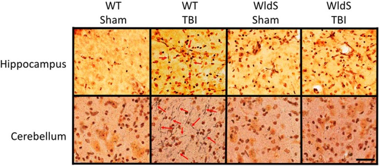
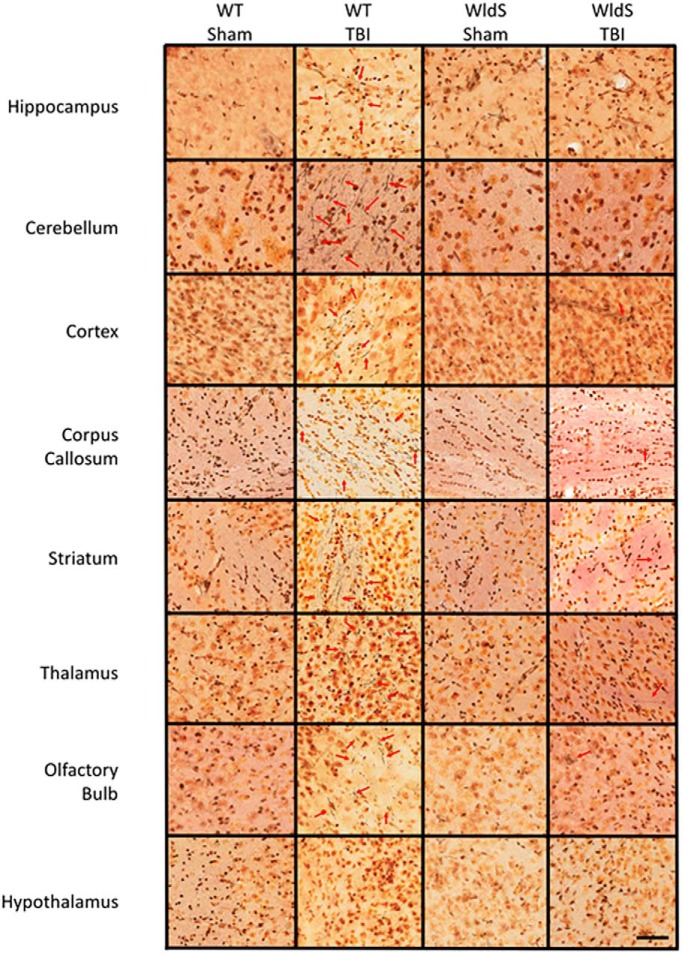
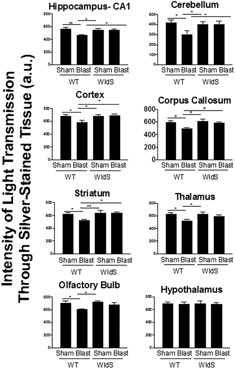
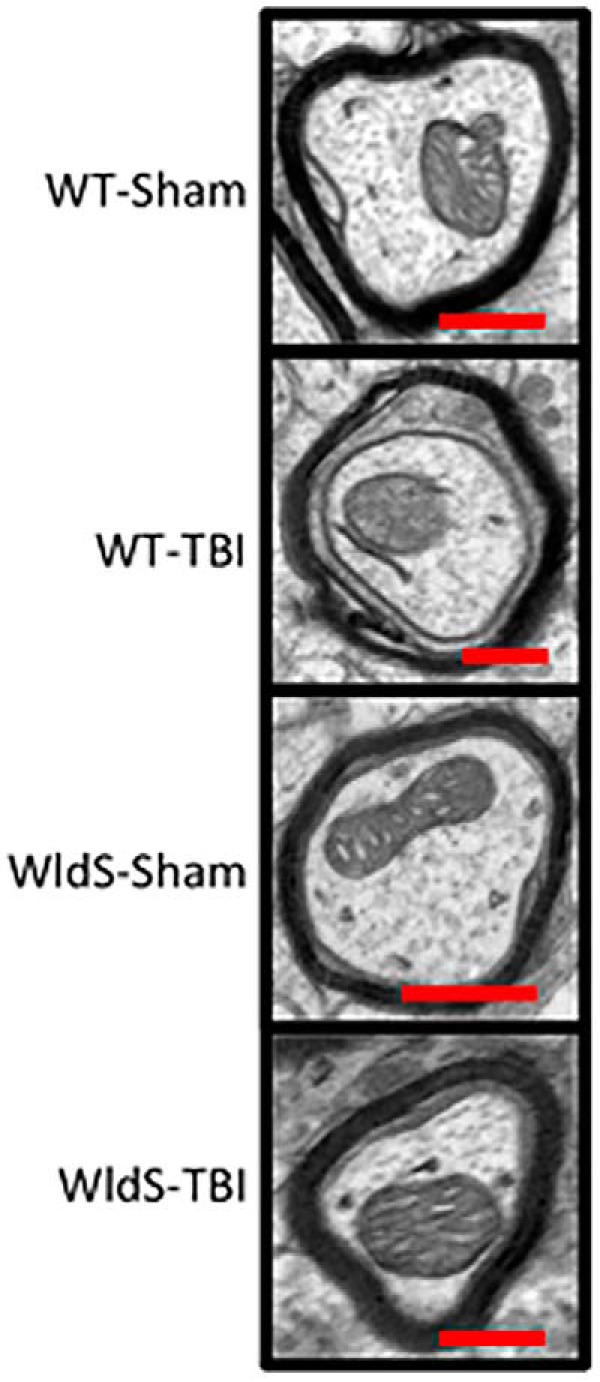
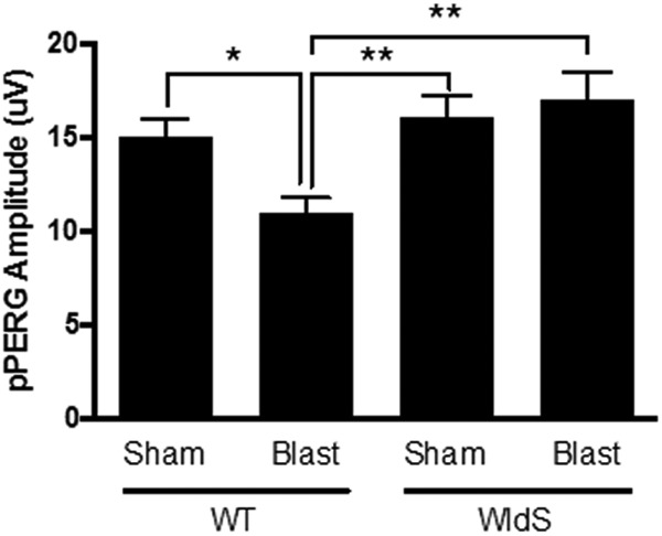
Axonal degeneration is a prominent feature of many forms of neurodegeneration, and also an early event in blast-mediated traumatic brain injury (TBI), the signature injury of soldiers in Iraq and Afghanistan. It is not known, however, whether this axonal degeneration is what drives development of subsequent neurologic deficits after the injury. The Wallerian degeneration slow strain (WldS) of mice is resistant to some forms of axonal degeneration because of a triplicated fusion gene encoding the first 70 amino acids of Ufd2a, a ubiquitin-chain assembly factor, that is linked to the complete coding sequence of nicotinamide mononucleotide adenylyltransferase 1 (NMAT1). Here, we demonstrate that resistance of WldS mice to axonal degeneration after blast-mediated TBI is associated with preserved function in hippocampal-dependent spatial memory, cerebellar-dependent motor balance, and retinal and optic nerve-dependent visual function. Thus, early axonal degeneration is likely a critical driver of subsequent neurobehavioral complications of blast-mediated TBI. Future therapeutic strategies targeted specifically at mitigating axonal degeneration may provide a uniquely beneficial approach to treating patients suffering from the effects of blast-mediated TBI.

Keywords: WldS mouse; axonal degeneration; neurodegeneration; nicotinamide adenine dinucleotide; traumatic brain injury.


Conflict of interest statement

Authors report no conflict of interest.

Figures

Figure 1.
Figure 1.
WldS mice are protected from memory deficits after blast-mediated TBI. A, Latency to escape progressively decreases over the 4-day training period in all groups. B, Blast-mediated TBI wild-type (WT) mice spend less than half the time as sham-injury WTs in the escape area (5-cm radius around the escape hole) during the probe test of memory. Blast-mediated TBI WldS mice spend a amount of time in the escape area comparable to that of sham-injury WT and sham-injury WldS mice. C, The average locomotion speed during the probe trial was similar in all groups. D, The total distance traveled during the probe trial was similar in all groups. Each group consisted of 25 male congenic C57/Bl6 mice, aged 12–14 weeks. Data were collected and scored in an automated manner blind to treatment group. Data are represented as mean ± SEM. Significance was determined by two-way ANOVA with Bonferroni post hoc analysis. p-values labeled as **<0.01 and ****<0.0001 compared with blast-injured WT animals.
Figure 2.
Figure 2.
WldS mice are protected from motor coordination deficits after blast-mediated TBI. Blast-injured wild-type (WT) mice showed an increased number of foot slips relative to sham-injury WT mice 28 days after blast-mediated TBI. Blast-injured WldS mice show a similar number of foot slips as sham-injury WT and sham-injury WldS mice. Each group consisted of 25 male congenic C57/Bl6 mice, aged 12–14 weeks. Data was manually collected and scored blind to treatment group. Data are represented as mean ± SEM. Significance was determined by two-way ANOVA with Bonferroni post hoc analysis. Significance was determined by two-way ANOVA with Bonferroni post hoc analysis. p-values labeled as ****<0.0001 compared to sham-injured WT animals.
Figure 3.
Figure 3.
WldS hippocampus and cerebellum are protected from axonal degeneration after blast-mediated TBI. High-power representative pictures with 40× objective from hippocampal CA1 stratum radiatum and cerebellum show prominent silver staining of degenerating axons (red arrows) in blast-injured wild-type mice 12 days after injury, with little to no axonal degeneration in WldS mice after the same injury. Images shown are representative of typical images from five animals in each group. Scale bar = 2.5 mm.
Figure 4.
Figure 4.
WldS mice are broadly protected throughout the brain from axonal degeneration after blast-mediated TBI. As in hippocampus and cerebellum, protection was also noted in WldS cortex, corpus callosum, olfactory bulb, striatum, and thalamus. Noticeably, the hypothalamus is resistant to axonal degeneration in wild-type mice after blast-mediated TBI. Images shown are representative of typical images from five animals in each group. Scale bar = 2.5 mm.
Figure 5.
Figure 5.
Optical densitometry of light transmitted through silver-stained brain regions from all animals in each group was used to quantify the protective effect. Signal was quantified for 18 sections for each of the five animals, spaced 480 mm apart. Here, a greater value indicates that more light was able to pass unimpeded through the section by virtue of less silver staining, which reflects less axonal degeneration. Data are represented as mean ± SEM. p-value *<0.05 and **<0.01 determined by two-way ANOVA with Bonferroni post hoc analysis compared with blast-injured WT animals.
Figure 6.
Figure 6.
Transmission electron microscopy 12 days after injury shows normal myelin and axonal mitochondrial structures in the CA1 stratum radiatum of sham-injury wild-type mice, with no differences seen in sham- and blast-injury WldS mice. Blast-injured wild-type mice show degeneration of the myelin sheath along, as well as abnormal outer membrane and internal cristae structures within neuronal mitochondria.
Figure 7.
Figure 7.
WldS mice are protected from pPERG deficits after blast-mediated TBI. pPERG serves as an early indicator of future chronic damage to the visual system, including retinal cell death. Wild-type (WT) mice exhibit ∼25% decrease in pPERG 4 weeks after blast injury, whereas both sham- and blast-injury WldS mice exhibit pPERG levels equivalent to sham-injury WT mice. Each group consisted of 25 male congenic C57/Bl6 mice, aged 12–14 weeks. Data were collected and scored in an automated manner blind to identification of the group and are represented as mean ± SEM. p-value *<0.05 and **<0.01 determined by-two way ANOVA with Bonferroni post hoc analysis, compared to blast-injured WT animals.

References

    1. Araki T, Sasaki Y, Milbrandt J (2004) Increased nuclear NAD biosynthesis and SIRT1 activation prevent axonal degeneration. Science 305:1010–1013. 10.1126/science.1098014 - DOI - PubMed
    1. Blaya MO, Bramlett HM, Naidoo J, Pieper AA, Dietrich WD (2014) Neuroprotective efficacy of a proneurogenic compound after traumatic brain injury. J Neurotrauma 31:476–486. 10.1089/neu.2013.3135 - DOI - PMC - PubMed
    1. Baldock RA, Poole I (1993) Video camera calibration for optical densitometry. J Microsc Oct. 172(Pt1):49–54. - PubMed
    1. Clark RS, Vagni VA, Nathaniel PD, Jenkins LW, Dixon CE, Szabó C (2007) Local administration of the poly(ADP-ribose) polymerase inhibitor INO-1001 prevents NAD+ depletion and improves water maze performance after traumatic brain injury in mice. J Neurotrauma 24:1399–1405. 10.1089/neu.2007.0305 - DOI - PubMed
    1. Cockerham GC, Goodrich GL, Weichel ED, Orcutt JC, Rizzo JF, Bower KSA, Schuchard RA (2009) Eye and visual function in traumatic brain injury. J Rehabil Res Dev 46:811–818. - PubMed

Publication types

MeSH terms

Substances