. 2016 Aug 2;1(4):e00046-16.
doi: 10.1128/mSystems.00046-16. eCollection 2016 Jul-Aug.

The Oral and Skin Microbiomes of Captive Komodo Dragons Are Significantly Shared with Their Habitat

Affiliations

The Oral and Skin Microbiomes of Captive Komodo Dragons Are Significantly Shared with Their Habitat

Embriette R Hyde et al. mSystems. .

Abstract

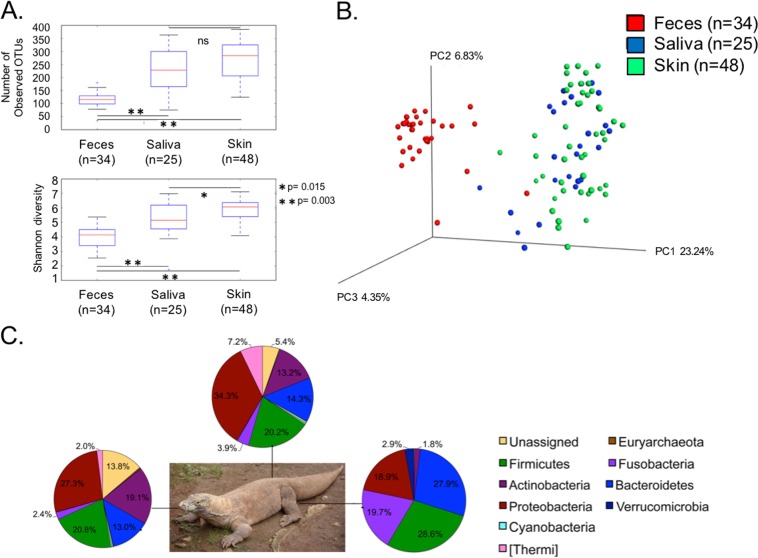
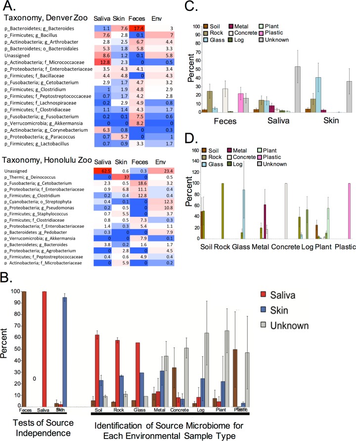
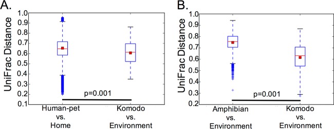
Examining the way in which animals, including those in captivity, interact with their environment is extremely important for studying ecological processes and developing sophisticated animal husbandry. Here we use the Komodo dragon (Varanus komodoensis) to quantify the degree of sharing of salivary, skin, and fecal microbiota with their environment in captivity. Both species richness and microbial community composition of most surfaces in the Komodo dragon's environment are similar to the Komodo dragon's salivary and skin microbiota but less similar to the stool-associated microbiota. We additionally compared host-environment microbiome sharing between captive Komodo dragons and their enclosures, humans and pets and their homes, and wild amphibians and their environments. We observed similar host-environment microbiome sharing patterns among humans and their pets and Komodo dragons, with high levels of human/pet- and Komodo dragon-associated microbes on home and enclosure surfaces. In contrast, only small amounts of amphibian-associated microbes were detected in the animals' environments. We suggest that the degree of sharing between the Komodo dragon microbiota and its enclosure surfaces has important implications for animal health. These animals evolved in the context of constant exposure to a complex environmental microbiota, which likely shaped their physiological development; in captivity, these animals will not receive significant exposure to microbes not already in their enclosure, with unknown consequences for their health. IMPORTANCE Animals, including humans, have evolved in the context of exposure to a variety of microbial organisms present in the environment. Only recently have humans, and some animals, begun to spend a significant amount of time in enclosed artificial environments, rather than in the more natural spaces in which most of evolution took place. The consequences of this radical change in lifestyle likely extend to the microbes residing in and on our bodies and may have important implications for health and disease. A full characterization of host-microbe sharing in both closed and open environments will provide crucial information that may enable the improvement of health in humans and in captive animals, both of which experience a greater incidence of disease (including chronic illness) than counterparts living under more ecologically natural conditions.

Keywords: Komodo dragon; SourceTracker; built environment; human microbiome; microbiome.


Figures

FIG 1
FIG 1
(A) Box-and-whisker plots illustrate the median, maximum, minimum, and first and third quartiles of the distribution of the number of observed OTUs and the Shannon diversity index for Komodo dragon fecal (n = 34), saliva (n = 25), and skin (n = 48) microbial communities. A nonparametric t test with Monte Carlo permutations was used to calculate significant differences in diversity between groups. ns, not significant. (B) An unweighted UniFrac-based PCoA plot reveals that fecal microbial communities cluster separately from skin and saliva microbial communities, which are more similar to each other. Principal components 1 to 3 (PC1 to PC3) are shown. (C) Pie charts overlaid onto a Komodo dragon picture illustrate the mean relative abundances of phyla present in Komodo dragon saliva (n = 25), skin (n = 34), and feces (n = 48).
FIG 2
FIG 2
(A) Heat maps illustrate the percent abundances of the most abundant genera (all OTUs taxonomically classified to the same genus were collapsed into a single genus summary) present in saliva, skin, feces, and environmental (Env) samples collected from the Denver and Honolulu zoos. The deepest taxonomic classification achieved is listed for each genus. The heat map colors indicate percent abundance (red [high abundance] to blue [low abundance]). (B) Komodo dragon SourceTracker analysis reveals that the microbial communities of many environmental sample types are sourced from skin, saliva, and feces rather than unknown sources (i.e., not from Komodo dragon skin, saliva, or feces). Data are plotted as the means ± standard errors of the means (error bars) of samples from Denver and Honolulu zoo Komodo dragons. (C) SourceTracker analyses with Komodo dragon fecal, salivary, and skin samples designated as sinks and environmental samples designated as sources reveals that a variety of environmental sources, rather than a single environmental source, contribute to the microbial communities of Komodo dragon feces, saliva, and skin. Unknown sources (i.e., not the environments sampled from the Komodo dragon enclosures) also contribute about 40% or more of the microbial community of saliva and skin samples (only 20% of fecal samples). (D) Independence tests reveal that about half of the environmental samples are not independent from other environmental samples. Data are the means ± standard errors of the means of Denver and Honolulu Komodo dragon and environmental samples.
FIG 3
FIG 3
(A) Amphibian SourceTracker analysis reveals that water is the only sample type that obtains a notable amount of its microbial community from amphibian skin; unknown sources (i.e., not amphibian skin) are the main microbiome contributors to soil and sediment. (B) Independence tests reveal that amphibian skin is independently specific to species. (C) Designating environment the source and amphibian skin the sink reveals that water is the only environmental type that contributes largely to the microbial communities on amphibian skin, with unknown sources also largely contributing to the amphibian skin microbiome. (D) Independence tests reveal that each environment type is also independent from each other environment type. Data are the means ± standard errors of the means (error bars).
FIG 4
FIG 4
Box-and-whisker plots illustrate the UniFrac distance between Komodo dragon and Komodo dragon environment samples and human/pet and home samples (A) and amphibian and amphibian environment samples (B).

References

    1. McFall-Ngai M, Hadfield MG, Bosch TCG, Carey HV, Domazet-Lošo T, Douglas AE, Dubilier N, Eberl G, Fukami T, Gilbert SF, Hentschel U, King N, Kjelleberg S, Knoll AH, Kremer N, Mazmanian SK, Metcalf JL, Nealson K, Pierce NE, Rawls JF, Reid A, Ruby EG, Rumpho M, Sanders JG, Tautz D, Wernegreen JJ. 2013. Animals in a bacterial world, a new imperative for the life sciences. Proc Natl Acad Sci U S A 110:3229–3236. doi: 10.1073/pnas.1218525110. - DOI - PMC - PubMed
    1. Tung J, Barreiro LB, Burns MB, Grenier J-C, Lynch J, Grieneisen LE, Altmann J, Alberts SC, Blekhman R, Archie EA. 2015. Social networks predict gut microbiome composition in wild baboons. Elife 4:1–18. doi: 10.7554/eLife.05224. - DOI - PMC - PubMed
    1. Uenishi G, Fujita S, Ohashi G, Kato A, Yamauchi S, Matsuzawa T, Ushida K. 2007. Molecular analysis of the intestinal microbiota of chimpanzees in the wild and in captivity. Am J Primatol 69:367–376. doi: 10.1002/ajp.20351. - DOI - PubMed
    1. Scupham AJ, Patton TG, Bent E, Bayles DO. 2008. Comparison of the cecal microbiota of domestic and wild turkeys. Microb Ecol 56:322–331. doi: 10.1007/s00248-007-9349-4. - DOI - PubMed
    1. Villers LM, Jang SS, Lent CL, Lewin-Koh SC, Norosoarinaivo JA. 2008. Survey and comparison of major intestinal flora in captive and wild ring-tailed lemur (Lemur catta) populations. Am J Primatol 70:175–184. doi: 10.1002/ajp.20482. - DOI - PubMed