

Structural Characterization of Monomers and Oligomers of D-Amino Acid-Containing Peptides Using T-Wave Ion Mobility Mass Spectrometry
- PMID: 27822705
- PMCID: PMC5177490
- DOI: 10.1007/s13361-016-1523-9
Structural Characterization of Monomers and Oligomers of D-Amino Acid-Containing Peptides Using T-Wave Ion Mobility Mass Spectrometry
Abstract
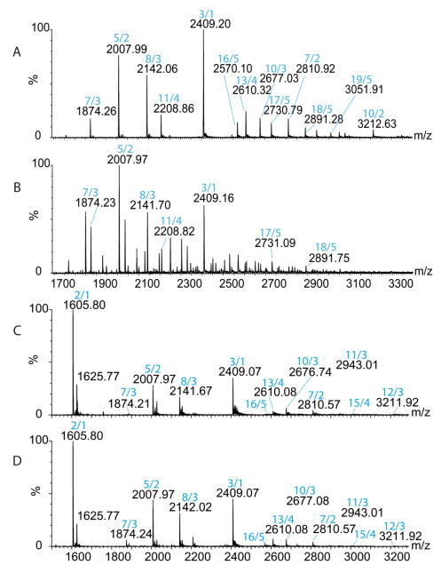
The D-residues are crucial to biological function of D-amino acid containing peptides (DAACPs). Previous ion mobility mass spectrometry (IM-MS) studies revealing oligomerization patterns of amyloid cascade demonstrated conversion from native soluble unstructured assembly to fibril ß-sheet oligomers, which has been implicated in amyloid diseases, such as Alzheimer's disease and type 2 diabetes. Although neuropeptides are typically present at very low concentrations in circulation, their local concentrations could be much higher in large dense core vesicles, forming dimers or oligomers. We studied the oligomerization of protonated and metal-adducted achatin I and dermorphin peptide isomers with IM-MS. Our results suggested that dimerization, oligomerization, and metal adduction augment the structural differences between D/L peptide isomers compared to protonated monomers. Dimers and oligomers enhanced the structural differences between D/L peptide isomers in both aqueous and organic solvent system. Furthermore, some oligomer forms were only observed for either D- or L-isomers, indicating the importance of chiral center in oligomerization process. The oligomerization patterns of D/L isomers appear to be similar. Potassium adducts were detected to enlarge the structural differences between D/L isomers. Graphical Abstract ᅟ.
Keywords: CCS; Collision cross-section; Conformational differences; D-amino acid containing peptides; DAACPs; Dimer; IM-MS; Ion mobility mass spectrometry; Metal adducts; Monomer; Native state; Oligomer; Oligomerization pattern; Organic solvent.
Figures


References
-
- Montecucchi PC, de Castiglione R, Piani S, Gozzini L, Erspamer V. Amino acid composition and sequence of dermorphin, a novel opiate-like peptide from the skin of Phyllomedusa sauvagei. International journal of peptide and protein research. 1981;17:275–283. - PubMed
-
- Soyez D, Van Herp F, Rossier J, Le Caer JP, Tensen CP, Lafont R. Evidence for a conformational polymorphism of invertebrate neurohormones. D-amino acid residue in crustacean hyperglycemic peptides. J Biol Chem. 1994;269:18295–18298. - PubMed
-
- Yasuda A, Yasuda Y, Fujita T, Naya Y. Characterization of crustacean hyperglycemic hormone from the crayfish (Procambarus clarkii): multiplicity of molecular forms by stereoinversion and diverse functions. General and comparative endocrinology. 1994;95:387–398. - PubMed
-
- Kamatani Y, Minakata H, Kenny PT, Iwashita T, Watanabe K, Funase K, Sun XP, Yongsiri A, Kim KH, Novales-Li P, Novales TNKGC, Takeuchi H, Nomoto K. Achatin-I an endogenous neuroexcitatory tetrapeptide from Achatina fulica Ferussac containing a D-amino acid residue. Biochem Biophys Res Commun. 1989;160:1015–1020. - PubMed
MeSH terms
Substances
Grants and funding
LinkOut - more resources
Full Text Sources
Other Literature Sources
