. 2016 Nov 8:7:13331.
doi: 10.1038/ncomms13331.

Genomic analyses identify recurrent MEF2D fusions in acute lymphoblastic leukaemia

Affiliations

Genomic analyses identify recurrent MEF2D fusions in acute lymphoblastic leukaemia

Zhaohui Gu et al. Nat Commun. .

Abstract

Chromosomal rearrangements are initiating events in acute lymphoblastic leukaemia (ALL). Here using RNA sequencing of 560 ALL cases, we identify rearrangements between MEF2D (myocyte enhancer factor 2D) and five genes (BCL9, CSF1R, DAZAP1, HNRNPUL1 and SS18) in 22 B progenitor ALL (B-ALL) cases with a distinct gene expression profile, the most common of which is MEF2D-BCL9. Examination of an extended cohort of 1,164 B-ALL cases identified 30 cases with MEF2D rearrangements, which include an additional fusion partner, FOXJ2; thus, MEF2D-rearranged cases comprise 5.3% of cases lacking recurring alterations. MEF2D-rearranged ALL is characterized by a distinct immunophenotype, DNA copy number alterations at the rearrangement sites, older diagnosis age and poor outcome. The rearrangements result in enhanced MEF2D transcriptional activity, lymphoid transformation, activation of HDAC9 expression and sensitive to histone deacetylase inhibitor treatment. Thus, MEF2D-rearranged ALL represents a distinct form of high-risk leukaemia, for which new therapeutic approaches should be considered.


Figures
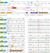
Figure 1
Figure 1. MEF2D rearrangements in ALL.
(a) Protein structure of wild-type MEF2D and fusion partners, (b) protein structure of each fusion protein and (c) electropherograms showing the results of RT–PCR and Sanger sequence (see also Supplementary Fig. 1). Four MEF2D-BCL9 isoforms were identified in 16 cases, with breakpoints involving exons 5 or 6 of MEF2D fused in frame to exons 9 or 10 of BCL9. Rearrangements involving MEF2D and other four fusion partners were identified in six samples. MEF2D-DAZAP1 and MEF2D-CSF1R fusions have previously been reported. Protein region annotation: MADS box, MEF2 (myocyte enhancer factor 2)-like/Type II subfamily of MADS; HJURP_C, Holliday junction regulator protein family C-terminal repeat. Ig domain, immunoglobulin domain; Ig4_SCFR_like, fourth Ig-like domain of stem cell factor receptor; PTK catalytic domain, tyrosine kinase catalytic domain.
Figure 2
Figure 2. MEF2D-rearranged ALL cases have a distinct gene expression profile.
(a) Principal component analysis of samples subjected to unstranded RNAseq data, showing clustering of MEF2D-rearranged cases. A full list of cases and rearrangements identified by RNAseq is provided in Supplementary Data 1. (b) The gene expression signature for MEF2D-rearranged ALL was determined by comparing 19 MEF2D fusion samples (excluding 2 cases that failed quality-control threshold for gene expression analysis and one MEF2D-CSF1R rearranged case) and 199 control samples through Wald's test and Benjamini–Hochberg adjustment. Genes with adjusted P-value <0.01 are provided in Supplementary Data 3. The top 50 upregulated and top 50 downregulated genes (based on the adjusted P-value) were selected to perform supervised clustering and shown in the order from most significant upregulated genes (top) to the most downregulated ones (bottom). Stranded-PE100, total stranded paired-end 100 bp RNAseq; unstranded-PE100, unstranded mRNA paired-end 100 bp RNAseq; unstranded-PE75, unstranded mRNA paired-end 75 bp RNAseq. MEF2D, fusions involving MEF2D; JAK2/TYK2, fusions involving JAK2 or TYK2; PAX5, fusions with PAX5 as the N-terminal fusion partner with non-kinase genes; ABL1/2, fusions with ABL1 or ABL2 observed in Ph-like ALL; CRLF2, IGH-CRLF2 or P2RY8-CRLF2 fusions; ZNF384, fusions involving ZNF384 as the C-terminal fusion partner; EPOR, fusions deregulating EPOR observed in Ph-like ALL; PDGFRB, fusions involving PDGFRB; IGH, IGH fusions except IGH-CRLF2 and IGH-EPOR; MLL, fusions involving KMT2A(MLL) gene.
Figure 3
Figure 3. Poor outcome of MEF2D-rearranged ALL.
The event-free (a,c) and overall survival (b,d) outcome of MEF2D-rearranged cases is compared with that of B-precursor ALL cases lacking known recurring chromosomal rearrangements (a,b) and key genetic subtypes (c,d) of B-ALL in the COG AALL0232 study.
Figure 4
Figure 4. Luciferase and drug-sensitivity assays.
(a) Luciferase assay to test MEF2 binding activities. The average levels of normalized luciferase activities±s.d. from six replicates are indicated. MEF2D-rearranged isoforms are more potent transcriptional activators than wild-type and truncated MEF2D. (b). Colony-forming assay for retroviral vectors transduced lineage negative bone marrow cells. Colony number counted from three replicate dishes is shown in mean±s.d. Western blotting was performed to verify expression of proteins by each construct (Supplementary Fig. 18). RLU, relative luminescence units; MIG, MSCV-IRES-GFP, represents empty vector; MEF2D-WT, vectors express wild-type MEF2D; MEF2DΔ, truncated MEF2D; BCL9-WT, wild-type BCL9; M/B-1 through M/B-3, three most common MEF2D-BCL9 fusion isoforms; M/H, MEF2D-HNRNPUL1. (c) Ex vivo drug sensitivity assays from two xenografted samples. Four chemical compounds including ABT-199 (selective BCL-2 inhibitor), vincristine and two HDAC inhibitors (panobinostat and vorinostat) were applied to measure growth inhibition curves in xenografted samples. The MEF2D rearrangements were confirmed in xenografted samples by RT–PCR and three more xenografted samples were also tested by the four drugs (Supplementary Fig. 16). Error bars represent mean±s.d.

References

    1. Hunger S. P. & Mullighan C. G. Redefining ALL classification: toward detecting high-risk ALL and implementing precision medicine. Blood 125, 3977–3987 (2015). - PMC - PubMed
    1. Hunger S. P. & Mullighan C. G. Acute lymphoblastic leukemia in children. N. Engl. J. Med. 373, 1541–1552 (2015). - PubMed
    1. Roberts K. G. et al. Targetable kinase-activating lesions in Ph-like acute lymphoblastic leukemia. N. Engl. J. Med. 371, 1005–1015 (2014). - PMC - PubMed
    1. Moorman A. V. The clinical relevance of chromosomal and genomic abnormalities in B-cell precursor acute lymphoblastic leukaemia. Blood Rev. 26, 123–135 (2012). - PubMed
    1. Willis T. G. et al. Molecular cloning of translocation t(1;14)(q21;q32) defines a novel gene (BCL9) at chromosome 1q21. Blood 91, 1873–1881 (1998). - PubMed

Publication types