

Neuropathology of mood disorders: do we see the stigmata of inflammation?
- PMID: 27824355
- PMCID: PMC5314124
- DOI: 10.1038/tp.2016.212
Neuropathology of mood disorders: do we see the stigmata of inflammation?
Abstract
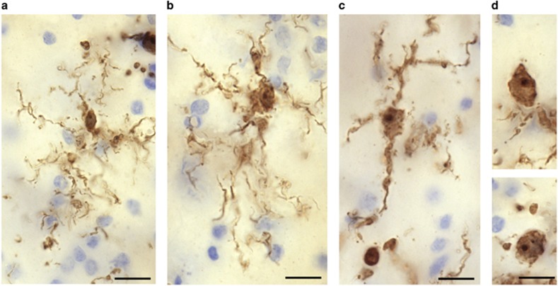
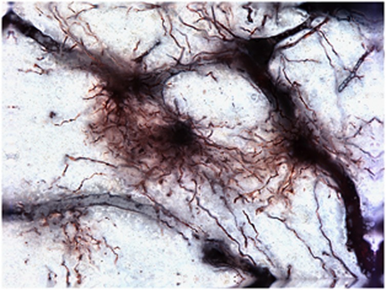
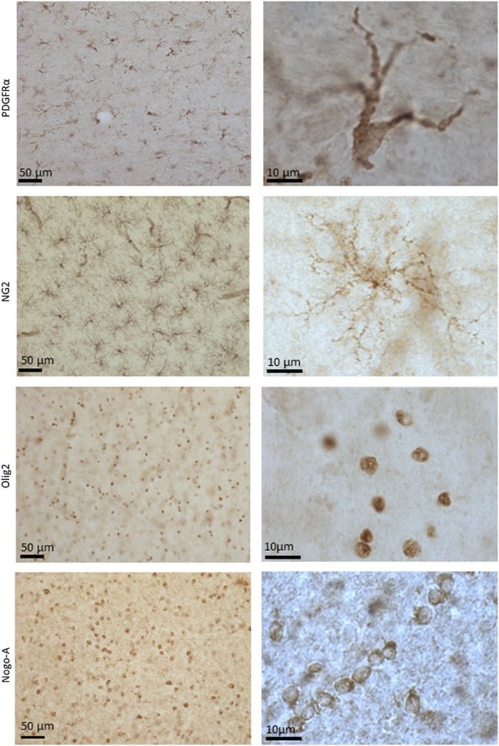
A proportion of cases with mood disorders have elevated inflammatory markers in the blood that conceivably may result from stress, infection and/or autoimmunity. However, it is not yet clear whether depression is a neuroinflammatory disease. Multiple histopathological and molecular abnormalities have been found postmortem but the etiology of these abnormalities is unknown. Here, we take an immunological perspective of this literature. Increases in activated microglia or perivascular macrophages in suicide victims have been reported in the parenchyma. In contrast, astrocytic markers generally are downregulated in mood disorders. Impairment of astrocytic function likely compromises the reuptake of glutamate potentially leading to excitotoxicity. Inflammatory cytokines and microglia/macrophage-derived quinolinic acid (QA) downregulate the excitatory amino acid transporters responsible for this reuptake, while QA has the additional effect of inhibiting astroglial glutamine synthetase, which converts glutamate to glutamine. Given that oligodendroglia are particularly vulnerable to inflammation, it is noteworthy that reductions in numbers or density of oligodendrocyte cells are one of the most prominent findings in depression. Structural and/or functional changes to GABAergic interneurons also are salient in postmortem brain samples, and may conceivably be related to early inflammatory insults. Although the postmortem data are consistent with a neuroimmune etiology in a subgroup of depressed individuals, we do not argue that all depression-associated abnormalities are reflective of a neuroinflammatory process or even that all immunological activity in the brain is deleterious. Rather, we highlight the pervasive role of immune signaling pathways in brain function and provide an alternative perspective on the current postmortem literature.
Conflict of interest statement
The authors declare no conflict of interest.
Figures

References
-
- Savitz JB, Price JL, Drevets WC. Neuropathological and neuromorphometric abnormalities in bipolar disorder: view from the medial prefrontal cortical network. Neurosci Biobehav Rev 2014; 42: 132–147. - PubMed
-
- Weinberger DR, Radulescu E. Finding the elusive psychiatric "Lesion" with 21st-century neuroanatomy: a note of caution. Am J Psychiatry 2016; 173: 27–33. - PubMed
-
- Dargel AA, Godin O, Kapczinski F, Kupfer DJ, Leboyer M. C-reactive protein alterations in bipolar disorder: a meta-analysis. J Clin Psychiatry 2015; 76: 142–150. - PubMed
-
- Munkholm K, Brauner JV, Kessing LV, Vinberg M. Cytokines in bipolar disorder vs. healthy control subjects: a systematic review and meta-analysis. J Psychiatr Res 2013; 47: 1119–1133. - PubMed
Publication types
MeSH terms
Substances
Grants and funding
LinkOut - more resources
Full Text Sources
Other Literature Sources
Medical
