Comparative Study
. 2016 Dec 13;7(50):83392-83408.
doi: 10.18632/oncotarget.13110.

CD19-specific triplebody SPM-1 engages NK and γδ T cells for rapid and efficient lysis of malignant B-lymphoid cells

Affiliations
Comparative Study

CD19-specific triplebody SPM-1 engages NK and γδ T cells for rapid and efficient lysis of malignant B-lymphoid cells

Christian B Schiller et al. Oncotarget. .

Abstract

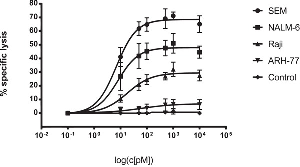
Triplebodies are antibody-derived recombinant proteins carrying 3 antigen-binding domains in a single polypeptide chain. Triplebody SPM-1 was designed for lysis of CD19-bearing malignant B-lymphoid cells through the engagement of CD16-expressing cytolytic effectors, including NK and γδ T cells.SPM-1 is an optimized version of triplebody ds(19-16-19) and includes humanization, disulfide stabilization and the removal of potentially immunogenic sequences. A three-step chromatographic procedure yielded 1.7 - 5.5 mg of purified, monomeric protein per liter of culture medium. In cytolysis assays with NK cell effectors, SPM-1 mediated potent lysis of cancer-derived B cell lines and primary cells from patients with various B-lymphoid malignancies, which surpassed the ADCC activity of the therapeutic antibody Rituximab. EC50-values ranged from 3 to 86 pM. Finally, in an impedance-based assay, SPM-1 mediated a particularly rapid lysis of CD19-bearing target cells by engaging and activating both primary and expanded human γδ T cells from healthy donors as effectors.These data establish SPM-1 as a useful tool for a kinetic analysis of the cytolytic reactions mediated by γδ T and NK cells and as an agent deserving further development towards clinical use for the treatment of B-lymphoid malignancies.

Keywords: antibody-dependent cellular cytotoxicity; gamma delta T cell; immunotherapy; leukemia; single chain triplebody.


Conflict of interest statement

CONFLICTS OF INTEREST

The authors report no conflicts of interest.

Figures

Figure 1
Figure 1. Structural characteristics and purification of triplebody SPM-1
A. CD19-specific scFv domains shown in light grey; CD16-specific scFv domain in dark grey. B. After purification with the 3-step chromatographic procedure described in Methods, SPM-1 eluted in size exclusion chromatography profiles (SEC) as a single monodisperse peak corresponding to the molecular mass of an SPM-1 monomer. Species of higher molecular mass (aggregates) were absent/below detection limit. C. Fractions from the SEC elution profile (eluting between the dashed lines in B) were analyzed by SDS-PAGE and stained with Coomassie blue. The preparation was of high purity; higher and lower molecular mass species were absent or only very minor contaminants.
Figure 2
Figure 2. SPM-1 mediates lysis of a panel of CD19-bearing cell lines derived from various types of B cell malignancies
Dose response profiles from Redirected Lysis (RDL) assays performed with SPM-1 or control proteins plus ex vivo expanded MNCs from healthy donors. Calcein release assays as described in Methods. SEM cells were derived from a pro-B ALL, NALM-6 from a pre-B ALL, RAJI from a Burkitt's Lymphoma, and ARH-77 from a multiple myeloma. MNCs were used at an 8 : 1 effector-to-target cell (E : T) ratio, corresponding to a net NK : target cell ratio of 2 : 1, because NK cells accounted for approx. 25 % of the expanded MNC population. SPM-1 concentrations in the reaction mixtures in pM units. Specific lysis plotted on the vertical axis was computed as explained in Methods. The control triplebody targeting HER2 failed to induce specific lysis at comparable concentrations as SPM-1, because this antigen was undetectable on the target cells used here. In combination with HER2-bearing targets this triplebody was active in positive control experiments, performed separately. Data points plotted here are mean specific lysis percentages ± standard error of the mean (SEM) from n = 4 to 5 separate experiments.
Figure 3
Figure 3. SPM-1 mediates stronger lysis of primary lymphoma- and leukemia blasts from newly diagnosed patients than the therapeutic antibody Rituximab (MabThera®)
Malignant cells from peripheral blood of newly diagnosed patients were used as targets in RDL and ADCC assays with SPM-1 and Rituximab, respectively. SPM-1 and Rituximab were present in the reaction volumes in the concentrations shown in pM units. NK cells were used at a net E : T ratio of 2 : 1, as defined for Fig. 2. Samples were from 1 patient with a mixed phenotype acute leukemia (MPAL(NOS); CD19+ CD20); 2 patients with B-CLL (CLL: chronic lymphocytic leukemia; CD19+ CD20LOW); 1 patient with a Non-Hogkin Lymphoma (NHL; CD19+ CD20HIGH) and 1 patient with newly relapsed B-CLL (CD19+ CD20DIM). This patient had undergone 6 previous treatments with Rituximab. Insufficient primary material from this patient was available to perform the HER2-16-HER2 control, which is indicated by an asterisk (*).
Figure 4
Figure 4. Triplebody SPM-1 performs equally well as other best-in-class CD19-specific agents in related molecular formats in comparative RDL/ADCC assays
A. Asterisks indicate positions of point mutations (substitutions S239D and I332E) in the Fc region of the Fc-engineered antibody 4G7SDIE. Single chain fragment variable (scFv) units used in the minibody and the triplebody are labeled. One minibody carried the non-engineered Fc-domain, the other the same 2 mutations S239D and I332E shown above for 4G7 SDIE plus a third substitution A330L (third asterisk). B. RDL analysis of SPM-1 (filled black circles) compared with the best-in-class antibody 4G7SDIE (open triangles), the Fc-engineered minibody (open circles) and the non-engineered minibody (black squares). Target cells: SEM (top) and Namalwa (bottom).
Figure 5
Figure 5. Primary γδ T cells are activated for cytolysis by exposure to SPM-1 plus target cells
A. Fresh γδ T cells were prepared from healthy donors as described in Methods and exposed to SPM-1 with or without NALM-6 or SEM target cells. Degranulation of γδ T cells was monitored indirectly by cytofluorimetric measurement of the surface antigen density of the degranulation marker CD107a. B. Changes in intracellular TNFα concentrations in primary γδ T cells following exposure to SPM-1 plus/minus target cells were measured cytofluorimetrically as described in Methods. C. Changes in intracellular IFN-γ concentrations in primary γδ T cells occurring after exposure to SPM-1 plus/minus target cells were measured cytofluorimetrically as described in Methods. Of the 6 samples from different donors that were analyzed, 4 had a γδ T cell content above 2 % in their PBMC compartments and each of these responded to exposure to SPM-1 plus target cells by increased cytokine production and increased surface exposure of the degranulation marker in comparison to treatment with the control triplebody SPM-2 or exposure to the target cells alone without mediator proteins.
Figure 6
Figure 6. SPM-1 directs expanded γδ T cells from healthy donors for very rapid lysis of CD19-bearing MCF7-CD19 tm target cells, monitored in a real-time assay
For γδ T cell donors # 2, 3 and 4 specific lysis curves were calculated from the cell indices (CI) of MCF7-CD19 tm cells, measured over the time course of the reaction with 1 nM SPM-1 or control proteins. SPM-2: control triplebody 33-16-123 with scFv binding domains for the target antigens CD33 and CD123, which are not carried by the MCF7-CD19 tm cells. CI values measured after treatment with control triplebody SPM-2 were comparable to those obtained with γδ T cell controls alone. Measurements were performed with the help of the label-free impedance-based xCelligence assay as described in Methods. Decreasing CI values are a correlate of and indicative of target cell lysis. In the presence of triplebody SPM-1, CI values decreased to approximately 9-fold lower values than after treatment with γδ T cells alone (Patient 4: 0.1 versus 0.9). Furthermore, specific lysis upon treatment with SPM-1 was more rapid and efficient than after treatment with ADCC-optimized CD19-antibody 4G7SDIE.

References

    1. Carter RH, Wang Y, Brooks S. Role of CD19 signal transduction in B cell biology. Immunologic research. 2002;26:45–54. - PubMed
    1. Del Nagro CJ, Otero DC, Anzelon AN, Omori SA, Kolla RV, Rickert RC. CD19 function in central and peripheral B-cell development. Immunologic research. 2005;31:119–131. - PubMed
    1. Ishiura N, Nakashima H, Watanabe R, Kuwano Y, Adachi T, Takahashi Y, Tsubata T, Okochi H, Tamaki K, Tedder TF, Fujimoto M. Differential phosphorylation of functional tyrosines in CD19 modulates B-lymphocyte activation. European journal of immunology. 2010;40:1192–1204. - PubMed
    1. Tedder TF, Inaoki M, Sato S. The CD19-CD21 complex regulates signal transduction thresholds governing humoral immunity and autoimmunity. Immunity. 1997;6:107–118. - PubMed
    1. Hammer O. CD19 as an attractive target for antibody-based therapy. mAbs. 2012;4:571–577. - PMC - PubMed

Publication types

MeSH terms