. 2016 Nov 8;16(1):871.
doi: 10.1186/s12885-016-2898-5.

Growth hormone is increased in the lungs and enhances experimental lung metastasis of melanoma in DJ-1 KO mice

Affiliations

Growth hormone is increased in the lungs and enhances experimental lung metastasis of melanoma in DJ-1 KO mice

Chia-Hung Chien et al. BMC Cancer. .

Abstract

Background: Growth hormone (GH) mainly serves an endocrine function to regulate somatic growth, but also serves an autocrine function in lung growth and pulmonary function. Several recent studies have demonstrated the role of autocrine GH in tumor progression in some organs. However, it is not clear whether excessive secretion of GH in the lungs is related to pulmonary nodule formation.

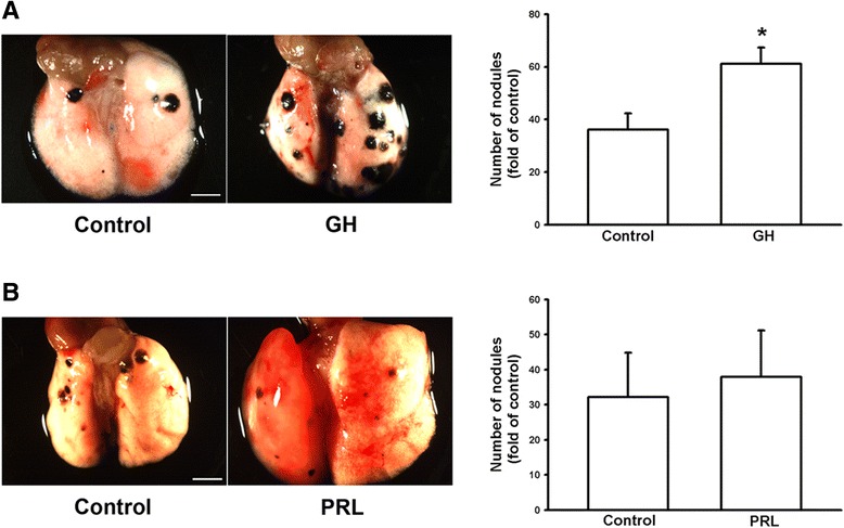
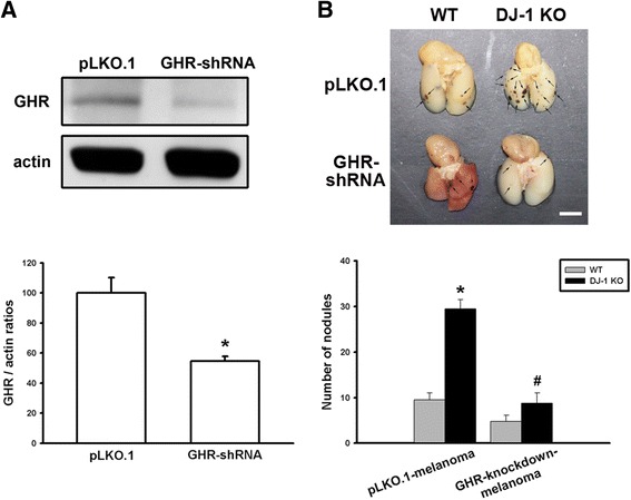
Methods: Firstly, the lung tissues dissected from mice were used for Western blotting and PCR measurement. Secondly, the cultured cells were used for examining effects of GH on B16F10 murine melanoma cells. Thirdly, male C57BL/6 mice were intravenously injected with B16F10 cells and then subcutaneously injected with recombinant GH twice per week for three weeks. Finally, stably transfected pool of B16F10 cells with knockdown of growth hormone receptor (GHR) was used to be injected into mice.

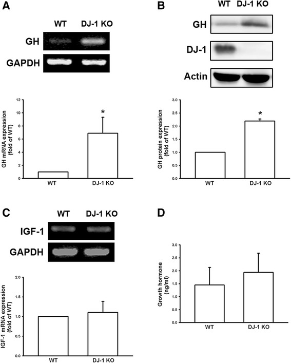
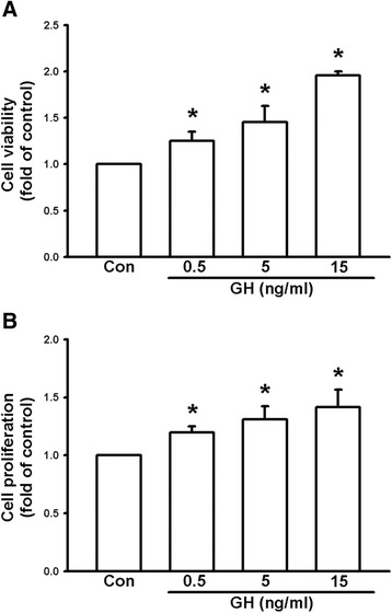
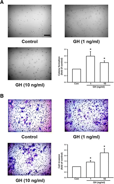
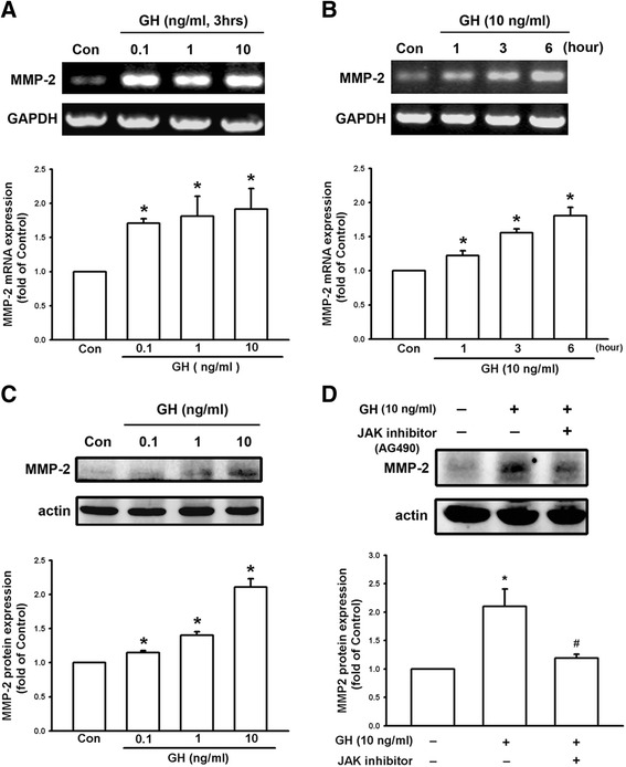
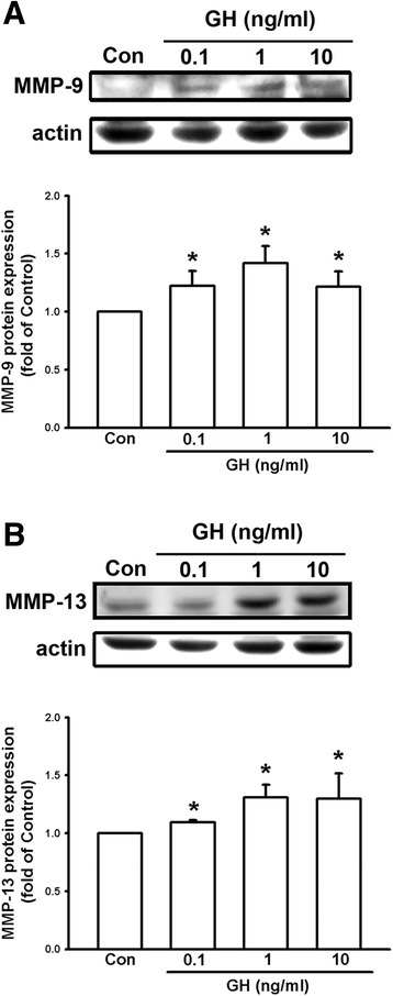
Results: We found that expression of GH was elevated in the lungs of DJ-1 knockout (KO) mice. We also examined the effects of GH on the growth of cultured melanoma cells. The results showed that GH increased proliferation, colony formation, and invasive capacity of B16F10 cells. In addition, GH also increased the expression of matrix metalloproteinases (MMPs) in B16F10 cells. Administration of GH in vivo enhanced lung nodule formation in C57/B6 mice. Increased lung nodule formation in DJ-1 KO mice following intravenous injection of melanoma cells was inhibited by GHR knockdown in B16F10 cells.

Conclusions: These results indicate that up-regulation of GH in the lungs of DJ-1 KO mice may enhance the malignancy of B16F10 cells and nodule formation in pulmonary metastasis of melanoma.

Keywords: Growth hormone; Knockout mice; Lung metastasis; Melanoma; Oncogenesis.


Figures

Fig. 1
Fig. 1
Increase of growth hormone expression in the lungs of DJ-1 KO mice. Lung tissues were isolated from WT and DJ-1 KO mice and used for semi-quantitative PCR (a, upper panel); real-time quantitative PCR (a, lower panel); and Western blotting (b). Note that there was an increase in GH mRNA and protein expression levels in pulmonary tissue of DJ-1 KO mice. c Insulin-like growth factor 1 (IGF-1) mRNA expression in pulmonary tissue and (d) serum levels of GH were not significantly different between WT and DJ-1 KO mice. Data are presented as mean ± SEM (n = 5 for each group); *, P < 0.05 compared to WT. WT: wild type; KO: knockout; GH: growth hormone
Fig. 2
Fig. 2
Growth hormone enhances survival and proliferation of B16F10 melanoma cells. a Cell viability measured using the MTT assay. Note that treatment of GH (0.5, 5, 15 ng/ml) increased cell viability of B16F10 cells in a concentration-dependent manner. b Cell proliferation evaluated using BrdU uptake analysis. Note that treatment of GH (0.5, 5, 15 ng/ml) enhanced cell proliferation in a concentration-dependent manner. Data are presented as mean ± SEM (n = 4 for each group); *, P < 0.05 compared to the control; BrdU, bromodeoxyuridine
Fig. 3
Fig. 3
Growth hormone enhances colony formation and invasive capacity of B16F10 melanoma cells. a Colony formation of B16F10 cells in soft agar with and without GH. Note that GH increased B16F10 cell colony formation in a concentration-dependent manner and colonies were photographed and counted. b B16F10 cells were seeded into a transwell with 8-μm pore polycarbonate filters and matrix gel. Cells penetrated the pores to the lower surface of filters and were stained with crystal violet and counted. The results showed that GH increased the invasive capacity of B16F10 cells in a concentration-dependent manner. Data are presented as mean ± SEM (n = 4 for each group); *, P < 0.05 compared to the control. Scale bar = 0.2 mm
Fig. 4
Fig. 4
Growth hormone increases the expression of MMP-2 in B16F10 cells through JAK signaling. a Expression of MMP2 mRNA was increased in B16F10 cells following 3 h of treatment with GH in a concentration-dependent manner. b MMP-2 expression was increased by 10 ng/ml in a time-dependent manner. c B16F10 cells treated with GH (0.1, 1, 10 ng/ml) for 6 h increased expression of MMP-2 protein in a concentration-dependent manner. d GH-induced increase of MMP-2 was inhibited by JAK inhibitor (AG490). Data are presented as mean ± SEM (n = 3 for each group); *, P < 0.05 compared to the control (Con); #, P < 0.05 compared to GH treatment alone; JAK, Janus kinase
Fig. 5
Fig. 5
Growth hormone enhances the expression of MMP-9 and MMP-13 in B16F10 cells. The protein levels of MMP-9 (a) and MMP-13 (b) in B16F10 cells were up-regulated in a concentration-dependent manner following GH treatment (0.1, 1, 10 ng/ml) for 6 h. Data are presented as mean ± SEM (n = 3 for each group); *, P < 0.05 compared to the control (Con)
Fig. 6
Fig. 6
Growth hormone enhances lung nodule formation in C57/B6 mice. B16F10 cells (6 × 104 cells) were injected into the femoral vein of C57/B6 mice. The mice were subcutaneously injected with GH (a) or prolactin (b) (5 mg/kg, twice/week, respectively). The lung nodules were photographed and counted three weeks later. Results showed an increase in the number of lung nodules in C57/B6 mice following GH administration and there was on difference after prolactin administration. Data are presented as mean ± SEM (n = 11–12 for each group in a; n = 5 for each group in b); *, P < 0.05 compared to the control. Scale bar = 3 mm
Fig. 7
Fig. 7
Elevated lung nodule formation in DJ-1 KO mice is suppressed following injection of GHR-knockdown melanoma cells. a Western blots showed the knockdown of GHR in B16F10 melanoma cells. Upper panel: representative blots of GHR and actin in cells stably transfected with empty plasmids (pLKO.1) or plasmids encoding GHR shRNA. Lower panel: bar chart showing statistical results of the Western blot. b B16F10 cells (6 × 104) transfected with pLKO.1 plasmids (pLKO.1-melanoma) or GHR shRNA plasmids (GHR-knockdown-melanoma) were intravenously injected into mice. Three weeks later, mice were sacrificed. Gross images (upper) showed the melanoma nodules (arrows in the panel) and bar chart (lower) showed the summarized results of lung nodule numbers in WT and DJ-1 KO mice. Note that melanoma nodule formation was enhanced in DJ-1 KO mice following injection of pLKO.1-melanoma cells, but was suppressed following injection of GHR-knockdown melanoma cells. Data are presented as mean ± SEM (n = 5 for each group); *, P < 0.05 compared to the control, WT mice with pLKO.1-melanoma; #, P < 0.05 compared to DJ-1 KO mice with pLKO.1-melanoma. Scale bars = 0.5 mm

References

    1. Le Naour F, Misek DE, Krause MC, et al. Proteomics-based identification of RS/DJ-1 as a novel circulating tumor antigen in breast cancer. Clin Cancer Res. 2001;7(11):3328–35. - PubMed
    1. Bonifati V, Oostra BA, Heutink P. Linking DJ-1 to neurodegeneration offers novel insights for understanding the pathogenesis of Parkinson's disease. J Mol Med (Berl) 2004;82(3):163–74. doi: 10.1007/s00109-003-0512-1. - DOI - PubMed
    1. Wood-Kaczmar A, Gandhi S, Wood NW. Understanding the molecular causes of Parkinson's disease. Trends Mol Med. 2006;12(11):521–8. doi: 10.1016/j.molmed.2006.09.007. - DOI - PubMed
    1. Nagakubo D, Taira T, Kitaura H, et al. DJ-1, a novel oncogene which transforms mouse NIH3T3 cells in cooperation with ras. Biochem Biophys Res Commun. 1997;231(2):509–13. doi: 10.1006/bbrc.1997.6132. - DOI - PubMed
    1. Pardo M, Garcia A, Thomas B, et al. The characterization of the invasion phenotype of uveal melanoma tumour cells shows the presence of MUC18 and HMG-1 metastasis markers and leads to the identification of DJ-1 as a potential serum biomarker. Int J Cancer. 2006;119(5):1014–22. doi: 10.1002/ijc.21942. - DOI - PubMed

Publication types