. 2016 Oct 25:10:482.
doi: 10.3389/fnins.2016.00482. eCollection 2016.

Analog Memristive Synapse in Spiking Networks Implementing Unsupervised Learning

Affiliations

Analog Memristive Synapse in Spiking Networks Implementing Unsupervised Learning

Erika Covi et al. Front Neurosci. .

Abstract

Emerging brain-inspired architectures call for devices that can emulate the functionality of biological synapses in order to implement new efficient computational schemes able to solve ill-posed problems. Various devices and solutions are still under investigation and, in this respect, a challenge is opened to the researchers in the field. Indeed, the optimal candidate is a device able to reproduce the complete functionality of a synapse, i.e., the typical synaptic process underlying learning in biological systems (activity-dependent synaptic plasticity). This implies a device able to change its resistance (synaptic strength, or weight) upon proper electrical stimuli (synaptic activity) and showing several stable resistive states throughout its dynamic range (analog behavior). Moreover, it should be able to perform spike timing dependent plasticity (STDP), an associative homosynaptic plasticity learning rule based on the delay time between the two firing neurons the synapse is connected to. This rule is a fundamental learning protocol in state-of-art networks, because it allows unsupervised learning. Notwithstanding this fact, STDP-based unsupervised learning has been proposed several times mainly for binary synapses rather than multilevel synapses composed of many binary memristors. This paper proposes an HfO2-based analog memristor as a synaptic element which performs STDP within a small spiking neuromorphic network operating unsupervised learning for character recognition. The trained network is able to recognize five characters even in case incomplete or noisy images are displayed and it is robust to a device-to-device variability of up to ±30%.

Keywords: HfO2; artificial synapse; memristor; resistive switching; spike time dependent plasticity; spiking neuromorphic network; synaptic plasticity; unsupervised learning.


Figures

Figure 1
Figure 1
DC characterization of the device. Transition from LRS to HRS is obtained with a DC sweep from 0 V to 1 V, transition from HRS to LRS is obtained with a DC sweep from 0 V to −0.7 V.
Figure 2
Figure 2
(A) Potentiation (orange) and depression (green) cycles using ramped trains of spikes. Spike time width 100 μs, 5 repetitions. Potentiation: ramps from −0.1 V to −0.65 V. Depression: ramps from 0.1 V to 1.2 V. Upper graph: device resistance after spikes; lower graph: spike amplitude. (B) Potentiation (orange) and depression (green) cycles using trains of 300 identical spikes. Potentiation: spike amplitude −0.55 V, time width 25 μs. Depression: spike amplitude 0.7 V, time width 20 μs. Upper graph: device resistance after spikes; lower graph: spike amplitude.
Figure 3
Figure 3
(A) Setup for Spike Time Dependent Plasticity and waveforms used as pre-spike (left) and post-spike (right) in STDP experiments. (B) Overlapping of pre-spike and post-spike to obtain a potentiation (left) and a depression (right). (C) Voltage-to-delay time mapping. Resulting voltage across the artificial synapse as a function of Δt.
Figure 4
Figure 4
(A) Potentiation and (B) depression dynamics with 250 identical spikes. Different voltage amplitudes and delay times are explored. The values of both voltage amplitude and Δt are written nearby each curve. Insets: detail of the first 12 spikes. (C,D) Spike Time Dependent Plasticity learning curve for different number of pre- post-spikes pair repetitions (Δt > 0 and Δt < 0). R0 is the initial HRS (C) and LRS (D).
Figure 5
Figure 5
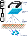
Proposed fully connected SNN. 25 pre-neurons are connected to 5 post-neurons through a layer of 125 artificial synapses. Each pixel of the images shown to the network are associated with a pre-neuron. Inset: images showed to the SNN during the training phase.
Figure 6
Figure 6
(A) Training session: spiking diagram of one epoch. The training character is shown at 0 s and the duration of an epoch is about 2.15 ms. (B) Image shown to the network (top left panel), synaptic weights after 200 epochs (top right panel), and detailed synaptic weight evolution during training session of character A (bottom panel). Black lines represent the synapses which are being depressed during the session and orange lines the ones potentiated. (C) Example of synaptic weight changes during a learning session. Each 5 × 5 matrix represents the group of 25 synapses contributing to the firing of neurons α to ϵ. Color bar on the right indicates the conductance range of the synapses. Increasing the number of epochs (from top to bottom), the SNN specializes each post-neuron to recognize a different character. (D) Distribution of the synaptic weights during the training session.
Figure 7
Figure 7
Variability in the behavior of 3 different devices for (A) potentiation and (B) depression when stimulated by trains of 300 identical spikes. Potentiation: voltage amplitude −0.55 V, time width 25 μs. Depression: voltage amplitude 0.75 V, time width 20 μs. Symbols indicate the mean value of 10 repetitions of the same train of spikes and the shaded area indicates the standard deviation.
Figure 8
Figure 8
Simulation of the training session including ±10% (A,B) and ±30% (C,D) of variability in synaptic behavior. (A,C) Detailed synaptic weight evolution during training session of all characters. Black lines represent the synapses which are being depressed during the session and orange lines the ones potentiated. Green lines indicate that the neuron did not fire in the corresponding epoch. (B,D) Distribution of the synaptic weights during the training session. (E) Recognition rate as a function of the number of epochs in the learning session. The blue circles represent the average recognition rate from 100 simulations where device-to-device variability is not taken into account. The red dotted line and the green dashed one indicate the best and worst results obtained in the simulations, respectively, whereas the other results lie in the shaded gray area. (F) Average recognition rate of 100 simulations as a function of the number of epochs in the learning session with device-to-device variability of 0% (blue circles), ±10% (red squares), and ±30% (green triangles). Error bars show the standard deviation of the results.

References

    1. Ambrogio S., Balatti S., Milo V., Carboni R., Wang Z. Q., Calderoni A., et al. (2016a). Neuromorphic learning and recognition with one-transistor-one-resistor synapses and bistable metal oxide RRAM. IEEE Trans. Electr. Dev. 63, 1508–1515. 10.1109/TED.2016.2526647 - DOI
    1. Ambrogio S., Balatti S., Nardi F., Facchinetti S., Ielmini D. (2013). Spike-timing dependent plasticity in a transistor-selected resistive switching memory. Nanotechnology 24:384012. 10.1088/0957-4484/24/38/384012 - DOI - PubMed
    1. Ambrogio S., Ciocchini N., Laudato M., Milo V., Pirovano A., Fantini P., et al. . (2016b). Unsupervised learning by spike timing dependent plasticity in phase change memory (PCM) synapses. Front. Neurosci. 10:56. 10.3389/fnins.2016.00056 - DOI - PMC - PubMed
    1. Berdan R., Serb A., Khiat A., Regoutz A., Papavassiliou C., Prodromakis T. (2015). A μ-controller-based system for interfacing selector-less RRAM crossbar arrays. IEEE Trans. Electr. Dev. 62, 2190–2196. 10.1109/TED.2015.2433676 - DOI
    1. Bi G.-Q., Poo M.-M. (1998). Synaptic modifications in cultured hippocampal neurons: dependence on spike timing, synaptic strength, and postsynaptic cell type. J. Neurosci. 18, 10464–10472. - PMC - PubMed