. 2016 Oct 25:7:389.
doi: 10.3389/fphar.2016.00389. eCollection 2016.

Fluoxetine Prevents Aβ1-42-Induced Toxicity via a Paracrine Signaling Mediated by Transforming-Growth-Factor-β1

Affiliations

Fluoxetine Prevents Aβ1-42-Induced Toxicity via a Paracrine Signaling Mediated by Transforming-Growth-Factor-β1

Filippo Caraci et al. Front Pharmacol. .

Abstract

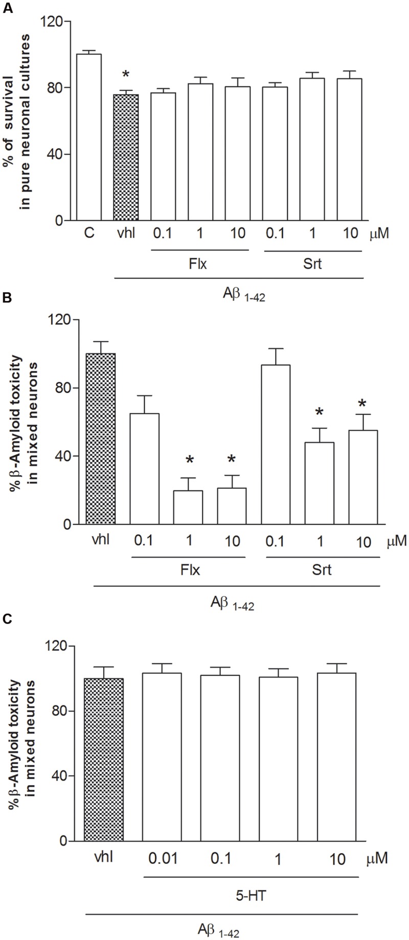
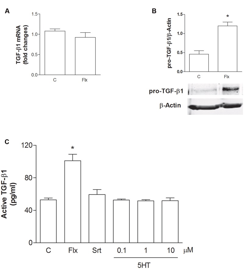
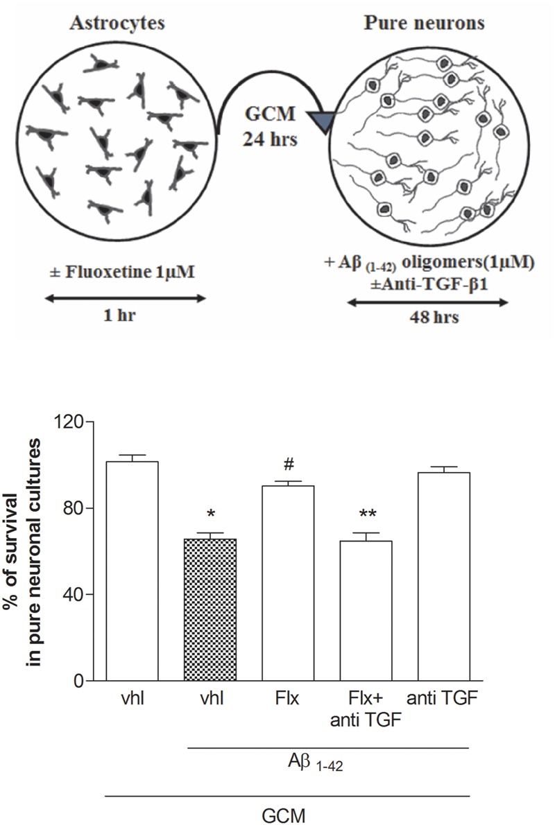
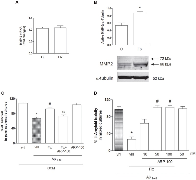
Selective reuptake inhibitors (SSRIs), such as fluoxetine and sertraline, increase circulating Transforming-Growth-Factor-β1 (TGF-β1) levels in depressed patients, and are currently studied for their neuroprotective properties in Alzheimer's disease. TGF-β1 is an anti-inflammatory cytokine that exerts neuroprotective effects against β-amyloid (Aβ)-induced neurodegeneration. In the present work, the SSRI, fluoxetine, was tested for the ability to protect cortical neurons against 1 μM oligomeric Aβ1-42-induced toxicity. At therapeutic concentrations (100 nM-1 μM), fluoxetine significantly prevented Aβ-induced toxicity in mixed glia-neuronal cultures, but not in pure neuronal cultures. Though to a lesser extent, also sertraline was neuroprotective in mixed cultures, whereas serotonin (10 nM-10 μM) did not mimick fluoxetine effects. Glia-conditioned medium collected from astrocytes challenged with fluoxetine protected pure cortical neurons against Aβ toxicity. The effect was lost in the presence of a neutralizing antibody against TGF-β1 in the conditioned medium, or when the specific inhibitor of type-1 TGF-β1 receptor, SB431542, was added to pure neuronal cultures. Accordingly, a 24 h treatment of cortical astrocytes with fluoxetine promoted the release of active TGF-β1 in the culture media through the conversion of latent TGF-β1 to mature TGF-β1. Unlike fluoxetine, both serotonin and sertraline did not stimulate the astrocyte release of active TGF-β1. We conclude that fluoxetine is neuroprotective against Aβ toxicity via a paracrine signaling mediated by TGF-β1, which does not result from a simplistic SERT blockade.

Keywords: Alzheimer’s disease; TGF-β1; antidepressants; cortical neurons; depression; fluoxetine; neuroprotection; β-amyloid.


Figures

FIGURE 1
FIGURE 1
Fluoxetine and sertraline prevent Aβ1-42-induced toxicity only in the presence of glial cells. Antidepressant drugs were applied at increasing concentrations (100 nM–10 μM) both to pure (A) and mixed cortical neurons (B) in co-treatment with Aβ1-42 oligomers (1 μM) for 48 h. (A) Aβ toxicity in pure neuronal cultures is expressed as percentage of neuronal survival (quantified by MTT assay). Values in pure cortical neurons are means ± SEM of six to nine determinations p < 0.05 vs. control (C) (One-way ANOVA + Bonferroni’s test). (B) Aβ toxicity in mixed neuronal cultures was assessed by cell counting after trypan blue staining. Cell count was performed in three random microscopic fields/well. Values in mixed cortical neurons are expressed as percentage of Aβ1-42 toxicity (vhl) and are means ± SEM of twelve determinations p < 0.05 vs. Aβ alone (One-way ANOVA + Bonferroni’s test). (C) Serotonin (10 nM–10 μM) was co-applied with Aβ1-42 oligomers (1 μM) to mixed neuronal cultures for 48 h. The effects of serotonin against Aβ toxicity in mixed neuronal cultures were assessed by cell counting after trypan blue staining.
FIGURE 2
FIGURE 2
Fluoxetine increases the expression of pro-TGF-β1 and the release of active TGF-β1 from cortical astrocytes. (A) TGF-β1 mRNA levels obtained by Real-time RT-PCR in cultured astrocytes transiently exposed to fluoxetine (Flx; 1 μM) for 6 h are shown. Values were normalized by endogenous GAPDH mRNA levels and are represented as means + SEM for n = 4 for two independent experiments. (B) Representative immunoblots of pro-TGF-β1 (about 55 kDa) in total protein extracts from rat cortical astrocytes exposed to fluoxetine (Flx; 1 μM) for 24 h. Bars refer to the means ± SEM of the densitometric values of pro-TGF-β1 bands normalized against β-actin. Each experiment was repeated four times; p < 0.05 vs. control by Student’s t-test. (C) Levels of active TGF-β1 in the medium of cultured astrocytes exposed for 24 h to fluoxetine (Flx; 1 μM), sertraline (Srt; 1 μM) or to increasing concentrations of serotonin (100 nM–10 μM) are shown. Values are means ± SEM of nine determinations; p < 0.05 (Student’s t-test) versus untreated control astrocytes.
FIGURE 3
FIGURE 3
Fluoxetine prevents Aβ toxicity via a paracrine mechanism mediated by TGF-β1. Pure cultures of rat cortical neurons were exposed to glial conditioned medium (GCM) collected from cortical astrocytes 24 h after a transient (1 hr) exposure to1 μM fluoxetine or vehicle. Neurons were then treated with Aβ1-42 oligomers (1 μM) for 48 h in the presence or absence of anti-TGF-β1 antibody. A schematic drawing of this experimental protocol is shown in the upper panel. Anti-TGF-β1 was added at a concentration of 2 μg/ml just before transferring of GCM into pure neuronal cultures. Aβ toxicity in pure neuronal cultures was assessed by MTT assay and is expressed as percentage of neuronal survival. Values are means ± SEM of 12–15 determinations p < 0.05 vs. control (GCM, vhl); #p < 0.05 vs. Aβ1-42 alone (GCM, Aβ1-42, vhl); ∗∗p < 0.05 vs. Aβ1-42 and fluoxetine (GCM, Aβ1-42, Flx) (One-way ANOVA + Bonferroni’s test).
FIGURE 4
FIGURE 4
The neuroprotective effects of fluoxetine against Aβ1-42-induced toxicity are mediated by TGF-β1. Mixed cortical cultures were challenged with Aβ1-42 oligomers (1 μM) for 48 h in the absence or presence of fluoxetine (1 μM) applied alone or combined with the selective inhibitor of Smad-dependent TGF-β1 signaling, SB431542 (SB; 10 μM) or with a neutralizing antibody specific for TGF-β1 (anti-TGF-β1) applied at a concentration of 2 μg/ml. Aβ toxicity in mixed neuronal cultures was assessed by cell counting after trypan blue staining. Cell counts was performed in three random microscopic fields/well. Values are expressed as percentage of Aβ1-42 toxicity and are means ± SEM of nine determinations p < 0.05 vs. Aβ alone (Aβ1-42, vhl) and #Aβ + fluoxetine (Aβ1-42, Flx, vhl) (One-way ANOVA + Bonferroni’s test).
FIGURE 5
FIGURE 5
Role of metalloproteinase-2 (MMP-2) in the neuroprotective effects of fluoxetine against Aβ toxicity. (A) MMP-2 mRNA levels in cultured astrocytes transiently exposed to fluoxetine (Flx) for 6 h are shown. Values were normalized by endogenous GAPDH mRNA levels and are represented as means +SEM for n = 4 for two independent experiments. (B) Representative immunoblots of active MMP-2 in total protein extracts from rat cortical astrocytes exposed to fluoxetine (Flx, 1 μM) for 24 h. Arrows indicate pro-MMP2 (72 kDa) and the active form at about 66 kDa. Bars refer to the means ± SEM of the densitometric values of MMP-2 bands normalized against α-tubulin. Each experiment was repeated four times p < 0.05 vs. control by Student’s t-test. (C) Pure cultures of rat cortical neurons were exposed to GCM collected from cortical astrocytes 24 h after a transient (1 hr) exposure to 1 μM fluoxetine with or without ARP-100 (50 nM), before treatment with Aβ1-42 oligomers (1 μM) for 48 h. Values are means ± SEM of 12–15 determinations p < 0.05 vs. control values obtained with GCM alone (GCM, vhl), #p < 0.05 vs. GCM+Aβ and ∗∗GCM+Aβ+FLX (One-way ANOVA + Bonferroni’s test). (D) Selective inhibition of MMP-2 prevents the neuroprotective effects of fluoxetine against Aβ toxicity. Mixed cortical cultures were challenged with Aβ1-42 oligomers (1 μM) for 48 h in the absence or presence of fluoxetine (1 μM) applied alone or combined with ARP100 (10–100 nM). Values are expressed as percentage of Aβ1-42 toxicity and are means ± SEM of twelve determinations p < 0.05 vs. Aβ alone and #Aβ+fluoxetine (Aβ1-42, Flx, vhl) (One-way ANOVA +Bonferroni’s test).

References

    1. Alboni S., Gibellini L., Montanari C., Benatti C., Benatti S., Tascedda F., et al. (2013). N-acetyl-cysteinepreventstoxicoxidativeeffectsinduced by IFN-α in human neurons. Int. J. Neuropsychopharmacol. 16 1849–1865. 10.1017/S1461145713000266 - DOI - PubMed
    1. Allaman I., Fiumelli H., Magistretti P. J., Martin J. L. (2011). Fluoxetine regulates the expression of neurotrophic/growth factors and glucose metabolism in astrocytes. Psychopharmacology (Berl) 216 75–84. 10.1007/s00213-011-2190-y - DOI - PubMed
    1. Annes J. P., Munger J. S., Rifkin D. B. (2003). Making sense of latent TGF beta activation. J. Cell Sci. 116 217–224. 10.1242/jcs.00229 - DOI - PubMed
    1. Ballard C., Corbett A., Chitramohan R., Aarsland D. (2009). Management of agitation and aggression associated with Alzheimer’s disease: controversies and possible solutions. Curr. Opin. Psychiatry 22 532–540. 10.1097/YCO.0b013e32833111f9 - DOI - PubMed
    1. Benatti C., Alboni S., Montanari C., Caggia F., Tascedda F., Brunello N., et al. (2011). Central effects of a local inflammation in three commonly used mouse strains with a different anxious phenotype. Behav. Brain Res. 224 23–34. 10.1016/j.bbr.2011.05.011 - DOI - PubMed

LinkOut - more resources