. 2016 Oct;4(19):372.
doi: 10.21037/atm.2016.09.32.

Anatomy essentials for laparoscopic inguinal hernia repair

Affiliations

Anatomy essentials for laparoscopic inguinal hernia repair

Xue-Fei Yang et al. Ann Transl Med. 2016 Oct.

Abstract

Laparoscopic inguinal hernia repair is performed more and more nowadays. The anatomy of these procedures is totally different from traditional open procedures because they are performed from different direction and in different space. The important anatomy essentials for laparoscopic inguinal hernia repair will be discussed in this article.

Keywords: Inguinal hernia; anatomy; laparoscopic repair.


Conflict of interest statement

The authors have no conflicts of interest to declare.

Figures

Figure 1
Figure 1
Representation of the myopectineal orifice.
Figure 2
Figure 2
Schematic of the principle of adult laparoscopic inguinal hernia repair.
Figure 3
Figure 3
Lower anterior abdominal wall, transverse fascia and spaces (the right lower anterior abdominal wall is taken as an example). The surgical area refers to the main surgical field of a laparoscopic inguinal hernia repair.
Figure 4
Figure 4
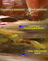
Representation of the superficial and deep transverse fasciae.
Figure 5
Figure 5
Representation of the incised internal spermatic fascia.
Figure 6
Figure 6
The preperitoneal retropubic space and the space of Bogros.
Figure 7
Figure 7
The deep transverse abdominal fascia is tightly attached to the anterior abdominal wall at the site lateral to the inferior epigastric blood vessel.
Figure 8
Figure 8
Bilateral inguinal area under laparoscopy.
Figure 9
Figure 9
Important anatomic landmarks in the extraperitoneal space.
Figure 10
Figure 10
Corona mortis.
Figure 11
Figure 11
Correct access to the space of Bogros.
Figure 12
Figure 12
Lateral femoral cutaneous nerve and genitofemoral nerve.
Figure 13
Figure 13
Representation of the right Cooper’s ligament, the iliopubic tract and the inguinal ligament.

References

    1. Pisanu A, Podda M, Saba A, et al. Meta-analysis and review of prospective randomized trials comparing laparoscopic and Lichtenstein techniques in recurrent inguinal hernia repair. Hernia 2015;19:355-66. 10.1007/s10029-014-1281-1 - DOI - PubMed
    1. Pahwa HS, Kumar A, Agarwal P, et al. Current trends in laparoscopic groin hernia repair: A review. World J Clin Cases 2015;3:789-92. 10.12998/wjcc.v3.i9.789 - DOI - PMC - PubMed

LinkOut - more resources