. 2016 Nov 9:6:36570.
doi: 10.1038/srep36570.

Mitochondrial Ca2+ uptake controls actin cytoskeleton dynamics during cell migration

Affiliations

Mitochondrial Ca2+ uptake controls actin cytoskeleton dynamics during cell migration

Julien Prudent et al. Sci Rep. .

Abstract

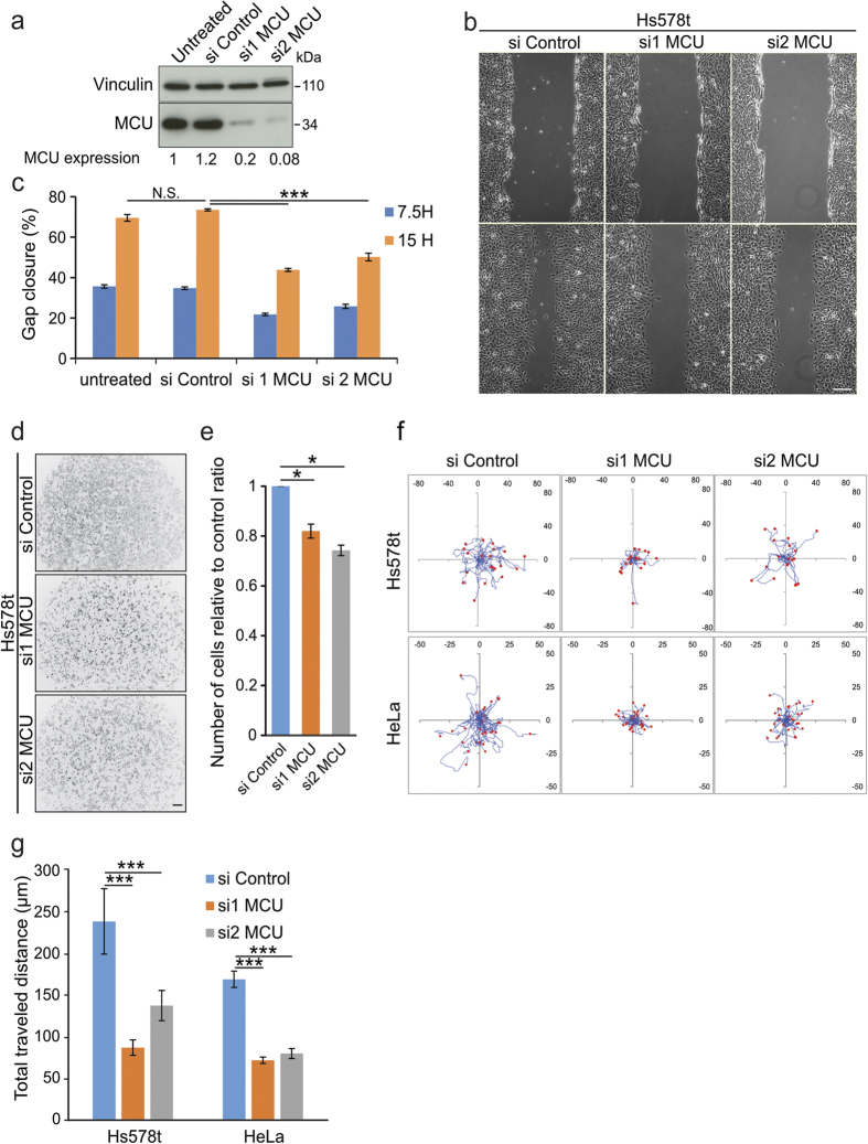
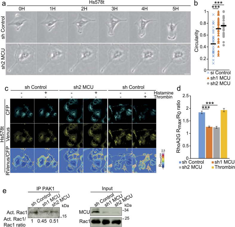
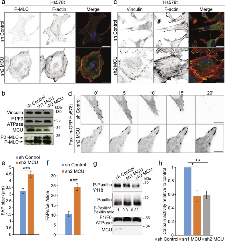
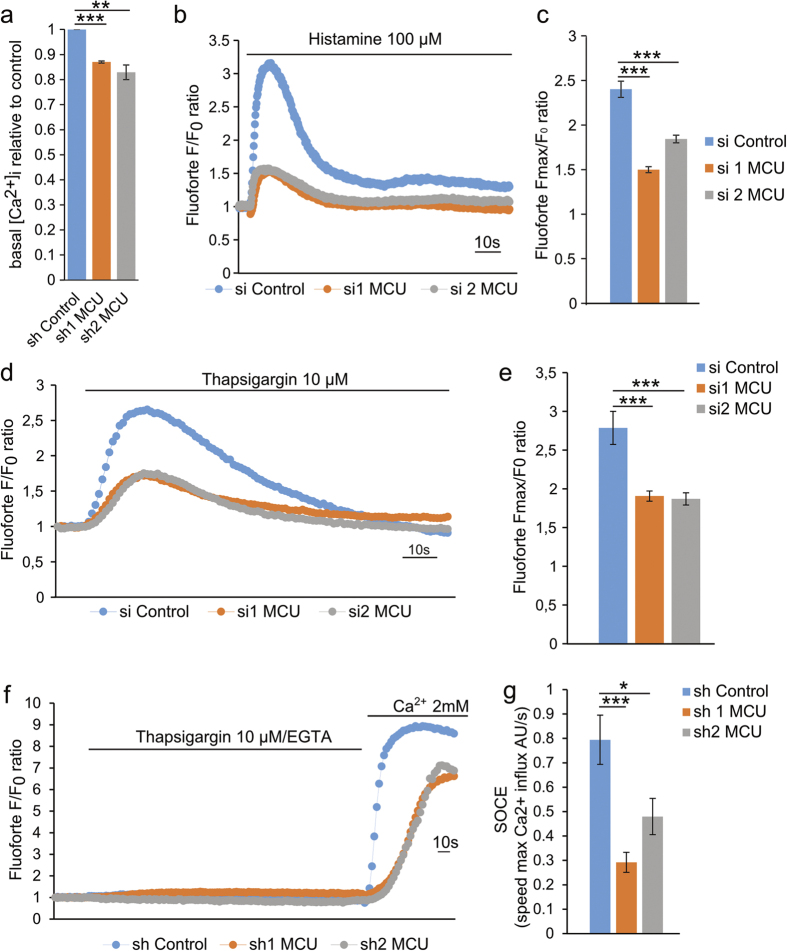
Intracellular Ca2+ signaling regulates cell migration by acting on cytoskeleton architecture, cell directionality and focal adhesions dynamics. In migrating cells, cytosolic Ca2+ pool and Ca2+ pulses are described as key components of these effects. Whereas the role of the mitochondrial calcium homeostasis and the Mitochondria Cacium Uniporter (MCU) in cell migration were recently highlighted in vivo using the zebrafish model, their implication in actin cystokeleton dynamics and cell migration in mammals is not totally characterized. Here, we show that mcu silencing in two human cell lines compromises their migration capacities. This phenotype is characterized by actin cytoskeleton stiffness, a cell polarization loss and an impairment of the focal adhesion proteins dynamics. At the molecular level, these effects appear to be mediated by the reduction of the ER and cytosolic Ca2+ pools, which leads to a decrease in Rho-GTPases, RhoA and Rac1, and Ca2+-dependent Calpain activites, but seem to be independent of intracellular ATP levels. Together, this study highlights the fundamental and evolutionary conserved role of the mitochondrial Ca2+ homeostasis in cytoskeleton dynamics and cell migration.


Figures

Figure 1
Figure 1. MCU knockdown induces cell migration defects.
(a) Immunoblot blot showing the decrease of MCU protein level in Hs578t cells transfected with two siRNAs (si1 and si2) targeting mcu transcript. Scrambled siRNA (si Control) was used as negative control. Vinculin was used as loading control. Quantitative ratios (MCU expression) between MCU and Vinculin were indicated below. (b) Representative images of the effect of mcu-silencing on Hs578t migration performed by wound healing assay. Compared to control cells, mcu-silenced cells present decreased capacities to close the gap. Scale bar: 200 μm. (c) Histogram representing the percentage of gap closure estimated at 7.5 and 15 hours post-wound from (b) (mean ± S.E.M; n = 4 independent experiments). N.S. = not significant; ***P < 0.001. (d) Representative Grayscale images of the effect of mcu-knockdown on Hs578t invasion performed with Boyden chamber assay using Fetal Bovine Serum (FBS) as chemoattractant. Compared to controls, less mcu-silenced cells have crossed the membrane. Scale bar: 200 μm. (e) Histogram representing the number of invading cells relative to control condition measured at 7.5 hours post-seeding from (d) (mean ± S.E.M; n = 3 independent experiments). *P < 0.05. (f) Representative single cell tracking experiments highlighting the cell paths (blue lines) of isolated Hs578t (red dots) or HeLa cells (red dots), silenced or not for mcu; acquisition time 24 hours. Compared to control cells, mcu-silenced cells have shorter migration paths. (g) Histogram deduced from the results displayed in (f) representing the distance of cell migration relative to control (mean ± S.E.M; 30 cells in each experiment; n = 3 independent experiments). ***P < 0.001. See also Supplementary Fig. S1.
Figure 2
Figure 2. Mitochondrial Ca2+ uptake impairment leads to cell polarization defects and decreased small GTPases activities.
(a) Representative transmission images of single cell tracking of Hs578t cells stably expressing shRNA control versus shRNA targeting mcu transcript. Cells expressing the control shRNA have forward-to-rear polarization, which was absent in shMCU cells. Scale bar: 20 μm. (b) Graph representing the quantification of circularity coefficients for sh Control (n = 34 cells), sh1 MCU (n = 32 cells) and sh2 MCU (n = 29 cells) (mean ± S.E.M; three independent experiments). Compared to control cells, shMCU cells present an increased circularity coefficient. ***P < 0.001. (c) Confocal time-lapse images showing the CFP (cyan), Venus (yellow) as well as Venus/CFP ratio (RVenus/CFP) of the RhoA2G biosensor dye following IP3R mobilization using Histamine 100 μM in Hs578t cells. Thrombin was used as positive control for RhoA activation. Ratio was visualized as a heat map using false colors. Images were acquired before and after 1 min treatment with Histamine or Thrombin. Following Histamine treatment, RhoA activation is higher in control cells compared to shMCU cells. Scale bar: 20 μm. (d) Histogram showing the Rmax/R0 ratio following Histamine or Thrombin stimulations in control versus mcu-silenced cells, from (c). (mean ± S.E.M.; 20 cells in each experiment; n = 3 independent experiments). **P < 0.01, ***P < 0.001. (e) Immunoblot showing the deacread Rac1 activation (Act. Rac1) in sh MCU compared to sh control cells. Activated Rac1 in Hs578t cells was purified using PAK1 beads and detected using specific Rac1 antibody. Total Rac1 levels were used to normalize the levels of activated Rac1 (numbers below). See also Supplementary Fig. S2.
Figure 3
Figure 3. Mitochondrial Ca2+ uptake is required for cytoskeleton and focal adhesion protein dynamics.
(a) Representative confocal images showing the accumulation of phospho-myosin light chain (P-MLC) signal in F-actin fibers in sh2 MCU cells compared to controls. P-MLC and F-actin were stained using P-MLC antibody and phalloidine rhodamine probe, respectively. Merge channels between P-MLC (green) and F-actin (red) were presented in the rightmost panels. Scale bar: 20 μm. (b) Immunoblot showing the increase of MLC phosphorylation on Serine19 (P-MLC) and Threonine18 and Serine19 (P2-MLC) positions in mcu-silenced cells. MCU antibody was used to show the efficiency of the shRNAs. F1F0 ATPase antibody was used as loading control. (c) Representative confocal images showing the accumulation of Vinculin in FAPs in sh2 MCU cells compared to control. Vinculin and F-actin were detected using anti-Vinculin antibody and phalloidine rhodamine probe, respectively. Merge channels between Vinculin (green) and F-actin (red) were presented in the rightmost panels. Scale bar: 20 μm. (d) Representative time-lapse confocal images showing the persistence of Paxillin-GFP signal in the FAPs of mcu-knockdown cells compared to control. Scale bar: 5 μm. (e) Histogram representing the increase of FAPs number estimated from Vinculin staining in sh2 MCU cells compared to control. (mean ± S.E.M; 10 cells for each condition; n = 3 independent experiments). ***P < 0.001. (f) Histogram representing the increase of FAP size estimated from Vinculin staining in sh2 MCU cells compared to control cells. (mean ± S.E.M; 10 cells for each condition; n = 3 independent experiments). ***P < 0.001. (g) Immunoblot showing the effect of sh1 and sh2 MCU on phospho-Paxillin Y118 protein levels (P-Paxillin Y118). Quantitative ratios between P-Paxillin Y118 and total Paxillin are indicated below. Mcu knockdown leads to decreased P-Paxillin Y118/total Paxillin ratio. MCU antibody was used to show the efficiency of the shRNAs. F1F0 ATPase antibody was used as loading control. (h) Histogram presenting the decrease of Calpain activity measured in mcu-silenced cells and compared to control cells (mean ± S.E.M; n = 3 independent experiments). *P < 0.05, **P < 0.01. See also Supplementary Fig. S3.
Figure 4
Figure 4. MCU knockdown results in intracellular Ca2+ impairment.
(a) Histogram showing the basal cytosolic Ca2+ levels ([Ca2+]i) of sh Control and sh1, sh2 MCU Hs578t cells using the cytosolic Ca2+ sensitive dye Fluoforte. Compared to control cells, mcu-silenced cells have lower basal cytosolic [Ca2+]i. Fluorescence intensities were reported as ratio to sh Control cells (mean ± S.E.M; n = 3 independent experiments; **P < 0.01, ***P < 0.001. (b) Representative trace of cytosolic Ca2+ rise following histamine (100 μM) stimulation using Fluoforte dye performed in control or mcu-silenced cells. Mcu-silencing led to a decrease in the ER-dependent cytosolic Ca2+ rise. Fluoforte fluorescence intensities were normalized to the baseline (F/F0 ratio). (c) Histogram depicting the maximum Ca2+-increase in the cytosol (Fmax/F0 ratio) in cells following histamine (100 μM) treatment, from (b). (mean ± S.E.M; n = 3 independent experiments). ***P < 0.001. (d) Representative trace of cytosolic Ca2+ rise following ER-Ca2+ leak induced by Thapsigargin (10 μM) stimulation. Mcu-silencing led to a decrease in the ER-dependent cytosolic Ca2+ rise. Fluoforte fluorescence intensities were normalized to the baseline (F/F0 ratio). (e) Histogram depicting the maximum Ca2+-release in the cytosol (Fmax/F0 ratio) in cells following thapsigargin (10 μM) treatment, from (d). (mean ± S.E.M; n = 3 independent experiments). ***P < 0.001. (f) Representative trace of the relative changes in the Fluoforte fluorescence intensities normalized to the baseline (F/F0 ratio). Following Thapsigargin (10 μM)-induced ER Ca2+ depletion, extracellular Ca2+ (2mM) was applied on the cells in order to measure SOCE efficiency corresponding to the maximum speed of Ca2+ influx. Mcu-silencing led to decrease SOCE efficiency compared to control cells. (g) Histogram depicting SOCE efficiency (speed max Ca2+ influx) measured in Hs578t cells stably expressing sh Control, sh1 MCU or sh2 MCU shRNAs, from (f) (mean ± S.E.M; n = 3 independent experiments) *P < 0.05; ***P < 0.001. See also Supplementary Fig. S4.
Figure 5
Figure 5. Pharmacological impairment of intracellular Ca2+ levels phenocopies mcu silencing.
(a) Representative cell paths (blue lines) of isolated Hs578t cells (red dots) treated with DMSO or the cell permeable Ca2+ chelator BAPTA-AM (5 μM); acquisition time 24 hours. BAPTA-AM led to decrease cell migration. (b) Histogram showing the decrease in the cell migration of BAPTA-AM treated cells (mean ± S.E.M; 30 cells in each experiment; n = 3 independent experiments). **P < 0.01. (c) Representative confocal images showing the accumulation of Vinculin in the FAPs in BAPTA-AM treated cells compared to DMSO treated controls. Vinculin and F-actin were stained using anti-Vinculin antibody and phalloidine rhodamine probe, respectively. Scale bar: 20 μm. (d) Histogram representing the Calpain activity measured in BAPTA-AM treated cells reported to control cells (mean ± S.E.M; n = 3 independent experiments). BAPTA-AM treatment led to a decrease Calpain activity. ***P < 0.001. (e) Schematic model showing the role of MCU in the control of Ca2+ homeostasis and cell migration. Mcu-silencing using si or shRNA strategies leads to SOCE impairment with subsequent decrease of the cytosolic Ca2+ levels ([Ca2+]c). This intracellular calcium reduction causes a decrease in the Rho family of GTPases and Calpains activities, which may alter the cytoskeleton dynamics and the migration of the cell. See also Supplementary Fig. S5.

References

    1. Friedl P. & Wolf K. Tumour-cell invasion and migration: diversity and escape mechanisms. Nat. Rev. Cancer 3, 362–374 (2003). - PubMed
    1. Parsons J. T., Horwitz A. R. & Schwartz M. a. Cell adhesion: integrating cytoskeletal dynamics and cellular tension. Nat. Rev. Mol. Cell Biol. 11, 633–643 (2010). - PMC - PubMed
    1. Gardel M. L., Schneider I. C., Aratyn-Schaus Y. & Waterman C. M. Mechanical integration of actin and adhesion dynamics in cell migration. Annu. Rev. Cell Dev. Biol. 26, 315–333 (2010). - PMC - PubMed
    1. Mitra S. K., Hanson D. A. & Schlaepfer D. D. Focal adhesion kinase: in command and control of cell motility. Nat. Rev. Mol. Cell Biol. 6, 56–68 (2005). - PubMed
    1. Giannone G. et al. Lamellipodial Actin Mechanically Links Myosin Activity with Adhesion-Site Formation. Cell 128, 561–575 (2007). - PMC - PubMed

Publication types