

Meta-analysis of brain iron levels of Parkinson's disease patients determined by postmortem and MRI measurements
- PMID: 27827408
- PMCID: PMC5101491
- DOI: 10.1038/srep36669
Meta-analysis of brain iron levels of Parkinson's disease patients determined by postmortem and MRI measurements
Abstract
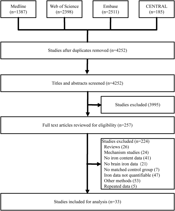
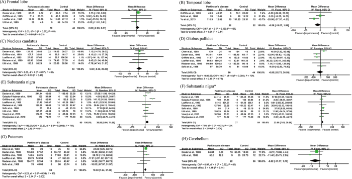
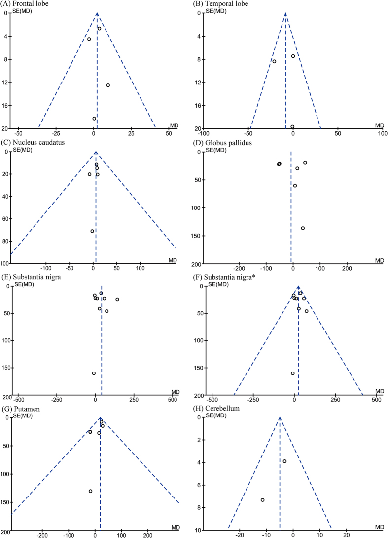
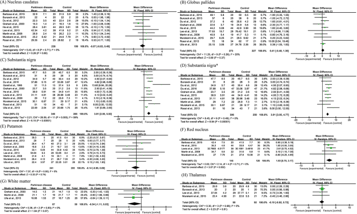
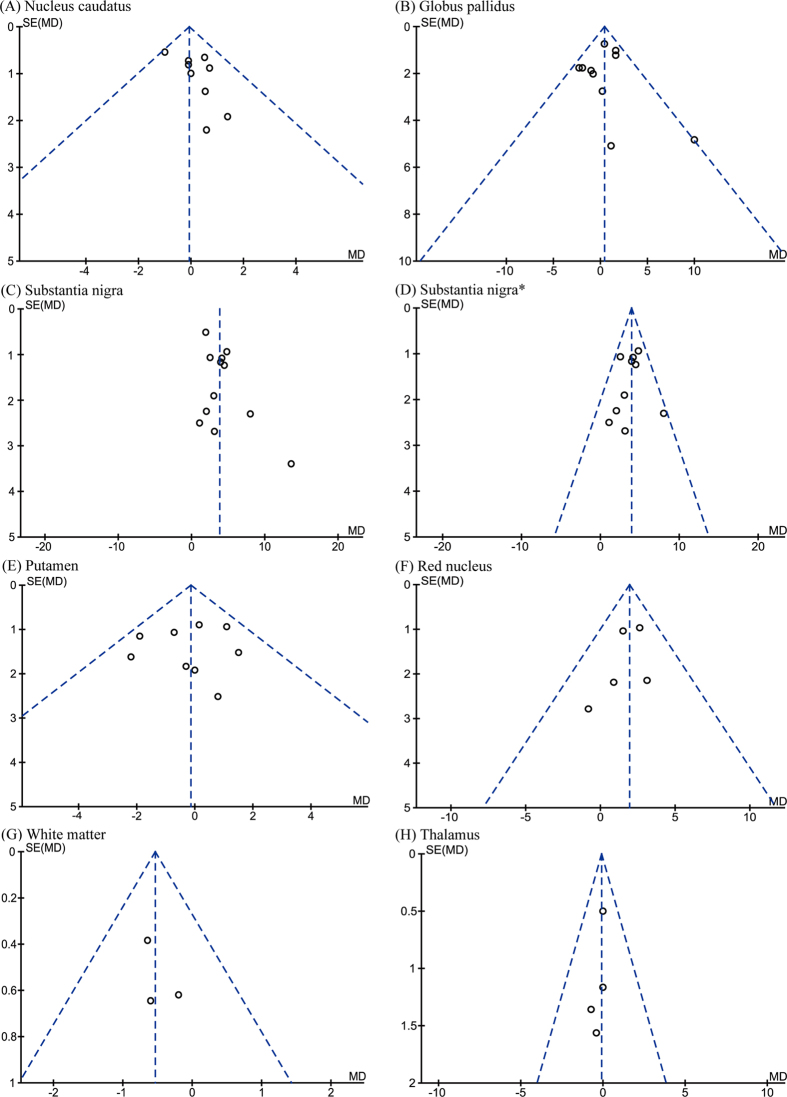
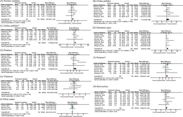
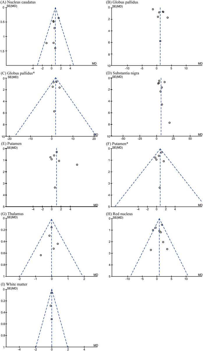
Brain iron levels in patients of Parkinson's disease (PD) are usually measured in postmortem samples or by MRI imaging including R2* and SWI. In this study we performed a meta-analysis to understand PD-associated iron changes in various brain regions, and to evaluate the accuracy of MRI detections comparing with postmortem results. Databases including Medline, Web of Science, CENTRAL and Embase were searched up to 19th November 2015. Ten brain regions were identified for analysis based on data extracted from thirty-three-articles. An increase in iron levels in substantia nigra of PD patients by postmortem, R2* or SWI measurements was observed. The postmortem and SWI measurements also suggested significant iron accumulation in putamen. Increased iron deposition was found in red nucleus as determined by both R2* and SWI, whereas no data were available in postmortem samples. Based on SWI, iron levels were increased significantly in the nucleus caudatus and globus pallidus. Of note, the analysis might be biased towards advanced disease and that the precise stage at which regions become involved could not be ascertained. Our analysis provides an overview of iron deposition in multiple brain regions of PD patients, and a comparison of outcomes from different methods detecting levels of iron.
Figures
References
-
- Dexter D. T. et al.. Alterations in the levels of iron, ferritin and other trace metals in Parkinson’s disease and other neurodegenerative diseases affecting the basal ganglia. Brain 114 (Pt 4), 1953–1975 (1991). - PubMed
-
- Dexter D. T. et al.. Increased nigral iron content and alterations in other metal ions occurring in brain in Parkinson’s disease. J Neurochem 52, 1830–1836 (1989). - PubMed
-
- Riederer P. et al.. Transition metals, ferritin, glutathione, and ascorbic acid in parkinsonian brains. J Neurochem 52, 515–520 (1989). - PubMed
-
- Hardy P. A. et al.. Correlation of R2 with total iron concentration in the brains of rhesus monkeys. J Magn Reson Imaging 21, 118–127 (2005). - PubMed
Publication types
MeSH terms
Substances
LinkOut - more resources
Full Text Sources
Other Literature Sources
Medical
