. 2016 Nov 9:6:36740.
doi: 10.1038/srep36740.

Lactate is a potent inhibitor of the capsaicin receptor TRPV1

Affiliations

Lactate is a potent inhibitor of the capsaicin receptor TRPV1

Jeanne de la Roche et al. Sci Rep. .

Abstract

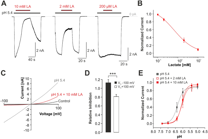
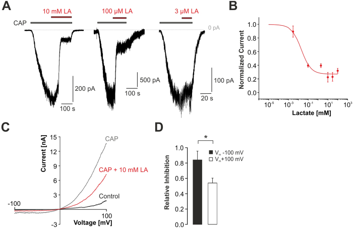
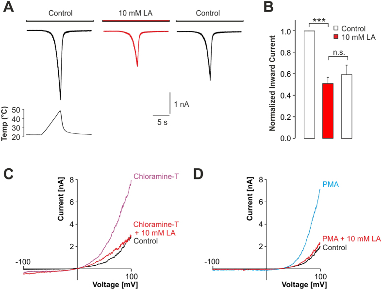
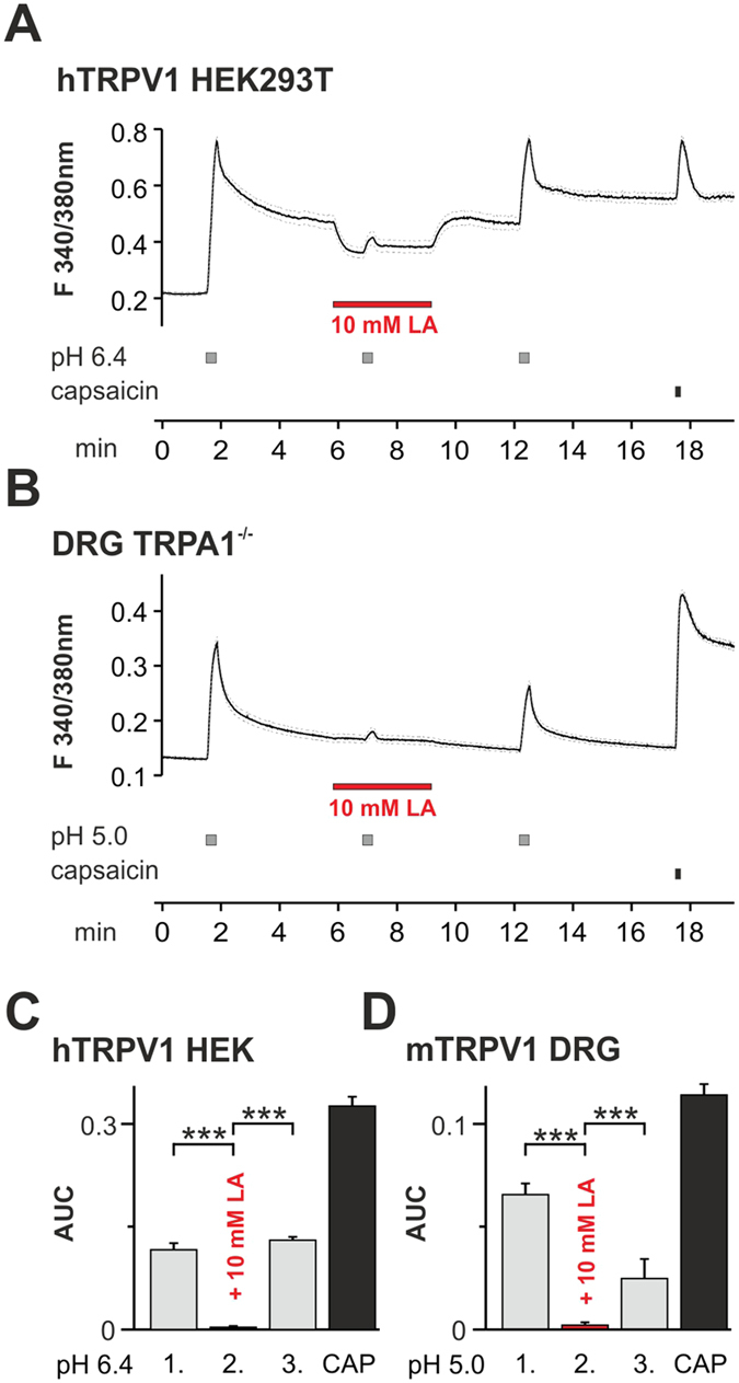
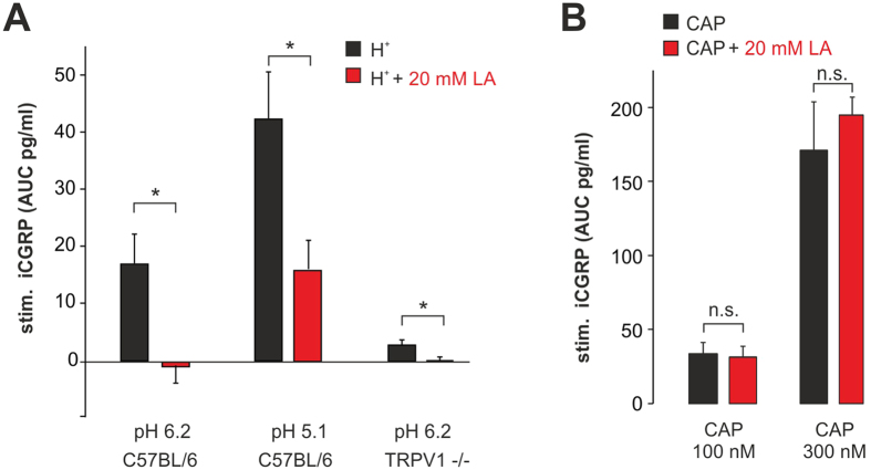
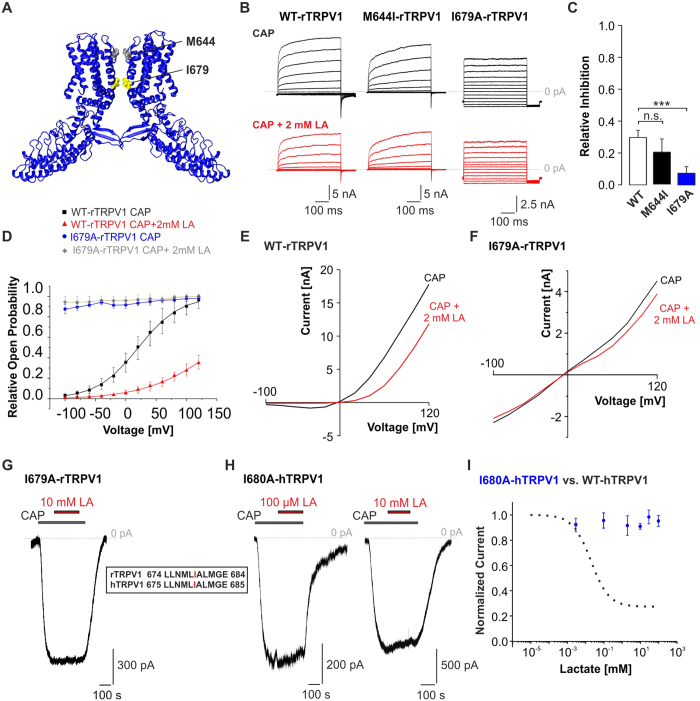
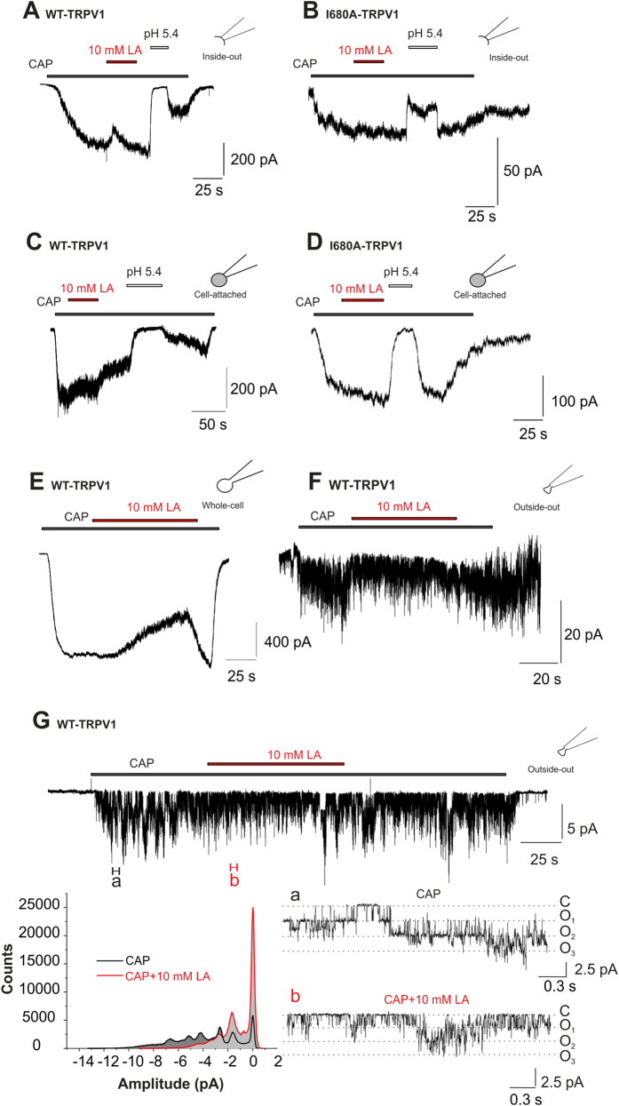
Tissue ischemia results in an accumulation of lactate and local or systemic lactic acidosis. In nociceptive sensory neurons, lactate was reported to sensitize or activate the transient receptor potential ion channel TRPA1 and acid-sensing ion channels (ASICs). However, it is unclear how lactate modulates the TRPV1 regarded as the main sensor for acidosis in sensory neurons. In this study we investigated the effects of lactate (LA) on recombinant and native TRPV1 channels and on TRPV1-mediated release of neuropeptides from mouse nerves. TRPV1-mediated membrane currents evoked by protons, capsaicin or heat are inhibited by LA at concentrations ranging from 3 μM to 100 mM. LA inhibits TRPV1-mediated proton-induced Ca2+-influx in dorsal root ganglion neurons as well as proton-evoked neuropeptide release from mouse nerves. Inhibition of TRPV1 by LA is significantly stronger on inward currents as compared to outward currents since LA affects channel gating, shifting the activation curve towards more positive potentials. The mutation I680A in the pore lower gate displays no LA inhibition. Cell-attached as well as excised inside- and outside-out patches suggest an interaction through an extracellular binding site. In conclusion, our data demonstrate that lactate at physiologically relevant concentrations is a potent endogenous inhibitor of TRPV1.


Figures

Figure 1
Figure 1. Inhibition of proton-evoked TRPV1 currents by LA.
(A) Representative recordings demonstrating inhibition of a pH 5.4-induced inward current by LA at different concentrations. Cells were held at −60 mV and the co-application of LA and pH 5.4 was started once the pH 5.4-induced inward current had reached a steady-state. (B) Concentration-dependency of LA-induced inhibition of proton-evoked inward currents. Current amplitudes were normalized to those before LA application. The solid line represents a fit with the Hill equation. (C) Membrane currents of TRPV1 monitored during 500-ms long voltage ramps from −100 mV to +100 mV in presence of control solution, pH 5.4 or pH 5.4 + 10 mM LA. (D) Mean relative inhibition of pH 5.4-induced inward currents at −100 mV and outward currents at +100 mV by 10 mM LA. The average relative inhibition = 1 − I/I0 for each concentration was determined by normalization; where I = current in presence of LA, I0 = current in absence of LA. Inward currents were significantly stronger inhibited as compared to outward currents. (E) Concentration-dependent activation of TRPV1 by protons applied alone or in combination with 2 or 10 mM LA. Peak current amplitudes were normalized to the largest current amplitude in the respective cells and plotted against the corresponding pH-values. The solid lines represent fits with the Hill equation. Data are presented as mean ± S.E.M. Statistical differences are indicated by ***p < 0.001.
Figure 2
Figure 2. Inhibition of capsaicin-evoked TRPV1 currents by LA.
(A) Representative recordings demonstrating inhibition of a capsaicin-induced inward current by different concentrations of LA. Cells were held at −60 mV and the co-application of LA and capsaicin (CAP, 50 nM) was started once the capsaicin-induced inward current had reached a steady-state. (B) Concentration-dependency of LA-induced inhibition of capsaicin-evoked inward currents. Current amplitudes were normalized to those before LA application. The solid line represents a fit with the Hill equation. (C) Membrane currents of TRPV1 monitored during 500-ms long voltage ramps from −100 mV to +100 mV in presence of control solution, capsaicin or capsaicin +10 mM LA. (D) Mean relative inhibition of capsaicin-induced inward currents at −100 mV and outward currents at +100 mV by 10 mM LA. Inward currents were significantly stronger inhibited as compared to outward currents. Data are presented as mean ± S.E.M. Statistical differences are indicated by *p < 0.05.
Figure 3
Figure 3. Inhibition of heat- and pro-algesic-evoked TRPV1 inward currents by LA.
(A) Representative traces of inward currents evoked by heat ramps from ~23 °C to ~43 °C in control solution (left trace), in presence of 10 mM LA (middle trace) and after 1 min washout of LA (right trace). (B) Mean inward currents of heat-evoked TRPV1 currents normalized to control currents before LA application. Data are presented as mean ± S.E.M. Statistical differences are indicated by ***p < 0.001. (C,D) Membrane currents of TRPV1 monitored during 500-ms long voltage ramps from −100 mV to +100 mV in presence of control solution and after treatment with 500 μM chloramine-T (C) or 1 μM PMA (D) before and after application 10 mM LA.
Figure 4
Figure 4. Inhibition of TRPV1-mediated proton-evoked Ca2+-influx by LA.
(A) Average effects of 10 mM LA on Ca2+-influx induced by pH 6.4 in cells expressing hTRPV1 as determined by ratiometric imaging. Cells were challenged with 3 subsequent 20s long applications of pH 6.4 within intervals of 5 min. 10 mM LA was co-applied with pH 6.4 during the second application. Capsaicin (0.3 μM) was applied to confirm expression of TRPV1. Dashed lines represent standard error of the mean. (B) Average effects of 10 mM LA on Ca2+-influx induced by pH 5.0 in DRG neurons from TRPA1−/− knockout mice; same protocol as in (A). (C,D) Average responses to the 3 applications of pH 6.4 on hTRPV1 (C) and pH 5.0 on DRG neurons (D) expressed as area under the curve for increase in intracellular calcium calculated for all capsaicin-responsive cells. Data are presented as mean ± S.E.M. Statistical differences are indicated by ***p < 0.001.
Figure 5
Figure 5. LA inhibits proton-evoked CGRP-release from sciatic nerves.
(A) Mean CGRP release evoked by pH 6.2 and pH 5.1 without and with 20 mM LA in nerves from wild type (C57BL/6) and TRPV1-knockout mice (TRPV1−/−). (B) Mean CGRP-release evoked by 100 and 300 nM capsaicin (CAP) without and with 20 mM LA at pH 7.4. Average stimulated release of CGRP as measured by CGRP ELISA (stim. iCGRP) expressed as pg/ml. Data are presented as mean ± S.E.M. Statistical differences are indicated by *p < 0.05 and n.s. (not significant).
Figure 6
Figure 6. LA reduces the open probability of the TRPV1 lower gate.
(A) Cryo-electron microscopy structure of the rTRPV1 channel (in blue chain A and D) derived and modificated from pubmed 3J5p.pdb and sequence gi71164787. M644 in the selectivity filter of rTRPV1 is shown in grey, and I679 located in the lower gate of the channel pore in yellow. (B) Representative recordings of WT, M644I- or I679A-rTRPV1 currents evoked by 500 ms long steps from −100 to +140 mV in presence of capsaicin or capsaicin +2 mM LA. Note that the current kinetic of mutation I679A-rTRPV1 is dramatically changed and shows no inhibition by 2 mM LA. The dashed line represents 0 pA current. (C) Mean relative inhibition of WT, M644I- and I679A-rTRPV1 by 2 mM LA at +120 mV. I679A-rTRPV1, but not M644I-rTRPV1 shows a significantly reduced inhibition as compared to the WT-rTRPV1. (D) Voltage dependence of relative open probabilities of WT-rTRPV1+CAP (black) and mutant I679A-rTRPV1+CAP (blue) in absence of LA and WT-rTRPV1+CAP+LA (red) and mutant I679A-rTRPV1+CAP+LA (grey) in presence of 2 mM LA. Activation curve of WT-rTRPV1 is declined and shifted towards positive potentials in presence of LA. Open probabilities of I679A-rTRPV1 are voltage insensitive, indicating a constitutively opened lower gate (E,F). Membrane currents of WT-rTRPV1 (E) and I679A-rTRPV1 (F) monitored during 500 ms long voltage steps from −100 mV to +120 mV in presence of 50 nM capsaicin or capsaicin +2 mM LA. (G,H) Representative current traces of I679A-rTRPV1 (G) and I680A-hTRPV1 (H) challenged by 50 nM capsaicin in combination with 100 μM or 10 mM LA. (I) Dose-response curves for LA–induced inhibition of capsaicin-evoked inward currents of hTRPV1-WT (black dashed line; compare Fig. 2B) and hTRPV1-I680A (blue). The solid lines represent fits with the Hill equation (mono-exponential Hill-4 parameter logistic). Data are presented as mean ± S.E.M. Statistical differences are indicated by ***p < 0.001.
Figure 7
Figure 7. LA inhibits TRPV1 by an extracellular site of action.
(A,B) Representative current traces from inside-out patches of WT-hTRPV1 (A) and I680A-hTRPV1 (B) challenged with capsaicin (500 nM) alone or in combination with 10 mM LA or pH 5.4. While 10 mM LA fails to inhibit both WT- and I680A-hTRPV1 in this mode, pH 5.4 induces as rapid and reversible inhibition. (C,D) Representative current traces from cell-attached recordings of WT-hTRPV1 (C) and I680A-hTRPV1 (D) challenged with capsaicin (500 nM) alone or in combination with 10 mM LA or pH 5.4. While 10 mM LA fails to inhibit both WT- and I680A-hTRPV1 in this mode, pH 5.4 induces as rapid and reversible inhibition due to intracellular acidosis. (E,F) Representative current traces from whole cell (E) and outside-out (F) WT-TRPV1 currents of the same cell, challenged with 50 nM capsaicin ±10 mM lactate at −60 mV. In both cases, we can see a clear inhibition of capsaicin-induced currents by lactate. (G) Single channel recordings in outside-out configuration at −30 mV (upper trace) reveal an increased open probability of TRPV1 channels upon capsaicin application (a) which is shifted back to the closed state by lactate (b). Sections a,b represent 300 ms of the upper trace with an expanded time scale (a, black, 200 nM CAP; b, red, 200 nM CAP+10 mM LA). The amplitude histogram (left bottom) displays a lactate inhibition of capsaicin-induced events leading to an increase in the closed state of TRPV1 channels.

References

    1. Basbaum A. I., Bautista D. M., Scherrer G. & Julius D. Cellular and molecular mechanisms of pain. Cell 139, 267–284, doi: 10.1016/j.cell.2009.09.028 (2009). - DOI - PMC - PubMed
    1. Caterina M. J. et al. Impaired nociception and pain sensation in mice lacking the capsaicin receptor. Science 288, 306–313 (2000). - PubMed
    1. Price M. P. et al. The mammalian sodium channel BNC1 is required for normal touch sensation. Nature 407, 1007–1011, doi: 10.1038/35039512 (2000). - DOI - PubMed
    1. Price M. P. et al. The DRASIC cation channel contributes to the detection of cutaneous touch and acid stimuli in mice. Neuron 32, 1071–1083 (2001). - PubMed
    1. Holzer P. Acid-sensitive ion channels and receptors. Handb Exp Pharmacol , 283–332, doi: 10.1007/978-3-540-79090-7_9 (2009). - DOI - PMC - PubMed

Publication types

LinkOut - more resources